  |

|
 |
|
Case Report
| ||||||
| Anomalies origin of the right coronary artery from the left coronary sinus and coursing between the pulmonary artery and aorta associated with mitral stenosis | ||||||
| Uliks Ekmekçiu1, Mimoza Lezha2, Gjin Ndrepepa3 | ||||||
|
1Internist, Department of Cardiology, University Hospital Center "Mother Teresa" Tirana, Albania.
2Cardiologist, Department of Cardiology, University Hospital Center "Mother Teresa" Tirana, Albania. 3Cardiologist, Researcher, Deutches Herzzentrum, Technische Universität, Munich, Germany. | ||||||
| ||||||
|
[HTML Abstract]
[PDF Full Text]
[Print This Article]
[Similar article in Pumed] [Similar article in Google Scholar] 
|


| How to cite this article |
| Ekmekçiu U, Lezha M, Ndrepepa G. Anomalies origin of the right coronary artery from the left coronary sinus and coursing between the pulmonary artery and aorta associated with mitral stenosis. Int J Case Rep Images 2015;6(10):614–617. |
|
Abstract
|
|
Introduction:
Coronary arteries anomalies are congenital. Usually, they are asymptomatic. They are found during the coronary angiography or computed tomography angiography. The most common coronary anomaly is separated origin of left anterior descending coronary artery and left circumflex artery coronary artery. Usually, it is a benign anomaly.
Case Report: A 54-year-old male was admitted at the service of cardiology. Twenty-five years ago he was diagnosed with mitral stenosis and five years ago as having a thrombotic cerebrovascular accident. Electrocardiogram showed atrial fibrillation Trans-thoracic echocardiography showed calcified mitral stenosis, with an anatomical area of 1.1 cm2. The patient was treated with oral anticoagulants (acenocumarol), beta-blockers (atenolol), and diuretics (hydrochlorothiazide plus spironolactone). The patient underwent coronary angiography which showed a 75% stenosis of the right coronary artery. The origin and course of right coronary artery was abnormal and thus a CT angiography was performed. The CT angiography confirmed that the origin of the right coronary artery was from the left coronary sinus and that the artery coursed between the aorta and the pulmonary artery. Under these circumstances, the patient was transferred to cardiac surgery where the mitral valve replacement and coronary artery bypass graft surgery were performed. The in-hospital course was uneventful. The patient was free shortness of breath and chest pain (angina). One month later CT angiography was repeated. The patient remained symptom-free and in good health status. Conclusion: The case highlights this anomaly and its potential association with mitral stenosis. | |
|
Keywords:
Coronary artery anomaly, Mitral stenosis, Computed tomography angiography
| |
|
Introduction
| ||||||
|
Coronary artery anomalies are present at birth. They are usually asymptomatic. The prevalence is less 1% [1] [2] [3]. They are found during coronary angiography or computed tomography angiography. The most common coronary anomaly is the separated origin of left anterior descending coronary artery and left circumflex artery. Usually, it is a benign anomaly. Anomalous origin of the right coronary artery from the left aortic sinus is found in 0.03–0.9% of patients during coronary angiography. A higher incidence of coronary anomalies was observed in young victims of sudden death than in adults (4– 15% versus 1%). A higher incidence of coronary artery anomalies in young versus old people with sudden cardiac death has also been reported. Most of coronary artery anomalies are usually asymptomatic and are recognised during autopsy. A single coronary artery and coronary artery fistulas are found in 0.024% and 0.2% of the autopsies, respectively. Coronary artery anomalies are the second most common case of sudden death in young athletes, after hypertrophic cardiomyopathy. No race or sex differences have been reported [1] [2] [3] [4] [5]. | ||||||
|
Case Report
| ||||||
|
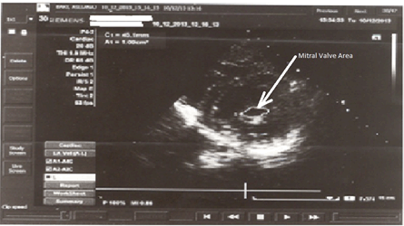
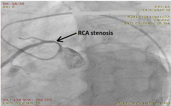
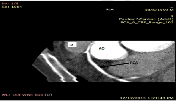
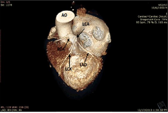
A 54-year-old male was admitted at the service of cardiology with the diagnosis of mitral stenosis. The patient complained of difficult breathing, chest pain and blue (cyanotic) fingers and lips during physical activities over the last 5–6 months. Twenty-five years ago he was diagnosed with mitral stenosis and five years ago as having a thrombotic cerebrovascular accident. The patient did not remember to have had any episode of rheumatic fever. At physical examination he was right hand monoparesis, irregular cardiac rhythm and an accentuated second heart tone at the mitral valve area. Blood pressure was 120/80 mmHg. Heart rate was 100 beats per minute. Electrocardiogram showed atrial fibrillation trans-thoracic echocardiography showed calcified mitral stenosis, with an anatomical area of 1.1 cm2, aortic regurgitation, left atrial dilatation and slight tricuspid valve regurgitation. Pulmonary artery systolic pressure was 30 mmHg (Figure 1). Blood tests were as follows: leukocytes 8700 cells/mm3, erythrocytes 5.610.000/mm3, hematocrit 45%, platelets 233,000/mm3, glucose 86 mg/dl, urea 30 mg/dl, creatinine 1.1 mg/dl, aspartate aminotransferase (AST) 29 UI/L, alanine-aminotransferase (ALT) 17 UI/L, total bilirubin 0.7 mg/dl, serum sodium 131 mmol/L, serum potassium 4.1 mmol/L, serum chlorine 97 mmol/L. The patient was treated with oral anticoagulants (acenocumarol), beta-blockers (atenolol), and diuretics (hydrochlorothiazide plus spironolactone). Coronary angiography which showed a 75% stenosis of the right coronary artery (Figure 2). The origin and course of right coronary artery was abnormal and thus a CT angiography was performed. The CT angiography confirmed that the origin of the right coronary artery was from the left coronary sinus and that the artery coursed between the aorta and the pulmonary artery (Figure 3) and (Figure 4). Under these circumstances, the patient was transferred to cardiac surgery where the mitral valve replacement and coronary artery bypass graft surgery (between aorta and right coronary artery) were performed. The in-hospital course was uneventful. The patient was free shortness of breath and chest pain (angina). One month later CT angiography was repeated. The patient remained symptom-free and in good health status. | ||||||
|
| ||||||
|
| ||||||
| ||||||
|
| ||||||
|
Discussion
| ||||||
|
Herein, we presented a rare case of a coronary artery anomaly associated with mitral stenosis. However, it remains unclear whether mitral stenosis was congenital or rheumatic in origin or whether there is any causal relationship between the coronary artery anomaly and mitral stenosis. Moreover, we did not find any reported case in which anomalies of the origin of right coronary artery were associated with mitral stenosis. The patients had dyspnea and chest pain. However, it remains unknown whether these symptoms were due to mitral stenosis or right coronary narrowing and subsequent myocardial ischemia (due to compression of the right coronary artery from aorta and pulmonary artery, particularly during effort) [1] [2]. Anatomic anomalies of coronary arteries include anomalies of origin, course, termination or their combination. The most common anomaly is the independent origin of the left anterior descending and left circumflex coronary arteries. Second is the anomalous origin of the left circumflex from the right coronary artery. The rarest anomaly is origin of the right coronary artery from the left coronary sinus. Coronary arteries are perpendicular to the aortic wall. Abnormal coronary arteries that get out ectopically usually course tangentially to the aortic wall and in close relationship to the aortic valve. Course between aorta and pulmonary artery may be associated with myocardial ischemia and sudden death. Although the reason for this variability is unknown, several mechanisms have been proposed:
This anomaly is associated with sudden death in young athletes during exercise. It is not known whether this pathology mediates an increased risk for early development of coronary artery disease. In our patient, the anomalous origin of the right coronary artery was not associated with atherosclerotic stenosis. The right coronary artery stenosis was mechanical (compressive) [1] [2] [3] [4] [5]. | ||||||
|
Conclusion
| ||||||
|
We presented here a very rare case of anomalies right coronary artery origin from the left aortic sinus coursing between aorta and pulmonary artery associated this with severe mitral stenosis. Although, the patient showed a 75% narrowing of the right coronary artery, the stenosis was not of atherosclerotic origin. The case highlights this anomaly and its potential association with mitral stenosis and non-atherosclerotic (compressive) narrowing of the right coronary artery. | ||||||
|
References
| ||||||
| ||||||

|
[HTML Abstract]
[PDF Full Text]
|
|
Author Contributions
Uliks Ekmekçiu – Substantial contributions to concept and design, Drafting the article, Revising it critically for important intellectual content, Final approval of the version to be published Mimoza Lezha – Substantial contributions to concept and design, Drafting the article, Revising it critically for important intellectual content, Final approval of the version to be published Gjin Ndrepepa – Substantial contributions to concept and design, Drafting the article, Revising it critically for important intellectual content, Final approval of the version to be published |
|
Guarantor of submission
The corresponding author is the guarantor of submission. |
|
Source of support
None |
|
Conflict of interest
Authors declare no conflict of interest. |
|
Copyright
© 2015 Uliks Ekmekçiu et al. This article is distributed under the terms of Creative Commons Attribution License which permits unrestricted use, distribution and reproduction in any medium provided the original author(s) and original publisher are properly credited. Please see the copyright policy on the journal website for more information. |
|
|