| |
|
|
|
Clinical Image
| ||||||
| Spontaneous subcutaneous orbital emphysema following nose blowing | ||||||
| Ozgur Tatli1, Faruk Ozsahin2, Selim Yurtsever3, Gurkan Altuntas4 | ||||||
|
1Physician, MD, Kanuni Training and Research Hospital, Department of Emergency Medicine, Trabzon, Turkey.
2Physician, MD, Giresun State Hospital, Department of Emergency Medicine, Giresun, Turkey. 3Physician, MD, Arhavi State Hospital, Department of Emergency Medicine, Artvin, Turkey. 4Research Assistant, MD, Kanuni Training and Research Hospital, Department of Emergency Medicine, Trabzon, Turkey. | ||||||
| ||||||
|
[HTML Abstract]
[PDF Full Text]
[Print This Article]
[Similar article in Pumed] [Similar article in Google Scholar] 
|
| How to cite this article |
| Tatli O, Ozsahin F, Yurtsever S, Altuntas G. Spontaneous subcutaneous orbital emphysema following nose blowing. Int J Case Rep Images 2015;6(9):603–605. |
|
Case Report
|
|
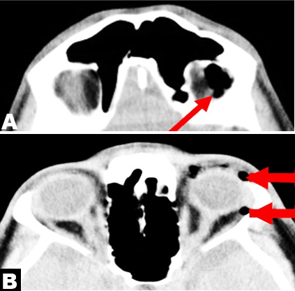
A 29-year-old male presented to the emergency department with sudden swelling and pain in the left eye. Pain accompanied by swelling had developed immediately after blowing his nose two days previously. On examination there was left periorbital swelling with crackling sound (crepitus) on palpation suggestive of subcutaneous orbital emphysema. The eye was painful at palpitation. Swelling was not accompanied by redness or elevated temperature. Bilateral ocular movements, pupillary reactions, funduscopic examination and visual acuity were normal. There was no diplopia or ptosis. The patient had no history of recent trauma to the head and penetrating the orbital area. Orbital computerized tomography revealed accumulations of air above the left lateral rectus muscle in the orbital ceiling and in the anterior palpebral area (Figure 1 A-B). There was no bone fracture. The sinuses were normal. Retro-orbital fat planes had a normal appearance. The patient was started on prophylactic antibiotherapy and kept under observation about 24 hours. As a prophylactic regimen cephalexin was started at a dosage of 500 mg every 12 hours for 5 days. The visual acuity in left eye was not dropped. Follow-up examinations showed no ophthalmological complications because of these normal findings, drainage and decompression were not performed. Swelling regressed entirely and clinical findings were normal by the 10th day of monitoring. |
|
|
|
Discussion
|
|
Orbital emphysema is a rare complication generally arising after trauma. Most cases involve an orbital bone fracture causing air ingress. It is a benign and temporary condition that usually develops in the first 24 h after paranasal sinus fractures, and spontaneous absorption generally takes place within two weeks [1]. Orbital emphysema is evaluated by means of palpation and radiological examination, particularly in patients with a history of trauma. Orbital fractures most frequently occur in the thinnest parts of the medial and inferior walls of the orbital bone, the ethmoid, maxillary and frontal sinuses, respectively [2]. The orbital bones are more flexible in children, and thus fracture less [3]. With paranasal sinus mucosa destruction, the fracture line has a valve-like effect and air enters the orbita [4]. Clinical symptoms such as swelling in the eye, closing of the eyelids, extraorbital subcutaneous emphysema, subconjunctival hemorrhage, sensitivity and pain may appear [5] . Rarely, occlusion of the central retina may develop with orbital compartment syndrome resulting from orbital emphysema. Air entering the orbital space may lead to occlusion and sight loss by creating a mass effect on the central retinal artery. Proptosis and diplopia may also occur. There are various sub-classifications within orbital emphysema. In genuine orbital emphysema, the air is localized behind the orbital septum whose integrity is not compromised immediately after orbital fracture [6]. Air can enter the orbital soft tissue during coughing, sneezing or nose blowing. There is no internationally agreed treatment modality for orbital emphysema. However, Hunts et al. published a study on the treatment of the condition in 1994 [7]. Patients should be advised not to blow their noses. Nasal decongestants, antibiotics and steroids may also be given [8]. If findings such as restricted ocular movement, disk edema or loss of sight occur in orbital emphysema, surgical procedures such as drainage and/or direct decompression may be performed. In diagnosis, the most important radiological imaging technique in determining fracture and the location of the air pockets is orbital tomography [9] . |
|
Conclusion
|
|
Diagnosis of orbital emphysema is generally made with accurate anamnesis, physical examination and orbital tomography. Orbital emphysema is a complication that may frequently arise following orbital traumas, though it should not be forgotten that it can also have non-traumatic causes. Orbital emphysema developing after nose blowing is one such rare condition. |
|
References
|
|
|
[HTML Abstract]
[PDF Full Text]
|
|
Author Contributions
Ozgur Tatli – Substantial contributions to conception and design, Acquisition of data, Revising it critically for important intellectual content, Final approval of the version to be published Faruk Ozsahin – Substantial contributions to conception and design, Acquisition of data, Revising it critically for important intellectual content, Final approval of the version to be published Selim Yurtsever – Substantial contributions to conception and design, Acquisition of data, Revising it critically for important intellectual content, Final approval of the version to be published Gurkan Altuntas – Substantial contributions to conception and design, Acquisition of data, Revising it critically for important intellectual content, Final approval of the version to be published |
|
Guarantor of submission
The corresponding author is the guarantor of submission. |
|
Source of support
None |
|
Conflict of interest
Authors declare no conflict of interest. |
|
Copyright
© 2015 Ozgur Tatli et al. This article is distributed under the terms of Creative Commons Attribution License which permits unrestricted use, distribution and reproduction in any medium provided the original author(s) and original publisher are properly credited. Please see the copyright policy on the journal website for more information. |
|
|