| |
|
|
|
Clinical Image
| ||||||
| Apocrine hidrocystoma of eye lid mimicking as sebaceous cell carcinoma | ||||||
| Undrakonda Vivekanand1, Heng Siang Ting2, Shankar Nag Vattipulusu3, BM Yashodhara4 | ||||||
|
1Professor, Department of Ophthalmology, Alluri Sitarama Raju Academy of Medical Sciences, Eluru, West Godavari District, 534005, Andhra Pradesh, India.
2House Officer, TuankuJa'afar Hospital, Seremban, Malaysia. 3Junior Resident, Department of Ophthalmology, Alluri Sitarama Raju Academy of Medical Sciences, Eluru, West Godavari District, 534005, Andhra Pradesh, India. 4Department of Medicine, Melaka Manipal Medical College, Melaka, Malaysia. | ||||||
| ||||||
|
[HTML Abstract]
[PDF Full Text]
[Print This Article]
[Similar article in Pumed] [Similar article in Google Scholar] 
|
| How to cite this article |
| Vivekanand U, Ting HS, Vattipulusu SN, Yashodhara BM. Apocrine hidrocystoma of eye lid mimicking as sebaceous cell carcinoma. Int J Case Rep Images 2015;6(9):600–602. |
|
Case Report
| ||||||
|
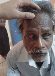
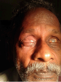
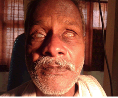
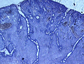
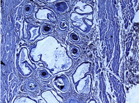
We report a case of a 58-years-old male who presented with a painless right upper eyelid swelling with mucoid discharge since two years. Ocular examination revealed phthisis bulbi in both eyes with nil perception of light. A nodular, reddish swelling of 1.5x2 cm was present in right eyelid involving middle one-third of upper lid margin (Figure 1). No such tumors were found in any other parts of the body of the patient. Wide excisional biopsy with Cutler-Beard lid reconstruction surgery was performed under local anesthesia (Figure 2) and (Figure 3). Histopathology (Figure 4A-B) revealed epidermis with underlying adnexa and basal incontinence of pigment, dermis showed multilocular cystic spaces lined by double layer secretory cells. Inner most cells were cuboidal with eosinophilic cytoplasm, outer layer cells were columnar suggestive of apocrine hidrocystoma of eyelid. | ||||||
| ||||||
| ||||||
| ||||||
| ||||||
|
| ||||||
|
| ||||||
|
Discussion
| ||||||
|
The eyelids are specialized structures of the ocular adnexa made up of various cells and tissues which can give to a variety of tumors of benign to malignant nature and inflammatory lesions. The management of each of these is variable. Papilloma, seborrheic keratosis, keratoacanthoma, inverted follicular keratosis, neurofibroma, pseudocarcinomatous hyperplasia and epidermal inclusion cyst are the common benign tumors. Basal cell carcinoma, squamous cell carcinoma, melanoma, sebaceous gland carcinoma and Merkel cell carcinoma are the malignant lesions of the eyelid. Tumors could also arise from sweat glands and some of them are: syringoma, eccrine hidrocystoma, apocrine hidrocystoma, syringocystadenoma papilliferum, pleomorphic adenoma and malignant sweat gland tumors. Likewise there are many tumors which could originate from vessels, hair follicles or sebaceous glands [1]. Apocrine hidrocystoma is a tumor originating from glands of Moll at the lid margin, usually solitary (sometimes multiple) cystic to nodular or papular lesions with bluish or black hue to iron deposit/lipofuscin/Tyndall effect in the cyst wall [2] [3]. Solitary tumors are treated by surgical excision. Recurrence of tumor following surgery is rare. Multiple lesions are treated with carbon dioxide laser and trichloroacetic acid or botulinum toxin. The differentials for this tumor are nevi, basal cell carcinoma, melanoma, milia and syringomyelia. The patient had a pinkish, fleshy lesion resembling sebaceous gland carcinoma which is a malignant lesion for which he underwent excision of tumor followed by Cutler-Beard reconstruction [4]. | ||||||
|
Conclusion
| ||||||
|
Apocrine hidrocystoma of eyelid may sometimes mimic as sebaceous cell carcinoma. Histopathology plays a crucial role in establishing a diagnosis. | ||||||
|
References
| ||||||
| ||||||
|
[HTML Abstract]
[PDF Full Text]
|
|
Author Contributions
Undrakonda Vivekanand – Substantial contributions to conception and design, Acquisition of data, Drafting the article, Final approval of version to be published Heng Siang Ting – Substantial contributions to conception and design, Acquisition of data, Drafting the article, Final approval of the version to be published Shankar Nag Vattipulusu – Substantial contributions to conception and design, Acquisition of data, Drafting the article, Final approval of the version to be published Yashodhara BM – Substantial contributions to conception and design, Acquisition of data, Drafting the article, Final approval of the version to be published |
|
Guarantor of submission
The corresponding author is the guarantor of submission. |
|
Source of support
None |
|
Conflict of interest
Authors declare no conflict of interest. |
|
Copyright
© 2015 Undrakonda Vivekanand et al. This article is distributed under the terms of Creative Commons Attribution License which permits unrestricted use, distribution and reproduction in any medium provided the original author(s) and original publisher are properly credited. Please see the copyright policy on the journal website for more information. |
|
|