| |
|
|
|
Case Report
| ||||||
| Rectosigmoid cancer recurrence surgically treated for bilateral pulmonary thromboembolism and liver metastases: A case report | ||||||
| Hiroshi Maekawa1, Hajime Orita2, Mutsumi Sakurada2, Tomoyuki Kushida2, Tomoaki Ito2, Koichi Sato2 | ||||||
|
1Assistant Professor, Department of Surgery, Juntendo University School of Medicine, Shizuoka Hospital, Izu-No-Kuni City, Shizuoka, Japan.
2Associate Professor, Department of Surgery, Juntendo University School of Medicine, Shizuoka Hospital, Izu-No-Kuni City, Shizuoka, Japan. | ||||||
| ||||||
|
[HTML Abstract]
[PDF Full Text]
[Print This Article]
[Similar article in Pumed] [Similar article in Google Scholar] 
|
| How to cite this article |
| Maekawa H, Orita H, Sakurada M, Kushida T, Ito T, Sato K. Rectosigmoid cancer recurrence surgically treated for bilateral pulmonary thromboembolism and liver metastases: A case report. Int J Case Rep Images 2015;6(8):502–506. |
|
Abstract
|
|
Introduction:
The survival time of recurrent cases of colorectal cancer has been prolonged by effective chemotherapy. However, side effects sometimes occur, which can be life-threatening condition.
Case Report: A 66-year-old female was admitted to our hospital complaining of dyspnea on effort and abdominal distension. Twenty-three months before admission, she had undergone anterior resection for rectosigmoid cancer. After anterior resection, adjuvant chemotherapy was performed. However, hepatic and pulmonary metastases were detected three months after starting adjuvant chemotherapy. She had been treated with chemotherapy for 22 months because of metastases before admission. Four months before admission, she had experienced dyspnea on effort caused of bilateral pulmonary embolism and the symptom had continued. Computed tomography scan showed two metastatic tumors in the liver, with diameters of 6.5 cm in S8 and 4.5 cm in S3, as well as bilateral pulmonary arterial embolism. From general condition, it was considered that surgical treatment could be tolerated. Bilateral pulmonary arterial endarterectomy and hepatic metastasectomy were performed, because the bilateral pulmonary thromboembolism and hepatic metastases were considered to be life-threatening. The postoperative course was uneventful and she was discharged from hospital on the 15th postoperative day. Conclusion: Synchronous endarterectomy and metastasectomy contributed to improve the patient's QOL. If this side effect is considered to be potentially fatal, the removal of the embolism is the treatment of choice. | |
|
Keywords:
Colorectal cancer, Recurrence, Chemotherapy, Pulmonary arterial thromboembolism
| |
|
Introduction
| ||||||
|
The survival time of recurrent cases of colorectal cancer has been prolonged by the progress of effective chemotherapy. Molecular-targeted therapies have also contributed to prolongation of the survival time. However, several side effects of chemotherapy and molecular-targeted therapy often occur, some of which are fatal. Thromboembolism occurs in approximately 8.3–20.7% of chemotherapy patients [1] [2]. This complication is also life-threatening. Here, we report a case of recurrence of rectal cancer surgically treated for bilateral pulmonary thromboembolism and hepatic metastases, which were both life-threatening conditions. In addition, the thromboembolism was supposed to be a side effect of chemotherapy. The surgical treatments contributed to improve the patient's quality of life. | ||||||
|
Case Report
| ||||||
|
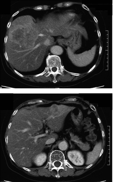
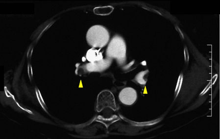
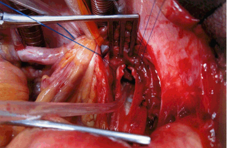
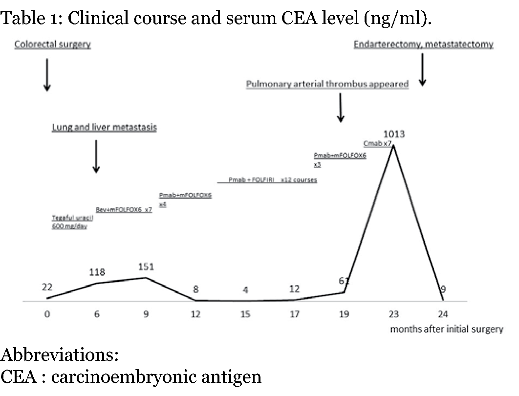
A 66-year-old female was admitted to our hospital because of dyspnea on effort and abdominal distension. Twenty-three months before admission, she had undergone anterior resection for rectosigmoid cancer. Pathological examination demonstrated that the lesion was T3N0M0 stage IIA. Adjuvant chemotherapy was performed with tegafur uracil per oral administration. Three months after starting chemotherapy, the serum level of carcinoembryonic antigen (CEA) was elevated to 118 ng/ml (the preoperative serum level of CEA was 22 ng/ml). CT examination was performed and revealed hepatic metastasis 2 cm in diameter (S8) and right pulmonary metastasis less than 1 cm in diameter. The patient was treated with bevacizumab (Bev)+mFOLFOX6 therapy. After seven courses of Bev+mFOLFOX6 administration, the serum level of CEA was elevated to 151 ng/ml. CT scan demonstrated two hepatic metastases of 3 cm in S8 and 1 cm in S3, and a tumor of 1.5 cm in lower segment of the right lung. Then, she was treated with another regimen, panitumumab (Pmab)+FOLFIRI therapy. After starting the Pmab+FOLFIRI, the serum level of CEA gradually decreased, but sometimes leukocytopenia occurred, so the dosages of the chemotherapeutic agents were decreased. After nine courses of Pmab+FOLFIRI therapy, the serum level of CEA was decreased to 12 ng/ml. The patient had grade 2 neurotoxicity. After 12 courses of Pmab+FOLFIRI therapy, CEA was increased to 61 ng/ml, although the liver and lung metastases were stable. Subsequently, then the patient experienced dyspnea on effort; CT scan revealed emboli in the right pulmonary artery, but we could not confirm their existence. The chemotherapy regimen was changed from Pmab+FOLFIRI therapy to cetuximab (Cmab) alone, but grade 3 leukocytopenia prevented us from completing the chemotherapy. After 22 months of the anterior resection, liver metastases were enlarged to 6.5 cm (S8) and 4.5 cm (S3) (Figure 1). CT also demonstrated bilateral pulmonary arterial embolism and two right pulmonary metastases 1.5 cm and 1 cm in sized (Figure 2) Both liver metastases and pulmonary arterial embolism were considered to be life-threatening, although the lung metastases were small and not immediately life-threatening. Her cardiovascular, lung, and renal function was good. Indocyanine green residual rate (0.5 mg/kg of body weight) at 15 minutes was 19.5%. We could not rule out the pulmonary arterial embolism were tumor embolism. Finally, she had treated with chemotherapy for 22 months. And 23 months after initial surgery, bilateral pulmonary arterial endarterectomy and hepatic metastasectomy were performed simultaneously (Figure 3). Pathological examination revealed that the embolism was due to a thrombus composed of fibrin. The postoperative course was uneventful and the patient was discharged home on the 15th postoperative day (Figure 4). The patient started to be administered warfarin, and two months later, pulmonary metastasectomy was successfully performed. She never experienced dyspnea on effort and could walk up stairs. However, she could not continue the chemotherapy further because of grade 2 neurotoxicity and leukocytopenia. The patient complained of dyspnea for cancer pleuritis at 6 months after the removal of pulmonary embolism and hepatic metastases. She died after seven months of operation. However, she could survive in a good condition for several months. | ||||||
| ||||||
| ||||||
| ||||||
| ||||||
|
Discussion
| ||||||
|
The metastasis of colorectal cancer is indicated for surgical treatment if curative resection is possible. However, metastatic cases are not surgically treated, because metastases are often seen in several sites or including several organs. Chemotherapy is often chosen for the treatment of metastatic cases of colorectal cancer. The progress of chemotherapy has contributed to prolongation of the life span. However, adverse events caused by chemotherapy sometimes prevent the continuation of effective chemotherapy. Thromboembolism is one of the severe adverse events caused by chemotherapy, and it sometimes requires intensive medication. Thromboembolism is a complication in approximately 8.3–20.7% of colorectal cancer cases [1] [2]. In addition, its frequency is elevated under chemotherapy [3]. It has been reported that the addition of anti-VEGF to chemotherapy increases the risk of thromboembolism [4]. Venous thromboembolic cases upon the addition of Bev to chemotherapy also have been reported. However, anti-EGFR, another molecular-targeted agent, may increase the risk of thrombosis because such thromboembolic cases have been reported [5] [6] [7] [8]. Heit et al. previously described that surgery, trauma, malignant neoplasm, chemotherapy, neurologic disease with paresis, central venous catheter or pacemaker, varicose veins, and superficial vein thrombosis were indicated as risk factors for deep vein thrombosis and embolism [9]. Our case had some of these factors namely, malignant neoplasm, central venous catheter insertion, and chemotherapy. Therefore, we should pay attention to the possibility of thrombosis when chemotherapy is performed and the patient complains of dyspnea on effort. This is because pulmonary embolism is one of the conditions associated with deep vein thrombosis and is sometimes life-threatening. Symptoms of pulmonary embolism are exertional dyspnea and exercise intolerance. These symptoms are often seen in patients treated with chemotherapy, especially in an anemic condition. In our case, the patient complained of exercise intolerance and this symptom became worse. We should have recognized that the patient was suffering from pulmonary thromboembolism at that time. If we had analyzed the serum level of d-dimer, we could have detected pulmonary thromboembolism earlier. Pulmonary thromboembolism may not be an indication for aggressive treatment, if the patient is in the life-terminal condition of malignancy and the embolism is not considered to be life-threatening, because active cancer is one of the persistent risk factors of recurrent venous thromboembolism [10]. However, in our case, bilateral pulmonary embolism was considered to be a life-threatening condition, and urgent treatment was necessary. Liver metastases were also considered to be severe conditions that were resistant to chemotherapy. We could not find previous reports of both pulmonary endarterectomy and metastasectomy being performed for patients with cancer recurrence in literature. We performed surgical treatments for both life-threatening diseases simultaneously. The patient could survive in a good condition for several months after surgical treatment without dyspnea. The surgical treatment for pulmonary arterial thromboembolism and hepatic metastases was actually very stressful for the patient. However, if the patient can tolerate surgical treatment, it can provide a good quality of life. In our case, the surgical treatment for pulmonary arterial thromboembolism and hepatic metastases was considered to have contributed to an improved quality of life. | ||||||
|
Conclusion
| ||||||
|
Synchronous endarterectomy and metastasectomy contributed to improve the patient's quality of life. If this side effect is considered to be potentially fatal, the removal of the embolism is the treatment of choice. | ||||||
|
References
| ||||||
| ||||||
|
[HTML Abstract]
[PDF Full Text]
|
|
Author Contributions
Hiroshi Maekawa – Conception and design, Acquisition of data, Analysis and interpretation of data, Drafting the article, Critical revision of the article, Final approval of the version to be published Hajime Orita – Conception and design, Acquisition of data, Analysis and interpretation of data, Drafting the article, Critical revision of the article, Final approval of the version to be published Mutsumi Sakurada – Conception and design, Acquisition of data, Analysis and interpretation of data, Drafting the article, Critical revision of the article, Final approval of the version to be published Tomoyuki Kushida – Conception and design, Acquisition of data, Analysis and interpretation of data, Drafting the article, Critical revision of the article, Final approval of the version to be published Tomoaki Ito – Conception and design, Acquisition of data, Analysis and interpretation of data, Drafting the article, Critical revision of the article, Final approval of the version to be published Koichi Sato – Conception and design, Acquisition of data, Analysis and interpretation of data, Drafting the article, Critical revision of the article, Final approval of the version to be published |
|
Guarantor of submission
The corresponding author is the guarantor of submission. |
|
Source of support
None |
|
Conflict of interest
Authors declare no conflict of interest. |
|
Copyright
© 2015 Hiroshi Maekawa et al. This article is distributed under the terms of Creative Commons Attribution License which permits unrestricted use, distribution and reproduction in any medium provided the original author(s) and original publisher are properly credited. Please see the copyright policy on the journal website for more information. |
|
|
|
About The Authors
| |||
| |||
| |||
| |||
| |||
| |||
| |||