| |
|
|
|
Case Report
| ||||||
| Rupture of a parietal brain abscess | ||||||
| Kaissar Farah1, Kevin Beccaria1, Nacer Zakaria Mansouri1, Thomas Graillon1, Henry Dufour1, Stéphane Fuentes1 | ||||||
|
1Assistance publique des Hôpitaux de Marseille, Department of Neurosurgery, Hospital La Timone 264 rue Saint Pierre, Marseille CEDEX 05, France.
| ||||||
| ||||||
|
[HTML Abstract]
[PDF Full Text]
[Print This Article]
[Similar article in Pumed] [Similar article in Google Scholar] 
|
| How to cite this article |
| Farah K, Beccaria K, Mansouri NZ, Graillon T, Dufour H, Fuentes S. Rupture of a parietal brain abscess Int J Case Rep Images 2015;6(8):493–497. |
|
Abstract
|
|
Introduction:
Ipsilateral hydrocephalus is an unusual complication of brain abscess. It is caused by the rupture of the abscess in the ventricle or thrombosis of the plexus choroid.
Case Report: We report here the case of a 65-year-old male treated from a complicated acute unilateral hydrocephalus secondary to the formation of inflammatory veils and septa. The hydrocephalus was successfully managed by endoscopic technique under neuronavigation. Conclusion: The strategy adopted in our case is safe, effective, simple and lead to little to no complications. After a review of literature, we discuss the pathological and management aspects of this rare entity. | |
|
Keywords:
Brain abscess, Hydrocephalus, Temporal Horn entrapment, Ventricle
| |
|
Introduction
| ||||||
|
Brain abscess is a serious, life-threatening condition. Despite the prompt use of modern antibiotics in its management, it still represents a potential cause of mortality (5–23%) in literature [1] [2] [3] [4] [5]. Presentation symptoms are non specific, and many are due to edema surrounding the lesion. Most are due to increased intracranial pressure (headache, nausea/vomiting, lethargy). Hemiparesis and seizures develop in 30–50% of cases. Symptoms tend to progress more rapidly than neoplasms. The most feared complications of cerebral abscess are herniation secondary to mass effect, intraventricular rupture of brain abscess, hemorrhage into the abscess or hemispheric infarction due to internal carotid artery thrombosis [6]. Unilateral acute hydrocephalus also represents an exceptional complication of cerebral abscess' complications. Few cases about this specific complication are reported in literature [7]. We describe the case of a patient treated from a complicated acute unilateral hydrocephalus secondary to cerebral abscess, successfully managed by endoscopic technique. | ||||||
|
Case Report
| ||||||
|
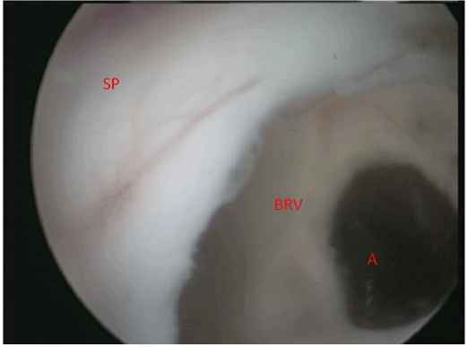
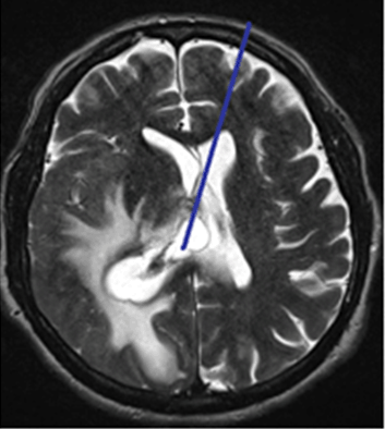
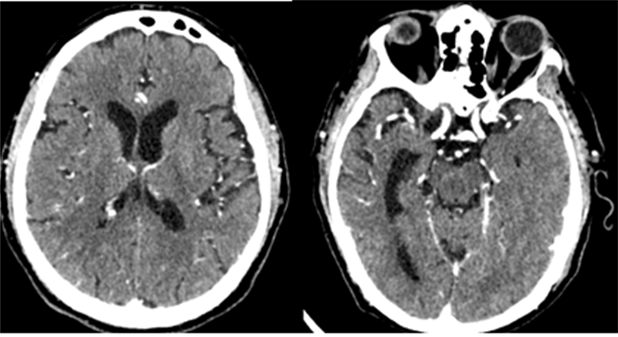
A 65-year-old male was initially admitted in the department for management of left hemiparesis and headaches, and was found to have a right parietal lesion. The rapid onsets of the symptoms during the week preceding the admission lead us to suspect a brain abscess. At admission, the patient presented with normal consciousness, left hemiplegia and left hemianopia, little concordant with initial brain MRI scan. Neither diffusion nor ADC sequences specific to intracranial infection was performed on the first MRI scan. Our initial hypothesis was high grade glioma. Unfortunately, neurological deterioration with disturbance of consciousness was observed within few hours. We decided then to perform a new MRI scan with diffusion and ADC sequences, which found a typical image of a right parietal brain abscess ruptured into the lateral right ventricle (Figure 1). Cerebral abscess brain drainage was urgently decided and performed. A 25 cm3 of foul-smelling pus was aspirated with a trocar. Partial excision of the brain abscess capsule was made. Multiple bacteriological samples were sent to the laboratory. Empirical antibiotic therapy by cefotaxim and metronidazole was initially prescribed and maintained for a total of six weeks after verification of bacterial samples. Microbiological tests found oral cavity flora: Streptococcus intermedius and Parvimonas micra. A cardiac transthoracic ultrasound was performed, eliminating infective endocarditis. Under antibiotic therapy, and introduction of corticosteroids two days after the initial surgery, we noticed improvement in general state, with absence of hyperthermia within 4 days. Through his hospitalization, the patient remarked improvement of his left hemiplegia, with reduced strength left hand motricity and beginning of motor recuperation of superior and inferior limbs. The muscle strength was rated 4/5. Biological inflammatory syndrome was normalized within 10 days; the C-reactive protein was 3 mg/L. A specialized dental examination and dental panoramic tomography revealed bad orodental state with six teeth to extract. The patient was then transferred to the neurological department for further treatment. Eleven weeks after the first surgery, the patient was re-admitted in our department for acute left hemiplegia. The CT scan and MRI scan found a right unilateral hydrocephalus with ipsilateral temporal horn entrapment secondary to secular partition of brain cerebral abscesses (Figure 2). Initial clinical examination reported confusion with left hemiplegia and left homonymous hemianopia. Operative Technique The operation was carried out with the patient in a semi-sitting position under general anesthesia. A left frontal burr-hole was made contralateral to the enlarged temporal horn and the endoscope with its stylet was introduced into the normal left frontal horn. The stylet was then removed and the optical system was inserted to visualize the ventricle. By gently moving the endoscope under neuronavigation control, a window was created in the septum pellucidum (Figure 3). The endoscope was passed through the communication and the contralateral enlarged temporal horn was explored; the cannulation of the compressed right temporal horn was performed successfully through the right atrium. No bleeding was observed. Then the endoscope was withdrawn from the fenestration, allowing immediate backflow of cerebrospinal fluid (CSF) (Figure 4). Clinical regression of the left hemiplegia with quasi-total motor recuperation was observed within few hours. The 24 hour control CT scan found a collapsed right temporal horn. The patient was retransferred to the reeducation center for further cares. At one month, systematic cerebral CT scan found significant decrease of the enlargement of the right temporal horn (Figure 5). Six months after the second operation, total motor recuperation was observed. The patient was able to walk with a cane. The initial left hemianopia recovered. | ||||||
|
| ||||||
|
| ||||||
|
| ||||||
|
| ||||||
| ||||||
|
Discussion
| ||||||
|
Ipsilateral hydrocephalus is an unusual complication of brain abscess [6]. The two main explanations are: plexus choroid thrombosis [8] or the formation of inflammatory septa [9]. The entity that we describe here was previously defined by Cairns [10]. It consists of obstruction of the atrium of one lateral ventricle so that the trapped or isolated temporal horn containing choroid plexus expands into a cyst. This gives rise to the symptoms of raised intracranial pressure as well as appropriate features of focal cerebral dysfunction such as homonymous hemianopia and contralateral hemiparesis. When the infection involves the ventricles, the patient may develop a number of thin veils or septa within the ventricles separating them into a number of compartments. These septa may progress after the infection has been overcome. They consist of thin translucent veils of tissue which are most often situated in the lateral ventricles just behind the foramen of Monro, though there may be multiple septa throughout the ventricular system. Their pathogenesis is uncertain but it has been suggested that they arise from tufts of glial tissue which grow out from areas of the ventricular wall which have been denuded of ependyma by the preceding ventriculitis. This so-called compartmentalized or multiloculated hydrocephalus is difficult to treat [11]. The two primary treatments of monoventricular hydrocephalus are CSF shunts or a neuroendoscopic approach [12]. Neuroendoscopy is a very useful and less invasive technique for treating many types of hydrocephalus, including isolated dilatation of one lateral ventricle or horn [13]. Due to the infectious environment, CSF shunts seems less appropriate, knowing that many complications might be encountered [14] [15] [16] [17]. | ||||||
|
Conclusion
| ||||||
|
Monoventricular hydrocephalus secondary to brain abscess are exceptional in literature. They are the consequence of either choroid plexus thrombosis or formation of inflammatory septa. Neuroendoscopic management is considered the "Gold Standard" in the treatment of obstructive hydrocephalus. The strategy adopted in our case is safe, effective, simple and lead to little to no complications. | ||||||
|
References
| ||||||
| ||||||
|
[HTML Abstract]
[PDF Full Text]
|
|
Author Contributions
Kaissar Farah – Substantial contributions to conception and design, Acquisition of data, Analysis and interpretation of data, Drafting the article, Revising it critically for important intellectual content, Final approval of the version to be published Kevin Beccaria – Substantial contributions to conception and design, Acquisition of data, Analysis and interpretation of data, Drafting the article, Revising it critically for important intellectual content, Final approval of the version to be published Nacer Zakaria Mansouri – Analysis and interpretation of data, Revising it critically for important intellectual content, Final approval of the version to be published Thomas Graillon – Analysis and interpretation of data, Revising it critically for important intellectual content, Final approval of the version to be published Henry Dufour – Analysis and interpretation of data, Revising it critically for important intellectual content, Final approval of the version to be published Stéphane Fuentes – Analysis and interpretation of data, Revising it critically for important intellectual content, Final approval of the version to be published |
|
Guarantor of submission
The corresponding author is the guarantor of submission. |
|
Source of support
None |
|
Conflict of interest
Authors declare no conflict of interest. |
|
Copyright
© 2015 Kaissar Farah et al. This article is distributed under the terms of Creative Commons Attribution License which permits unrestricted use, distribution and reproduction in any medium provided the original author(s) and original publisher are properly credited. Please see the copyright policy on the journal website for more information. |
|
|