  |

|
 |
|
Case In Images
| ||||||
| Hemangiopericytoma: A rare case report | ||||||
| Sonali Deshmukh1, Ketki P. Kalele1, Kaustubh P. Patil2, Abhishek Singh Nayyar3 | ||||||
|
1Department of Oral and Maxillofacial Pathology and Microbiology, V.Y.W.S Dental College & Hospital, Amravati, Maharashtra, India.
2Department of Periodontics and Oral Implantology, Dr. D.Y. Patil Dental College & Hospital, Pune, Maharashtra, India. 3Department of Oral Medicine and Radiology, Saraswati-Dhanwantari Dental College and Hospital and Postgraduate Research Institute, Parbhani, Maharashtra, India. | ||||||
| ||||||
|
[HTML Abstract]
[PDF Full Text]
[Print This Article]
[Similar article in Pumed] [Similar article in Google Scholar] 
|


| How to cite this article |
| Deshmukh S, Kalele KP, Patil KP, Nayyar AS. Hemangiopericytoma: A rare case report. Int J Case Rep Images 2015;6(8):517–522 |
|
Abstract
|
|
Introduction:
Hemangiopericytoma is a rare neoplasm which was first described by Stout and Murray in 1942 as a vascular tumor derived from the pericytes. They account for 2–3% of all soft tissue sarcomas in humans and they occur mainly in the musculoskeletal system. 15–30% of all hemangiopericytoma occur in the head and neck region. Only 5% are located in the sinonasal region, where they display a more benign behavior than in other parts of the body.
Case Report: Herein, we are presenting an extremely rare case report of hemangiopericytoma in a 32-year-old male with a brief overview regarding its epidemiology, macro- and microscopical characteristics, the clinicopathological findings and the treatment of this extremely rare vascular neoplasm. Conclusion: Conclusion rests with the inclusion of the possibility of such rare vascular tumors in the possible differential diagnoses of the various peripheral soft tissue lesions. An early diagnosis thus helps in the effective management of such lesions without further bringing changes on the subjacent and adjacent structures and tissues including bone. | |
|
Keywords:
Hemangiopericytoma, Pericytes, Vascular neoplasm
| |
|
Introduction
| ||||||
|
Hemangiopericytoma is a rare neoplasm which was first described by Stout and Murray in 1942 as a vascular tumor derived from the pericytes. These tumors most often show benign biologic behavior, however, very rarely, they may also present with malignant characteristics. These tumors are very rare in their occurrence with only 2–3% of all soft tissue neoplasms being comprised of hemangiopericytoma. Moreover, of all the reported hemangiopericytoma, about 15–30% occur in the head and neck region. 5% of these tumors are located in the sinonasal region where they possess a less aggressive behavior in comparison with tumors occurring elsewhere in the body [1]. Hemangiopericytoma can develop anywhere in the body wherever pericytes are present. The tumor holds specific significance due to its potential for malignant behavior. Some of the known sites for tumor development are the pelvic region, retroperitoneum and along the extremities, however, primary bone localization is not very common. Tumor manifestation signs usually depend on tumor's location, size and potential for malignancy [2]. Oral hemangiopericytoma are fast growing tumors, with a characteristic bluish red color. There is no age or gender predilection for these tumors. However, the lesions are rare reported before 20 years and after 70 years of age. Their consistency varies from being soft to rubbery and most commonly, the lesions are not associated with pain. The lesions are also often well-delineated from the surrounding mucosa. Chromosomal translocations t (12;19) and t (13;22) have been observed in lesional cells [2]. Tumors seen in perivascular areas especially deep seated tumors in muscles are larger tumors. The smaller tumors are usually benign. In contrast, the larger lesions are more aggressive and are feared for their malignant behavior, thereby, requiring more aggressive treatment with wide resection followed by postoperative radio and chemotherapies [3]. This case report describes a very rare case of hemangiopericytoma located in the oral cavity and also takes a glance on relevant literature about the same. | ||||||
|
Case Report
| ||||||
|
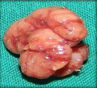
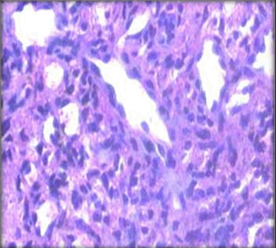
A 32-year-old male patient visited the department of oral medicine and radiology/outpatient department of our College with a chief complaint of swelling in relation to the lower right back tooth region in the lingual aspect since eight months. The history revealed that the swelling had started insidiously, not preceded by trauma, which was initially smaller in size and steadily increased in size since its onset. Patient had experienced mild discomfort causing difficulty during speech and mastication and it was not associated with any history of pus discharge. Patient was operated for the same swelling along with extraction of grossly carious 44 in a private set-up 3 months back. Lesion recurred and patient visited the same clinician again for the similar complaint. Medical, family, and personal histories were not relevant. General physical examination revealed no abnormalities. Extraoral examination revealed no gross asymmetry. No regional lymphadenopathy was evident. Intraoral examination revealed a solitary, painless, sessile, oval swelling in the mandibular lingual sulcus, measuring about 2x3 cm in its greatest dimensions, extending anteroposteriorly from the mesial aspect of 43 till the mesial aspect of 45 and superoinferiorly from the occlusal surface of teeth in the affected region till the attached gingivae in the same quadrant. The overlying mucosa was red in color. It was firm in consistency with well defined borders, lobulated in shape and smooth in texture (Figure 1). On palpation, it was fixed to the underlying gingiva and it slightly bled on probing. Clinical differential diagnosis included fibroma, peripheral giant cell granuloma and pyogenic granuloma. Radiological investigations revealed no distinct radiological characteristics. Routine hematological investigations revealed normal values. Under aseptic conditions, the lesion was surgically excised and the excised specimen was submitted for histopathological analysis (Figure 2). The Hematoxylin and Eosin stained sections showed parakeratinized stratified squamous epithelium with corrugated surface layer and underlying connective tissue stroma (Figure 3). The underlying connective tissue stroma showed lesional tissue which was highly cellular in nature with numerous vascular spaces lined by endothelial cells (Figure 4). Irregular branching of vascular spaces revealed characteristic 'staghorn' pattern (Figure 5). Surrounding these vascular spaces, there were proliferations of tightly packed oval and spindle cells showing indistinct cytoplasmic borders with moderate amount of cytoplasm and hyperchromatic nuclei (Figure 6). The histopathological impression was suggestive of hemangiopericytoma. The patient was followed-up for around one year and no recurrence was reported. | ||||||
|
| ||||||
| ||||||
| ||||||
| ||||||
| ||||||
|
| ||||||
|
| ||||||
|
| ||||||
|
| ||||||
|
Discussion
| ||||||
|
Hemangiopericytoma is a rare neoplasm that was originally described as a vascular tumor derived from the pericytes. Clinically, the lesion may be either sessile or pedunculated, and may demonstrate a surface lobularity with a telangiectatic appearance. Intraosseous cases have also been reported [2][3]. Hemangiopericytomas have been reported to occur in all age groups however, majority (40%) of the cases occur in the fifth and sixth decades [3] [4]. Hemangiopericytomas do not have any specific radiological characteristics. They may be either lytic or may represent focal sclerosis. They may also show a honeycomb or reticular pattern. Tumors may also cause cortical erosion, which alarms the presence of a malignancy. Along with routine radiography, even CT scan and MRI are not contributory in diagnosis. However, they are beneficial to differentiate benign tumors from those which are malignant. They are although also significant to determine the extent of the tumor. Angiography of the tumor reveals spider-like, radially branching vessels [3]. Histology of these tumors is although very characteristic. It depicts numerous vascular channels with plump endothelial cells. The stroma surrounding the vascular channels consists of proliferation of tightly packed ovoid and spindle shaped cells with hyperchromatic nuclei and a moderate amount of cytoplasm. The cells do not have distinct cytoplasmic borders. The tumor cells are seen surrounding the proliferating vascular channels. The irregular vascular channels show branches of varying sizes that give the characteristic 'staghorn' appearance to the histology of these tumors. Older lesions are less aggressive and they have less cellularity in comparison to the younger lesions. They may possess a large mucoid interstitial appearance which can appear as the one in mucoid type of lipoma or that of liposarcoma of myxoid type. Rarely, these tumors may also show focal areas of cartilage production and in such cases, have to be cautiously differentiated from mesenchymal chondrosarcomas [1] [4]. On gross examination, hemangiopericytomas appear grayish-white in appearance and may present as well circumscribed lesions with a lesser tendency to bleed unlike other common vascular tumors. The consistency varies from being solid to spongy, friable or granular. One variant of this tumor is congenital hemangiopericytoma which is also known as infantile hemangiopericytoma. Oropharyngeal mucosa is the predominant location for this very rare lesion. This tumor variant most often is present at birth, occurs more than one in number, and at various sites along the mucosa. It shows faster growth rate after birth. Recurrence rate is high for this variant of the tumor, although they are less prone to develop metastasis as is reflected in literature [4]. The histogenesis of hemangiopericytomas revolves around pericytes. Pericytes are a normal component of blood capillaries. These are contractile cells that are zipped around the endothelium of small blood vessels all over the vasculature. Thus, hemangiopericytomas can occur anywhere in the body. Rouget cells or mural cells are other names for pericytes. They are found embedded in the basement membrane and provide communication with the endothelial cells of blood vessels. There is dual mode of communication both via direct physical contact and via paracrine signaling. Pericytes help in regulation of blood flow along the capillary walls and are also involved in washing out of cellular waste by the process of phagocytosis. They stabilize and monitor the maturation of endothelial cells. Apart from their role along the capillary bed, pericytes are also a significant component of the neurovascular unit, which includes endothelial cells, astrocytes and neurons [2]. Pericytes and endothelial cells lie in close proximity to each other and thereby it is difficult to identify both of these cell types. However, these two cell types can easily be differentiated from one another on the basis of cytomorphology of the cell species with pericytes having prominent round nuclei compared to the flat elongated nuclei of the endothelial cells [3]. Ultrastructurally, pericytes have numerous finger - projections on their surface that are wounded around the capillary walls and which help in directing the blood flow. There is a co-ordination between the endothelial cells and pericytes and this balance is highly controlled by the numerous signaling pathways which function in an autocrine and paracrine manner [2] [4]. Pericytes lead to endothelial cell proliferation and their stabilization while endothelial cells, in turn, stimulate the precursor cells leading to pericytes' proliferation. Immunohistochemical analysis is of great help in distinguishing between hemangiopericytomas and the various vascular tumors from the closely mimicking connective tissue tumors. Molecular markers that are traditionally employed for establishing a definitive diagnosis in such cases include CD31, CD34, CD68 along with vimentin and also, certain cytokeratins. These markers however specifically used in the diagnosis of connective tissue tumors also aid in the recognition of mutated stem cells that surround the blood vessels [5]. Hemangiopericytomas can show varied biological behavior right from being a slow growing mass to a growth with an aggressive growth having characteristics of a malignancy. Sometimes, its behavior lies in between these two and such type of hemangiopericytoma is known to have borderline or intermediate type of behavior. Examination of abnormal/atypical mitoses, cellularity, pleomorphism, tendency to bleed and necrosis of the tumor to determine the malignant behavior in a study showed no signs of recurrence or metastasis even after prolonged periods [5] [6]. However, in contrast, Enzinger and Smith found that majority of the cases in their study developed recurrences at locations nearby the primary tumor before metastasizing. The most preferable organs for metastasis of this tumor were the lungs. Hemangiopericytomas, specially the malignant ones, are very notorious and in some cases, recurrence and also, metastasis develops after many years of treatment. Thus, it is recommended by a number of studies that patient should be kept essentially under a long-term follow-up even after radical and extensive resection of the tumor [6] [7]. The treatment of hemangiopericytomas lies primarily with wide base resection. Underestimating this entity is risky because of its wide behavior range; thus, a generous resection with extended wide margins becomes unavoidable. Similar procedure was advocated in our case. Adjuvant therapies such as radiation therapy are most often used in cases which are inoperable or which have reappeared after optimal treatment or for palliative care. Chemotherapy, however, is not routinely employed and its efficacy in the treatment is still not known [8] [9] [10]. The differential diagnosis of this lesion includes a broad spectrum of lesions ranging from fibroma, pyogenic granuloma, peripheral giant cell granuloma and fibrous histiocytoma to malignancies such as malignant fibrous histiocytoma and synovial sarcoma. Certain other rarer stromal sarcomas such as fibrosarcoma, mesenchymal chondrosarcoma, vascular leiomyoma and juvenile hemangioma are also considered in the differential diagnoses [11]. Fibrous histiocytoma shows a storiform or cartwheel pattern and a less prominent vascular network. Synovial sarcoma may show a biphasic cellular pattern and include fibrosarcoma-like areas. Mesenchymal chondrosarcoma cells are smaller than those of a hemangiopericytoma, and well-defined islands of cartilage are present [12][13]. Reticulin staining can be helpful in such cases to arrive-at a definitive diagnosis of hemangiopericytomas as it is a special stain that demonstrates lesional vessels lined by a single layer of endothelial cells with the pericytes covering the basement membrane of blood vessel (Figure 7) [13] [14] [15]. | ||||||
| ||||||
|
| ||||||
|
Conclusion
| ||||||
|
Conclusion rests with the inclusion of the possibility of such rare vascular tumors in the possible differential diagnoses of the various peripheral soft tissue lesions. An early diagnosis thus helps in the effective management of such lesions without further bringing changes on the subjacent and adjacent structures and tissues including bone. Also, histopathology of hemangiopericytomas plays a crucial role as the treatment of this lesion is dependent on the amount of cellular atypia and mitotic activity present in the lesion. The more bland lesions with minimal mitotic activity are treated by a wide base excision while the more active and dysplastic lesions are treated by radical excisions with or without adjunctive radio- and chemotherapies. | ||||||
|
References
| ||||||
| ||||||


|
[HTML Abstract]
[PDF Full Text]
|
|
Author Contributions
Sonali Deshmukh – Substantial contributions to conception and design, Acquisition of data, Analysis and interpretation of data, Drafting the article, Revising it critically for important intellectual content, Final approval of the version to be published Ketki P. Kalele – Analysis and interpretation of data, Revising it critically for important intellectual content, Final approval of the version to be published Kaustubh P. Patil – Analysis and interpretation of data, Revising it critically for important intellectual content, Final approval of the version to be published Abhishek Singh Nayyar – Analysis and interpretation of data, Revising it critically for important intellectual content, Final approval of the version to be published |
|
Guarantor of submission
The corresponding author is the guarantor of submission. |
|
Source of support
None |
|
Conflict of interest
Authors declare no conflict of interest. |
|
Copyright
© 2015 Sonali Deshmukh et al. This article is distributed under the terms of Creative Commons Attribution License which permits unrestricted use, distribution and reproduction in any medium provided the original author(s) and original publisher are properly credited. Please see the copyright policy on the journal website for more information. |
|
|