| |
|
|
|
Case In Images
| ||||||
| Late presentation of right aortic arch with large left sided Kommerell diverticulum | ||||||
| Shalini Koppisetty1, Giorgios Bis2, Amr E. Abbas3, Ravneet K. Dhillon4 | ||||||
|
1Research Assistant, Cardiology, Beaumont Health System, Royal Oak, Michigan, USA.
2Transitional Year, Radiology, Oakwood Hospital, Dearborn, Michigan, USA. 3Section Head, Echocardiography, Beaumont Health System, Royal Oak, Michigan, USA. 3Physician, Emergency Medicine, Henry Ford Hospital, West Bloomfield, Michigan, USA. | ||||||
| ||||||
|
[HTML Abstract]
[PDF Full Text]
[Print This Article]
[Similar article in Pumed] [Similar article in Google Scholar] 
|
| How to cite this article |
| Koppisetty S, Bis G, Abbas AE, Dhillon RK. Late presentation of right aortic arch with large left sided Kommerell diverticulum. Int J Case Rep Images 2015;6(8):511–516. |
|
Abstract
|
|
Introduction:
Kommerell diverticulum (KD), a rare embryological variant, is an aneurysm of aortic arch at the origin of an aberrant subclavian artery (ASA). It is usually asymptomatic and detected incidentally on imaging studies or postmortem. Common symptoms include dysphagia (dysphagia lusoria) and chest pain. The most common complications include rupture due to an enlarged diverticulum. Surgical correction of the diverticulum and repair of aorta and aberrant subclavian artery is the treatment of choice.
Case Report: A case of Kommerell diverticulum in a 68-year-old female with right aortic arch with large left sided KD was found incidentally on imaging studies. Conclusion: Subtle findings on a chest X-ray may help in diagnosis of KD. Vascular anomalies such as KD should be considered as differential diagnosis in patients with chronic cough or dysphagia when other causes have been ruled out. | |
|
Keywords:
Aberrant subclavian artery, Carotid body tumor, Dysphagia lusoria, Kommerell diverticulum, Right sided aortic arch
| |
|
Introduction
| ||||||
|
Anomalous origin of the right subclavian artery was first described in 1794 by David Bayford. He discovered it incidentally in a postmortem study of 62 years old patient who had a history of dysphagia. He then gave the term lusus naturae which means dysphagia by freak of nature. In 1936, Burchard Kommerell described an aortic diverticulum for the first time in a living patient [1]. Burchard described KD as saccular aneurysmal dilatation of aorta arch at the origin of an aberrant subclavian artery [2] [3]. | ||||||
|
Case Report
| ||||||
|
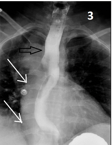
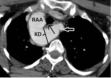
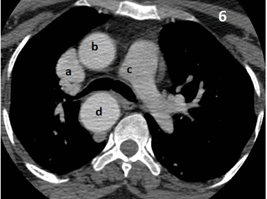
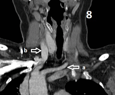
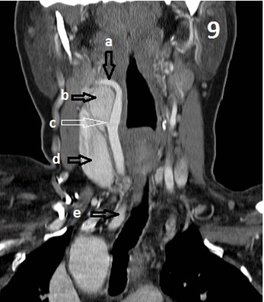
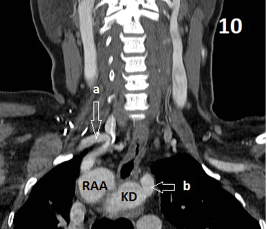
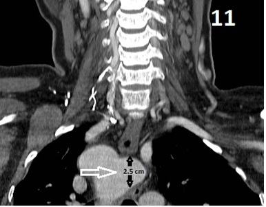
A 68-year-old female with history of hypertension (HTN), hyperlipidemia, diabetes mellitus (DM) presented to the emergency department with chief complaints of chest pain and sore throat of four days duration. Initially, she had intermittent chest pain for about two weeks, which was sharp, radiating to upper back and went away without any treatment. She also had complained of a long standing history of intermittent shortness of breath and dysphagia. She had no relief of symptoms and presented to the emergency department. Initial electrocardiogram (EKG) in the emergency department showed straightening of and biphasic T waves in anterior leads (V1, V3, V4, and V5) and T wave inversions on repeat EKG. Patient was admitted to the hospital for further management. During hospitalization her troponins continued to rise in the hospital resulting in cardiac catheterization which showed significant mid left anterior descending (LAD) stenosis, which was subsequently stented. Subsequent workup consisted of chest X ray, barium esophagram and a computed tomography (CT) scan of the neck. Chest X-ray showed prominent right aortic arch and slight deviation of trachea to left side (Figure 1) (Figure 2). Barium studies showed significant narrowing of esophagus and indentation of right lateral border of esophagus (Figure 3) (Figure 4). CT scan of soft tissue of neck revealed enhancing mass within carotid space concern for carotid body tumor, and right aortic arch with ALSA consistent with KD causing narrowing of the airway and esophagus at the thoracic inlet (Figure 5) (Figure 6) (Figure 7) (Figure 8) (Figure 9) (Figure 10) (Figure 11). The differential diagnosis considered were non-ST elevation myocardial infarction (NSTEMI), Streptococcal pharyngitis, right aortic arch with ALSA demonstrating a diverticulum of Kommerell with a diameter of 2.5 cm, and neoplasm of carotid body. The patient was treated as NSTEMI with aspirin, heparin and underwent cardiac catheterization. Coronary angiogram revealed obstruction of left anterior descending artery (>95%) and underwent successful percutaneous coronary transluminal angioplasty and placement of bare metal stent in LAD. A course of cephalexin was prescribed for her pharyngitis. The patient was discharged on regimen of atorvastatin, aspirin, toprol, lisinopril, and clopidogrel. The patient was referred to vascular surgeon for KD but refused surgical intervention at this point. | ||||||
| ||||||
| ||||||
|
| ||||||
| ||||||
|
| ||||||
|
| ||||||
|
| ||||||
|
| ||||||
|
| ||||||
|
| ||||||
| ||||||
|
Discussion
| ||||||
|
Kommerell diverticulum is a rare congenital aortic arch anomaly with an incidence of 0.5–2.0% in normal population. There are two variants that are commonly described in literature. Left-sided aortic arch with aberrant right subclavian artery (ARSA prevalence 0.5–2.0%) or right aortic arch with aberrant left subclavian artery (prevalence 0.05–0.1%) [3]. Kommerell diverticulum occurs in a number of anomalies of aortic arch system. An aberrant subclavian artery may be located posterior to esophagus (80%), runs between esophagus and trachea (15%), anterior to trachea (5%) [4]. A right-sided aortic arch is associated with deletion of 22q11 chromosome with 24% incidence [5]. Most of the patients with KD are asymptomatic, found incidentally on imaging studies of the chest. Common symptoms of KD include dysphagia, cough, chest pain and stridor due to extrinsic compression of esophagus or tracheobronchial tree by the KD or dilatation of an aberrant subclavian artery [1] [3]. Dysphagia is the most common symptom with KD, termed dysphagia lusoria [6]. Complications of KD include atherosclerosis, embolism, rupture and dissection [7]. The most valuable diagnostic studies include barium swallow esophagram, CT scan and cardiac magnetic resonance imaging (MRI) scan which demonstrate a diverticulum of Kommerell and compression of esophagus or trachea. Standard surgical treatment has not been established being a rare entity. Treatment is surgical resection of the diverticulum regardless of the size because of risk of rupture (19%) which is associated with high mortality [8]. The mean size associated with rupture was 5.8 cm [5]. | ||||||
|
Conclusion
| ||||||
|
Kommerell diverticulum (KD) is a rare cause of dysphagia with distinct radiological features. Subtle findings on a chest X-ray may help in the diagnosis. Vascular anomalies such as KD should be considered as differential diagnosis in patients with chronic cough or dysphagia when other causes have been ruled out. | ||||||
|
References
| ||||||
| ||||||
|
[HTML Abstract]
[PDF Full Text]
|
|
Author Contributions
Shalini Koppisetty – Substantial contribution to conception and design, Acquisition of data, Analysis and interpretation of data, Drafting of the article, Critical revision of the article, Final approval of the version to be published Giorgios Bis – Substantial contribution to conception and design, Acquisition of data, Analysis and interpretation of data, Drafting of the article, Critical revision of the article, Final approval of the version to be published Amr E. Abbas – Substantial contribution to conception and design, Acquisition of data, Analysis and interpretation of data, Drafting of the article, Critical revision of the article, Final approval of the version to be published Ravneet K. Dhillon – Substantial contribution to conception and design, Acquisition of data, Analysis and interpretation of data, Drafting of the article, Critical revision of the article, Final approval of the version to be published |
|
Guarantor of submission
The corresponding author is the guarantor of submission. |
|
Source of support
None |
|
Conflict of interest
Authors declare no conflict of interest. |
|
Copyright
© 2015 Shalini Koppisetty et al. This article is distributed under the terms of Creative Commons Attribution License which permits unrestricted use, distribution and reproduction in any medium provided the original author(s) and original publisher are properly credited. Please see the copyright policy on the journal website for more information. |
|
|