  |

|
 |
|
Case Report
| ||||||
| Primary hydatid cyst of gallbladder: A case report | ||||||
| Pronoy Kabiraj1, Shib Shankar Kuiri2, Utpal De3 | ||||||
|
1MS, Postgraduate Trainee, Department of Surgery, Bankura Sammilani Medical Colege, Kenduadih, Bankura, West Bengal, India.
2MS, Clinical Tutor, Department of Surgery, Bankura Sammilani Medical Colege, Kenduadih, Bankura, West Bengal, India. 3MS, Professor, Department of Surgery, Bankura Sammilani Medical Colege, Kenduadih, Bankura, West Bengal, India. | ||||||
| ||||||
|
[HTML Abstract]
[PDF Full Text]
[Print This Article]
[Similar article in Pumed] [Similar article in Google Scholar] 
|


| How to cite this article |
| Kabiraj P, Kuiri SS, De U. Primary hydatid cyst of gallbladder: A case report. Int J Case Rep Images 2015;6(7):440–443. |
|
Abstract
|
|
Introduction:
Hydatid cyst is a common clinical entity in India. Liver and lungs are the common organs involved. No organ is immune to infection.
Case Report: We report a case of primary hydatid cyst of gallbladder presenting as a gallbladder lump. Conclusion: In endemic regions hydatid cyst should be considered in patients presenting with gallbladder lump. | |
|
Keywords:
Echinococcus granulosus, Echinococcus multilocularis, Gallbladder, Hydatid cyst, Liver, Lungs
| |
|
Introduction
| ||||||
|
Hydatid cyst in humans is an accidental dead end infestation caused by Echinococcus granulosus. It is endemic in sheep rearing countries. In India, Jammu and Kashmir is an endemic region though reports of hydatid disease has been reported from almost whole of the Indian subcontinent [1]. Liver (75%) and lungs (15%) are common sites of infection in humans [1] [2]. Primary extrahepatic intra-abdominal hydatid cyst is a rare clinical entity. This article reports and reviews a case of primary gallbladder hydatid (Table 1). | ||||||
| ||||||
|
| ||||||
|
Case Report
| ||||||
|
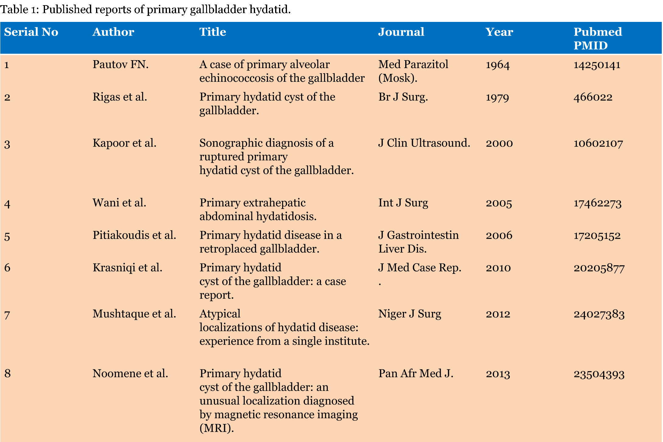
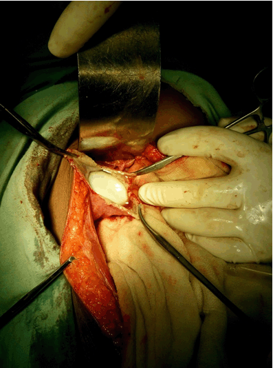
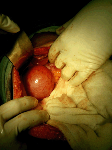
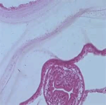
A 16-year-old female patient admitted suffering from intermittent mild pain in the right hypochondrium and epigastrium associated with nausea since six months. There was no history of fever or jaundice. Physical examination revealed mild tenderness in the right upper quadrant of the abdomen. On deep palpation a globular lump (4 cmx3 cm), moving with respiration was noted in the right hypochondrium. The upper margin of the lump was impalpable while the other margins were well defined. Murphy's sign was positive. Base line hematological examination was unremarkable. Plain abdominal X-ray was normal. The ultrasound examination showed that her gallbladder was distended with a localised thickening of its wall. A single cystic lesion was noted inside the gallbladder lumen giving it an impression of double gallbladder. There was no image of gallstone. An abdominal CT scan was performed showing inflammatory gallbladder wall with a cyst within cyst appearance (Figure 1). Hepatic parenchyma and other abdominal organs proved absolutely normal. Anti-Echinococcus antibodies were not found in serum. The diagnosis of primary hydatid cyst of gallbladder was made and surgery was decided. The patient underwent right subcostal laparotomy. Intraoperatively, the gallbladder was found to be tense and hugely distended (Figure 2). The wall appeared oedematous and inflamed. After proper precaution the gallbladder wall was opened (Figure 3). A single cyst with germinal layer was delivered intact. No other cysts were found in the liver and peritoneal cavity. Cholecystectomy was performed. It permitted a total removal of the cyst without rupture. A peroperative cholangiography searching daughter cysts in the common bile duct proved unremarkable. The patient's postoperative course was uneventful and she was discharged on tenth postoperative day after stitch removal. The patient was put on albendazole (10 mg/kg) in divided doses. The histopathology confirmed the presence of hydatid cyst (Figure 4). At sixth month follow-up, the patient was well and had no recurrence of hydatid disease. | ||||||
| ||||||
| ||||||
| ||||||
| ||||||
|
Discussion
| ||||||
|
Human hydatosis is a zoonosis caused byEchinococcus multilocularis and Echinococcus multilocularis (malignant hydatid). Echinococcus multilocularis predominates in humans. Cattles (sheep, goat, dogs) are primary (definitive, sexual cycle) host. Human beings are secondary dead end (asexual cycle) host [1][2] [3] [4] [5]. Human infection is caused by fecal-oral route or ingestion of contaminated vegetables or meat [1] [2] [3] [4] [5] [6] [7] [8] [9]. Cystic transformation of Echinococcus multilocularis in human organs results in varied symptoms. No organ is immune to echinococcal infection [5]. Liver (first filter) and Lungs (second filter) are the commonest organs involved worldwide. In children, due to unknown cause lung infection (41–70%) is more common than liver (42–48%) [5] [6] [7]. Other organs are involved secondary to pulmonary or hepatic involvement. Among the unusual primary sites, in order of frequency, central nervous system (4–5%), musculoskeletal system (0.5–4%) and cardiovascular system (0.5–2%) are involved [2] [5] [6]. The incidence of primary extrahepatic intra abdominal hydatid cyst is 6.5% [1] [2] [3] [4] [5] [6] [7] [8] [9]. Spleen (2–3%) is the predominant organ involved [1] [3] [4] [5]. Gallbladder is primarily involved in 0.6%. GB cysts may be located intraluminally or on the surface [1] [2] [3] [4] [5] [6] [7] [8] [9]. As a rule luminal cysts are single and parietal cysts are multiple containing daughter cyst [2] [5]. Luminal cyst may attain enormous size before they are symptomatic due to distensile capacity of the gallbladder. On the contrary parietal cysts produce early symptoms. The pathogenesis of gallbladder infection is not well documented. The hypothetical routes of spread include portal circulation, biliary, lymphatic and peritoneum [1] [2] [3] [4] [5] [6] [7] [8] [9]. Univariate presentation of gallbladder hydatid include acute cholecystitis with cholangitis, gallbladder lump, obstructive jaundice or peritonitis with shock due to rupture. A combination of the above symptoms is not infrequent. Gallbladder hydatid with daughter cysts in the common duct producing obstructive symptoms may give rise to a false positive Courvoisier's law [2] [5] [6] [7] [8] [9]. So in endemic areas this should be included in the differential diagnosis of patients with obstructive jaundice. Abdominal sonography is highly sensitive and specific for diagnosing hydatid cyst but accurate extrahepatic organ localization is at times doubtful [5] [8] [9]. Computed tomography, and magnetic resonance imaging, are adjuncts to sonography for definitive diagnosis. Serological (ELISA) and hematological (eosinophilia) tests may clinch the diagnosis along with radiology [4] [6] [7] [8] [9]. Cystoprostatectomy (cholecystectomy) is the treatment of choice [2] [3] [4] [5] [8] [9]. Apart from the usual per-operative precautions gentle handling of the gallbladder is of utmost importance. An intraluminal rupture may result in spillage into common bile duct. Manual handling instead of instrumental (application of Moynihan's clamps) holding of the gallbladder during dissection is warranted [1] [7]. Cystic duct should be identified early and stay suture passed in order to prevent accidental spillage of contents into CBD in the event of rupture of cyst. Careful ligation of cystic duct and artery together with removal of gallbladder from liver bed along with a portion of the liver tissue prevents rupture and spillage. Postoperative treatment with albendazole (10–15 mg/kg) in divided doses should be continued for a period of four months to prevent recurrence [1] [2] [3] [4] [5] [6] [7] [8] [9]. | ||||||
|
Conclusion
| ||||||
|
In endemic countries, hydatid cyst should be considered in the differential diagnosis in patients presenting with gallbladder lump. | ||||||
|
References
| ||||||
| ||||||

|
[HTML Abstract]
[PDF Full Text]
|
|
Author Contributions
Pronoy Kabiraj – Substantial contributions to conception and design, Acquisition of data, Analysis and interpretation of data, Revising it critically for important intellectual content, Final approval of the version to be published Shib Shankar Kuiri – Substantial contributions to conception and design, Drafting the article, Critical revision of the article, Final approval of the version to be published Utpal De – Substantial contributions to conception and design, Revising it critically for important intellectual content, Final approval of the version to be published |
|
Guarantor of submission
The corresponding author is the guarantor of submission. |
|
Source of support
None |
|
Conflict of interest
Authors declare no conflict of interest. |
|
Copyright
© 2015 Pronoy Kabiraj et al. This article is distributed under the terms of Creative Commons Attribution License which permits unrestricted use, distribution and reproduction in any medium provided the original author(s) and original publisher are properly credited. Please see the copyright policy on the journal website for more information. |
|
|