|

|
 |
|
Case Report
| ||||||
| Single coronary artery arising from the right coronary sinus with mid-left anterior descending artery segment courses through the ventricular myocardium: A rare entity | ||||||
| Andrea Romagnoli1, Irene Coco1, Dominique De Vivo1, Eros Calabria1, Giovanni Simonetti1 | ||||||
|
1MD,Department of Diagnostic Imaging, Molecular Imaging, Interventional Radiology and Radiotherapy, Tor Vergata University Hospital Foundation, University of Rome Tor Vergata, Italy.
| ||||||
| ||||||
|
[HTML Abstract]
[PDF Full Text]
[Print This Article]
[Similar article in Pumed] [Similar article in Google Scholar] 
|


| How to cite this article |
| Romagnoli A, Coco I, De Vivo D, Calabria E, Simonetti G. Single coronary artery arising from the right coronary sinus with mid-left anterior descending artery segment courses through the ventricular myocardium: A rare entity. Int J Case Rep Images 2015;6(7):431–435. |
|
Abstract
|
|
Introduction:
Congenital anomalies of the coronary artery have an incidence of 1% and isolated single coronary artery without other congenital cardiac anomalies has an approximate incidence of 0.024–0.066% in general population. This rare entity can be diagnosed incidentally during life, however may lead to symptomatic or asymptomatic myocardial infarction and sudden heart attack, even among young athletes. The prognosis varies according to the anatomic distribution, associated atheromatous disease and associated vascular anomalies.
Case Report: A 45 years old male with presentation of a single coronary artery from the right coronary sinus with subsequent coursing between the aorta and pulmonary trunk arteries and mid anterior descending artery segment course through the myocardium. Patient presented with non-typical angina symptoms, normal resting electrocardiograph, myocardial perfusion scintigraphy suspect to reduced left ventricular blood flow but negative angiographic evaluation. Conclusion: Thin bridges cannot be demonstrable angiography, so anatomic and panoramic CT-evaluation give more information especially if associated with functional evaluation. Knowledge of physiology, normal and variant anatomy, is most important in managing congenital and acquired disease, and variation in coronary arterial patterning is frequent. | |
|
Keywords:
Coronary artery anomaly, Cardiac anomalies, Heart attack, Isolated single coronary artery, Myocardial bridge, Ventricular myocardium
| |
|
Introduction
|
|
Congenital anomalies of the coronary artery have an incidence of 1% and isolated single coronary artery without other congenital cardiac anomalies has an approximate incidence of 0.024–0.066% in general population. This rare entity can be diagnosed incidentally during life, however may lead to symptomatic or asymptomatic myocardial infarction and sudden heart attack, even among young athletes [1]. The prognosis varies according to the anatomic distribution, associated atheromatous disease and associated vascular anomalies. |
|
Case Report
|
|
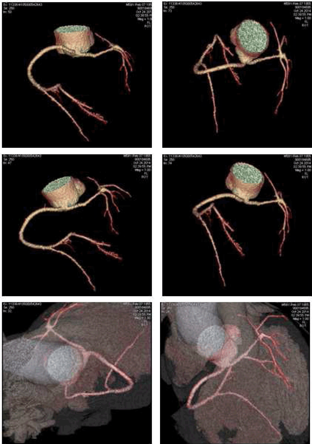
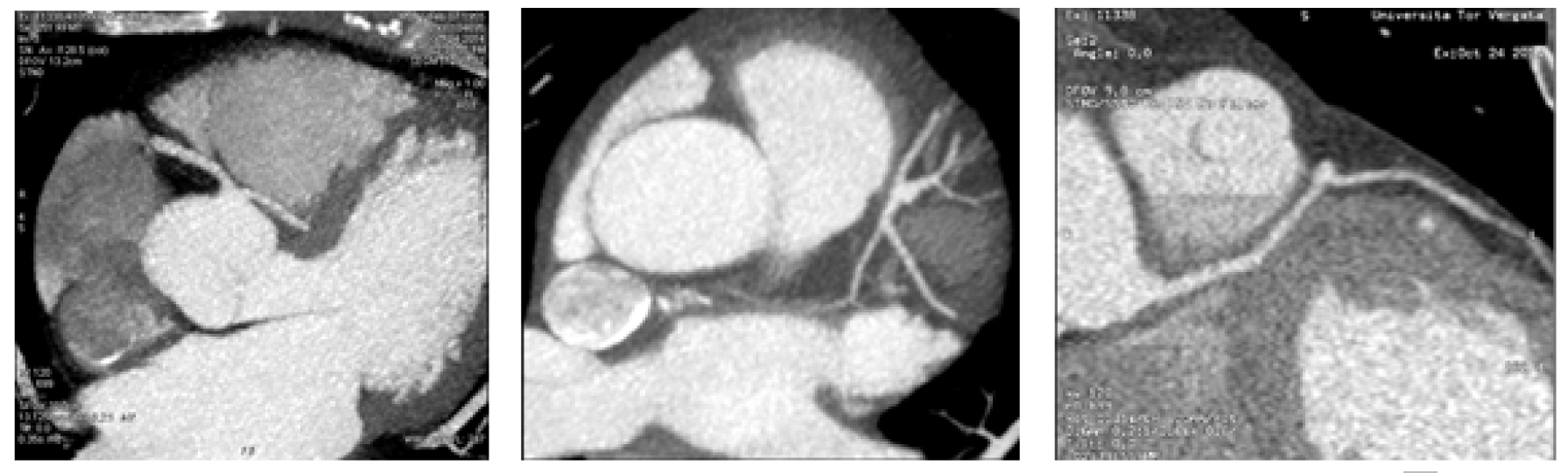
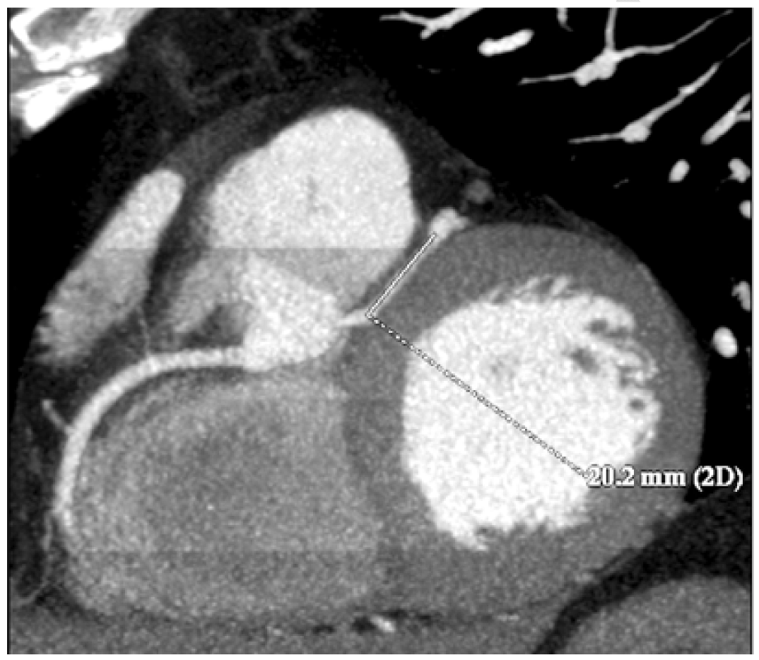
A 45-year-old male was admitted in our department with symptoms characterized by intermittent substernal chest discomfort non-provoked by exertion or emotional stress enlarging for over four months. The patient was a smoker of 7–8 packs of cigarettes per week for 10 years. Clinical history was negative for hypertension, diabetes mellitus, hyperlipidemia or family history of premature vascular disease and no risk factors for pulmonary embolism or respiratory distress. Physical examination showed normal blood pressure (BP: 128/88 mmHg), heart rate of 73 beats/min, no signs of vascular disease and normal cardiac examination. Initial laboratory tests showed no particular anomalies with normal blood levels of glucose and cholesterol (LDL: 103 mg/dl; HDL: 35 mg/dl; TG: 198 mg/dl). Resting electrocardiogram (ECG) showed normal sinus rhythm at 82 beats/min without anomalies in stress examination. Several echocardiographic examinations revealed a moderate enlargement of the left atrium; an apical form of left ventricular hypertrophy; a preserved left ventricular (LV) systolic function (LV ejection fraction, 57%); and moderate mitral valve regurgitation, with no abnormality of the mitral valve apparatus or of LV wall motion. Three months ago the patient had undergone coronary angiography evaluation which demonstrated an anomalous vascular anatomy with absence of coronary ostium in left cusp of the aortic valve and a single coronary artery (SCA) with origin by the right coronary cusp with one and only common trunk (CT) which gives rise to right coronary artery (RCA), left anterior descending (LAD) and diagonal artery, circumflex branch (CB) and the proximal left marginal artery (LMA). The RCA gives off the posterior descending artery (PDA) and the postero-lateral branch (PL). Significant flow-limiting stenosis was not detected with regular caliper representation of the three main coronary branches up to their distal segments. Contrast ventriculography confirmed the apical LVH and showed no wall motion abnormality. In our department, the patient underwent CT scanning, which confirmed the anomalous origin and course of coronary vessels showing also an inter-arterial course of the CT between the pulmonary artery and the aorta (Figure 1) (Figure 2). A 2-cm segment of the proximal-middle tract of the common trunk was embedded within the inter-ventricular septum, following an intramural course with a relative reduction of tunneled vessel (Figure 3). No significant atheromatous alterations have been highlighted, if we except for some parietal irregularities at LAD. Dipyridamole Tl-201 SPECT confirmed a perfusion abnormalities with reversible perfusion defects in the mid anteroseptal wall without an apical abnormality. The patient was admitted to cardiology center for further evaluation, treatment and periodical follow-up. |
|
|
|
|
|
|
|
Discussion
|
|
Isolated SCA without other congenital cardiac anomalies is very rare among the different variations of anomalous coronary patterns occurring in approximately 0.024% of the population [1]. Origin of the LCA in the right coronary sinus has been described at a frequency of 0.02% in autopsy series and from 0.05–0.19% in angiographic series [2]. The LCA either has a common ostium with the RCA, or arises independently to the ostium of RCA. These cases are classified according to the course of LCA into four categories: (1) inter-truncal or inter-atrial, between the aorta and pulmonary arteries; (2) anterior, in front of the right ventricular outflow; (3) posterior or retro-aortic, behind the aorta; (4) inter truncal-septal or trans-septal, through the supraventricular crest and inter-ventricular septum. The retro-aortic course of the LCA is an uncommon entity. This anomaly is serious and associated with sudden cardiac death and myocardial infarction, if anomalous LCA passes between the aorta and the pulmonary artery [3] [4]. The anterior as well as the posterior course have been considered to be clinically insignificant [4]. However, there have been isolated reports of ischemia or sudden death associated with the retro-aortic course of LMCA or one of its branches [5]. Although most patients with the anomalous LCA arising from the right sinus of Valsalva are asymptomatic, the therapeutic approach must be individualized in each subject. In asymptomatic subjects, the age of the patient and the type of anomalous artery should be carefully evaluated [6]. Usually, typical angina does not occur with SCA in the absence of coexisting coronary artery disease or aortic stenosis [1]. In our case, the patient's symptoms could be attributable to the intramyocardial segment of LAD. The myocardial bridge, occurs when the artery coursing within the myocardium, presents compression to the contraction of the heart muscle to systole, which is clinically silent most of the time [6]. A deepened critical analysis of many autopsy samples was first presented by Geiringer et al. in 1951: clinical interest and systematic research were triggered by the observation of myocardial bridging right along with myocardial ischemia [7]. The rate of angiographic bridging is <5%, linked to thin bridges that provide a light compression. Carrying out provocation tests in subjects presenting normal angiographic coronary arteries may enhance the systolic myocardial compression and could thereby demonstrate myocardial bridges in =40% of cases [8]. Myocardial bridges are preferably localized in the middle segment of the LAD [7]. One of the parallel LAD branches frequently keeps an intramural course [7]. Diagonal and marginal branches may be respectively involved in 18% and 40% of cases [6]. By the angiographic side, myocardial bridges are almost exclusively spotted in the LAD. They set at 1 to 10 mm depth showing typical length around 10 to 30 mm [7]. Ferreira et al. distinguished two types of bridging: (1) superficial bridges (75% of cases) crossing the artery in perpendicular way or taking an acute angle towards the apex, and (2) muscle bundles arising from the right ventricular apical trabeculae (25% of cases) that transversely, obliquely, or helically cross the LAD before they flow in the interventricular septum. Arterial segments could also be set in a deep interventricular gully. The segment proximal to the bridge frequently, because of hemodynamic forces, shows atherosclerotic plaque formation, although the tunneled segment is typically spared [9]. Neither low value proximal to the bridge stenosis, nor systolic compression of the tunneled segment are allowed by the way to explain a severe ischemia and its related symptoms. When the arterial occlusion was limited to the only systole, phasic coronary blood flow and distal coronary pressure were observed in a considerable delay, contributing to a smaller myocardial oxygen consumption and to the increase of the coronary sinus lactate concentration. Angina, myocardial ischemia, myocardial infarction, left ventricular dysfunction, myocardial stunning, paroxysmal AV blockade, as well as exercise-induced ventricular tachycardia and sudden cardiac death are reported as sequelae of the myocardial bridging.However, following the prevalence of myocardial bridging, these complications are rare. Patients may complain atypical or angina-like chest pain with unsure association between the severity of these symptoms and length/depth values of the tunneled segment, or the systolic compression degree [6]. The up-to-date gold standard for diagnosing myocardial bridges remains the coronary angiography presenting the typical "milking effect" and a "step down-step up" phenomenon induced by the systolic compression of the tunneled segment. By the way, these signs give insufficient information about the functional impact on myocardium. Proximal stenosis and myocardial bridging could only be identified by carrying out a percutaneous transluminal coronary angioplasty, because higher intravascular pressure values and reversed hypokinesis can reveal the myocardial bridging [10]. |
|
Conclusion
|
|
Coronary angiography evaluation has demonstrated an anomalous vascular anatomy with absence of coronary ostium in left cusp of the aortic valve. Computed tomography (CT) scanning has confirmed the anomalous origin and course of coronary vessels showing also an inter-arterial course of the CT between the pulmonary artery and the aorta. A 2-cm segment of the proximal-middle tract of the CT was embedded within the inter-ventricular septum, following an intramural course with a relative reduction of tunneled vessel. Thin bridges can be not demonstrable angiography, so anatomic and panoramic CT-evaluation give more information especially if associated with functional evaluation. Knowledge of physiology, normal and variant anatomy, is most important in managing congenital and acquired disease, and variation in coronary arterial patterning is frequent. |
|
References
|
|

|
[HTML Abstract]
[PDF Full Text]
|
|
Author Contributions
Andrea Romagnoli – Substantial contributions to conception and design, Revising it critically for important intellectual content, Final approval of the version to be published Irene Coco – Acquisition of data, Drafting the article, Final approval of the version to be published Dominique De Vivo – Acquisition of data, Revising it critically for important intellectual content, Final approval of the version to be published Eros Calabria – Analysis and interpretation of data, Drafting the article, Final approval of the version to be published Giovanni Simonetti – Substantial contributions to conception and design, Revising it critically for important intellectual content, Final approval of the version to be published |
|
Guarantor of submission
The corresponding author is the guarantor of submission. |
|
Source of support
None |
|
Conflict of interest
Authors declare no conflict of interest. |
|
Copyright
© 2015 Andrea Romagnoli et al. This article is distributed under the terms of Creative Commons Attribution License which permits unrestricted use, distribution and reproduction in any medium provided the original author(s) and original publisher are properly credited. Please see the copyright policy on the journal website for more information. |
|
|
|
About The Authors
| |||
| |||
| |||
| |||
| |||
| |||