| |
|
|
|
Case Report
| ||||||
| Coincident retroperitoneal and sigmoid colon liposarcoma: A rare occurrence | ||||||
| Ayvaz Ulaş Urganci1, Erkan Oymaci1, Enver Vardar3, Ebru Akincilar1, Ömer Engin1 | ||||||
|
1MD, Department of General Surgery, Izmir Buca Goverrment Hospital, Turkey.
2MD, Department of Gastroenterology Surgery, Izmir Bozyaka Training and Reserch Hospital, Turkey. 3As. Prof. and Clinic Chief, Department of Pathology, Izmir Bozyaka Training and Reserch Hospital, Turkey. | ||||||
| ||||||
|
[HTML Abstract]
[PDF Full Text]
[Print This Article]
[Similar article in Pumed] [Similar article in Google Scholar] 
|
| How to cite this article |
| Urganci AU, Oymaci E, Vardar E, Akincilar E, Engin Ö. Coincident retroperitoneal and sigmoid colon liposarcoma: A rare occurrence. Int J Case Rep Images 2015;6(7):411–415. |
|
Abstract
|
|
Introduction:
Retroperitoneal liposarcomas are rare malignancies. There are a limited number of liposarcoma cases in gastrointestinal system in literature. There is no known etiological factor in the pathogenesis of liposarcomas yet. The treatment is total resection.
Case Report: A 59-year-old male patient detected synchronous liposarcoma both in retroperitoneum and in colon and treated with en block resection. We discussed our case with review of literature. Conclusion: We think that even though it could not be detected in preoperative examinations, this rare togetherness should be considered intraoperatively. | |
|
Keywords:
Colon liposarcoma, Gastric liposarcoma, Retroperitoneal liposarcoma
| |
|
Introduction
| ||||||
|
Soft tissue sarcomas are less than 1% among all types of cancer [1]. One-third of malignant tumors originating from retroperitoneum are sarcomas and liposarcomas are the most frequently observed retroperitoneal sarcoma types [2]. Fifteen percent of adult soft tissue sarcomas are located in retroperitoneum. Forty-one percent of retroperitoneal sarcomas are liposarcomas and malignant tumors with mesenchymal origin [2]. Moreover, liposarcomas with colon origin are quite rare and there are limited numbers of cases reported in literature [3]. There is no known etiological factor in the pathogenesis of liposarcomas yet. There are five types of liposarcomas: well differentiated, myxoid, round cell, pleomorphic, and undifferentiated, out of these types pleomorphic and undifferentiated types have poorer prognosis due to high recurrence and metastasis rates [1] [4]. Retroperitoneal liposarcomas usually grows slowly without manifesting any symptoms, and they form a gross abdominal mass [1] [2]. Their treatments consist of en block R0 (absence of residual tumor) resection. Adjuvant treatment modalities are controversial [2] [5]. The complication risks of these tumors may be reduced by multidisciplinary approach, careful diagnosis and treatment approach; and survival rates and life qualities of the patients may be improved. In this study, we aimed to present a case of rare retroperitoneal sarcoma and synchronous colon liposarcoma. | ||||||
|
Case Report
| ||||||
|
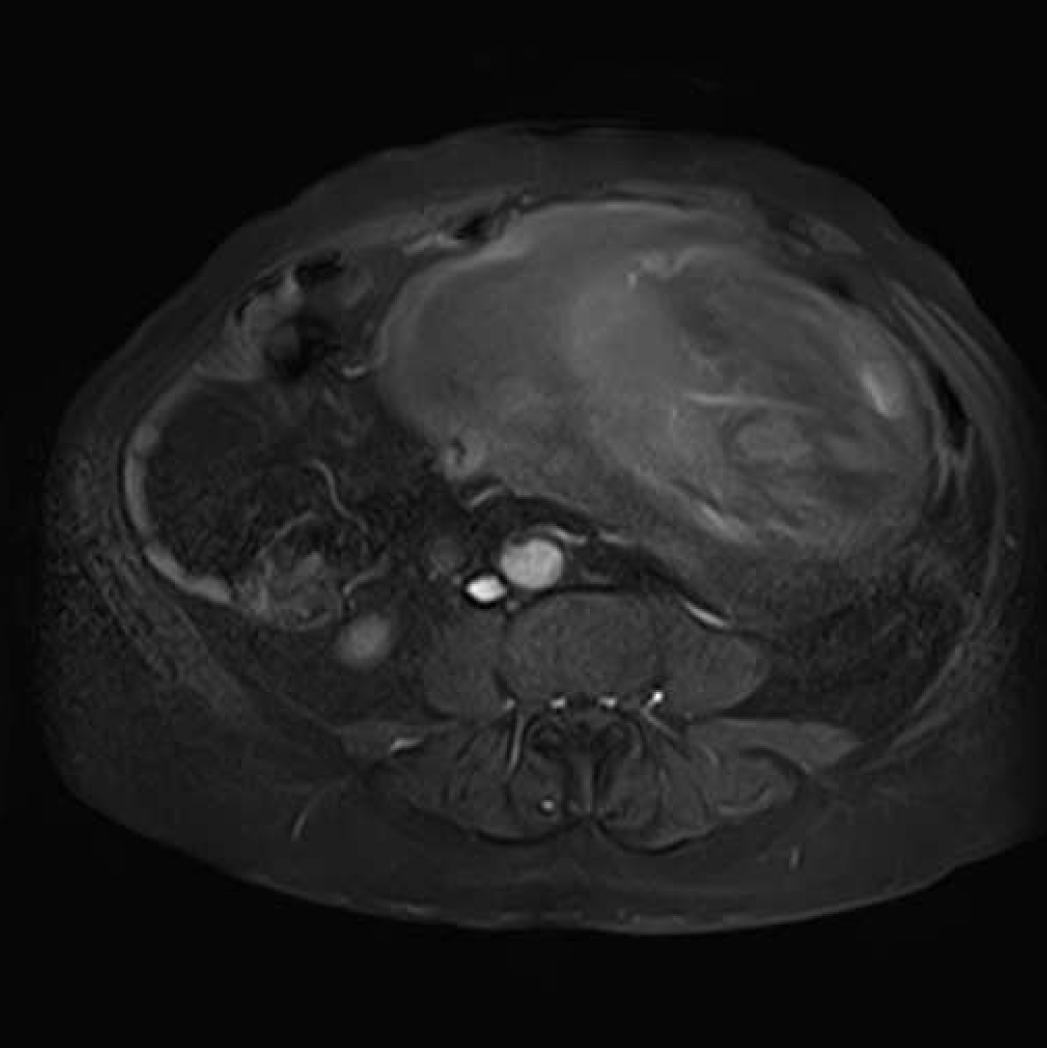
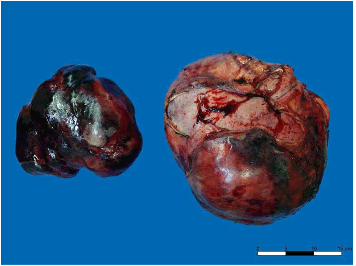
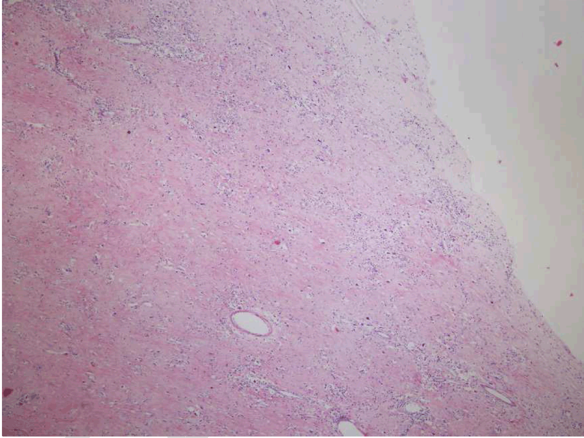
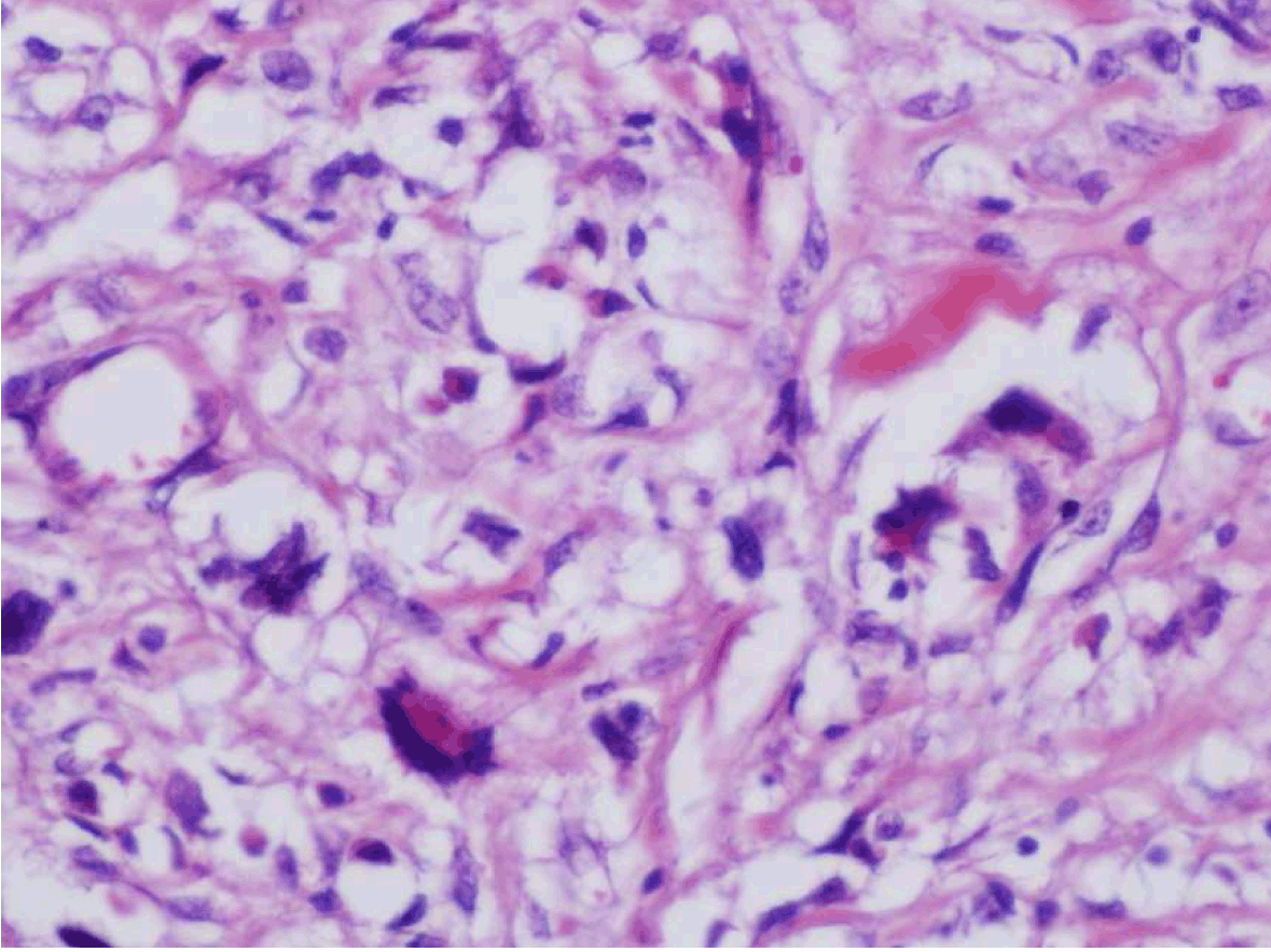
A 59-year-old male patient admitted after he realized a swelling in his abdomen with asthenia, fatigue and abdominal pain complaints for the last few months. In the physical examination, vital findings were normal. In palpation, a mass was detected filling lower left quadrant, and extending to the right of midline and umbilicus. In laboratory examinations, white blood cell (WBC):15.400/mm3, Albumin:2.1 mg/dl and HCV RNA (+). Carcinoembryonic antigen (CEA), CA 15-3, CA 19-9, CA 125 were found to be within normal ranges. In magnetic resonance imaging (MRI) scan, a mass lesion with the size of 25x19 cm, filling the lower half of abdomen, having cystic necrotic areas internally. The mass was well circumscribed and pushed the intestinal segments and mesenteric tissues towards periphery (Figure 1). No finding indicating metastatic disease was found. The patient was taken for operation after preoperative preparation. An approximately 30x30 cm mass originating from left retroperitoneum, pushing sigmoid colon and intestinal segments to the upper right abdominal quadrant was detected in the operation. The mass had no organ invasion but left testicular vessels and left ductus deferens could not be prepared. The mass was excised totally (Figure 2). Afterwards in the exploration, a second lesion, approximately 20x20 cm in mass, that was originating from sigmoid colon wall was detected (Figure 2). It was excised with Wedge resection. Both excised masses were reported to be pleomorphic liposarcoma in the pathological examination of the piece (Figure 3) (Figure 4) (Figure 5). CD-34 was detected to be (+), CD-68 was (-) in pleomorphic cells, S-100(-), actin was (-), desmin was (-) in the mass originating from sigmoid colon. Actin was found to be (-), S-100 was (-), and CD-68 was (-) in pleomorphic cells in retroperitoneal mass. Surgical margins of the resected pieces were reported to be negative. Postoperative period of the patient was problem-free. The patient was mobilized on the 1st postoperative day. Bowel movements were started on the 2nd postoperative day, and oral food was given to the patient on the 3rd postoperative day. Abdominal drains were removed in 6th and 7th postoperative days. The patient was discharged on the 9th postoperative day. The patient was discussed at tumor committee. Follow-up without any additional treatment was decided. No findings indicating metastasis or recurrence were detected in the three months follow-up of the patient. | ||||||
| ||||||
| ||||||
| ||||||
| ||||||
| ||||||
|
Discussion
| ||||||
|
Most frequent intra-abdominal mesenchymal tumors are liposarcomas. They usually manifest with non-specific abdominal pain and abdominal mass [2]. The patients may usually have mild abdominal symptoms, weight loss and usually they are generally diagnosed in late period. In our case, the present symptoms were recognized in later period and the patient was admitted with a big mass. Rarely, neurological findings due to mass pressure, may accompany [6]. Metastasis may be detected in approximately 11% of the patients and metastases are frequently occur in lungs and liver [6]. Despite admitting in later period, no distant metastases were detected in the patient. Abdominal computed tomography (CT) scan and MRI scan may show the association of the mass with adjacent organs and vascular structures, and may provide information about histological type. There are publications recommending abdominal CT scan or MRI scan as ideal method [6] [7] [8]. In our case, the structure of mass and its association with surrounding tissues and adjacent organs were assessed in detail before the operation with MRI. In our day, the only treatment for retroperitoneal liposarcomas is en bloc R0 resection [2] [5][6] [9]. There is not enough evidence regarding radiotherapy and chemotherapy modalities [2] [5][6] [9]. The most important prognostic factors are R0 resection and tumor histology [3] [9]. Multifocality is a rare but important prognostic parameter [6]. Liposarcomas are assessed in five sub-groups such as well differentiated, dedifferentiated, myxoid, round cell and pleomorphic based on their cytogenic and morphologic anomalies [1] [4]. Well differentiated and myxoid liposarcomas are seen much more frequently among primary tumors, and their prognoses are better and metastasis rates are lower than other types. On the other hand, dedifferentiated liposarcomas are seen more frequently in recurrent tumors. The rates of dedifferentiated or pleomorphic subtypes, are higher in recurrent tumors than primary ones [2] . Very frequent local recurrence (66%) and organ invasion were the leading mortality causes of the disease [2] [5][9]. In wide series, five-year disease-free survival after en-block resection was given as 18% [10]. Recommend treatment is re-operation in recurrent cases. De-bulking and radiotherapy may be recommended in cases for which R0 resection cannot be performed [2] [9]. A second mass was detected in intra-operative observation in our case despite it could not be recognized in preoperative examinations. Liposarcomas are rarely seen in gastrointestinal system. While there are liposarcoma cases detected as multiple foci in retroperitoneum in literature, we did not encounter any case who had synchronous liposarcoma in gastrointestinal system and retroperitoneum. Recurrence is an important criterion for survival in these patients, and the probability of synchronous tumor must also be considered. | ||||||
|
Conclusion
| ||||||
|
In conclusion, while retroperitoneal liposarcomas are rarely seen as single tumors, synchronous colon liposarcoma occurring together is very rare in the literature. We think that even though it could not be detected in preoperative examinations, this rare togetherness should be considered intraoperatively. | ||||||
|
References
| ||||||
| ||||||
|
[HTML Abstract]
[PDF Full Text]
|
|
Author Contributions
Ayvaz Ulaş Urganci – Substantial contributions to conception and design, Acquisition of data, Analysis and interpretation of data, Drafting the article, Revising it critically for important intellectual content, Final approval of the version to be published Erkan Oymaci – Substantial contributions to conception and design, Acquisition of data, Analysis and interpretation of data, Drafting the article, Revising it critically for important intellectual content, Final approval of the version to be published Enver Vardar – Analysis andinterpretation of data, Drafting the article, Revising it critically for important intellectual content, Final approval of the version to be published Ebru Akincilar – Acquisition of data, Drafting the article, Final approval of the version to be published Ömer Engin – Acquisition of data, Drafting the article, Final approval of the version to be published |
|
Guarantor of submission
The corresponding author is the guarantor of submission. |
|
Source of support
None |
|
Conflict of interest
Authors declare no conflict of interest. |
|
Copyright
© 2015 Ayvaz Ulaş Urganci et al. This article is distributed under the terms of Creative Commons Attribution License which permits unrestricted use, distribution and reproduction in any medium provided the original author(s) and original publisher are properly credited. Please see the copyright policy on the journal website for more information. |
|
|
|
About The Authors
| |||
| |||
| |||
| |||
| |||
| |||