|

|
 |
|
Case Report
| ||||||
| An unusual association of von Hippel–Lindau disease and posterior nutcracker syndrome: Management of neurological and urological aspects in a young male | ||||||
| Nadhir Karmani1, Faouzi Mallat2, Wissem Hmida2, Khaled Ben Ahmed2, Oussama Karmani3, Sidiya Oueld Chavey3, Amel Ben Abdallah3, Faouzi Mosbah2, Hedi Krifa1 | ||||||
|
1Neurosurgery Department, SahloulHospital, Sousse, Tunisia.
2Urology Department, SahloulHospital, Sousse, Tunisia. 3Radiology Department, SahloulHospital, Sousse, Tunisia. | ||||||
| ||||||
|
[HTML Abstract]
[PDF Full Text]
[Print This Article]
[Similar article in Pumed] [Similar article in Google Scholar] 
|


| How to cite this article |
| Karmani N, Mallat F, Hmida W, Ahmed KB, Karmani O, Chavey SO, Abdallah AB, Mosbah F, Krifa H. An unusual association of von Hippel–Lindau disease and posterior nutcracker syndrome: Management of neurological and urological aspects in a young male. Int J Case Rep Images 2015;6(7):396–402. |
|
Abstract
|
|
Introduction:
Von Hippel–Lindau (VHL) disease is a complex and systemic entity that can be discovered by neurological complications. Its association with other serious malformative disease like nutcracker syndrome (NCS) was not described, and its complex presentation must not make some other serious problem easily missed or neglected.
Case Report: A 27-year-old male patient with 3 intramedullary hemangioblastoma of six months of insidious evolution complicated by medullary compression. The lesions were completely removed with excellent neurological postoperatory outcome. Thinking of VHL disease, our investigations were expanded and revealed multiple vermian and cerebellar hemangioblastomas, renal masses, and multiple pancreatic cysts. In addition, an incidentally found of posterior NCS during the abdominal computed tomography (CT) done for VHL disease investigations. Our attitude to both neurosurgical and urological problems (the serious presentation of VHL disease with multiple locations of hemangioblastoma and right renal carcinomas) and for the missed symptomatic posterior nutcracker syndrome were discussed. Conclusion: The complex presentation of the VHL disease must not makesome other serious problems easily missed or neglected like renal carcinoma or nutcracker syndrome as seen in our case. | |
|
Keywords:
Hemangioblastoma, Nutcracker syndrome, Renal carcinomas, Total removal, Von Hippel–Lindau (VHL) disease
| |
|
Introduction
| ||||||
|
Hemangioblastoma is a rare tumour of the central nervous system characterised by a high vascularization. It accounts for 1.5–2.5% of spinal cord tumors. In the case of von Hippel–Lindau (VHL) disease, hemangioblastoma occurs in 20–30% of patients and is often multiple [1] [2]. Surgical experience is often limited because of the rarity of this tumor. Microsurgical removal is recommended in symptomatic patients or in cases with tumor growth during follow-up [3] [4]. The association of VHL disease with other serious malformative disease like nutcracker syndrome was not described. Herein, we present the case of young male having complicated presentation VHL disease with an incidentally radiological found of a missed symptomatic posterior nutcracker syndrome. | ||||||
|
Case Report
| ||||||
|
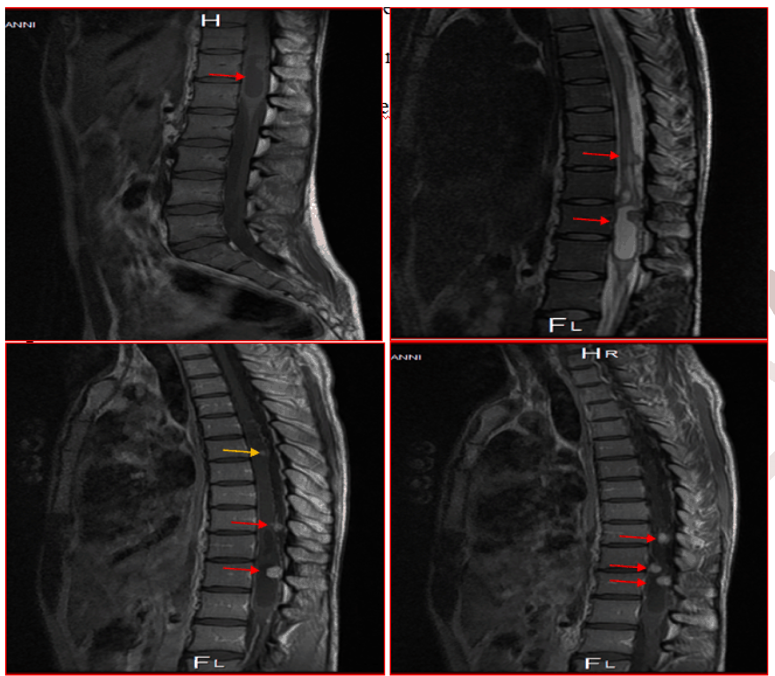
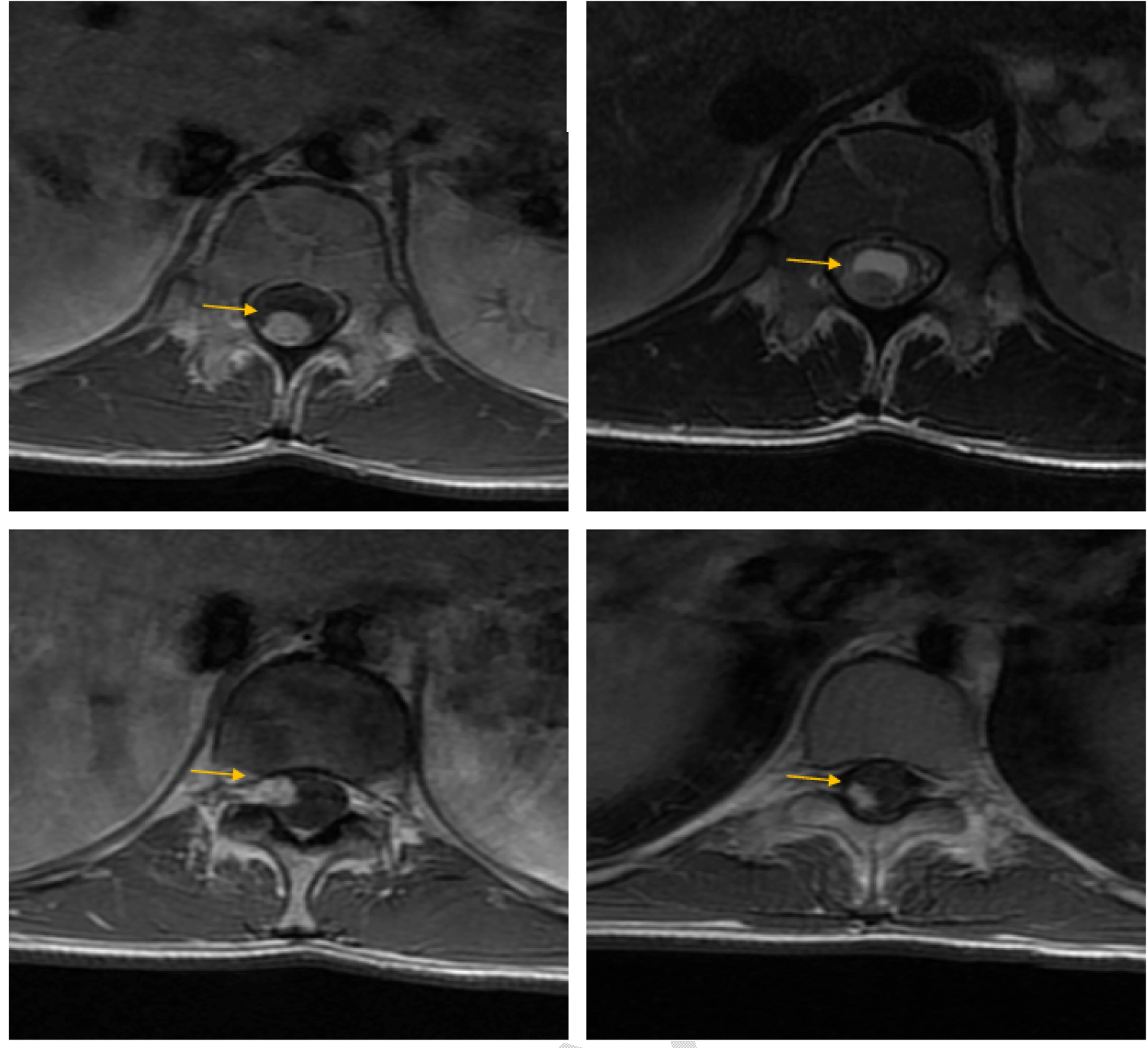
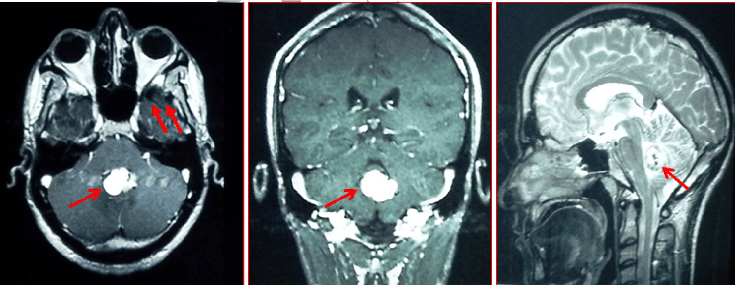
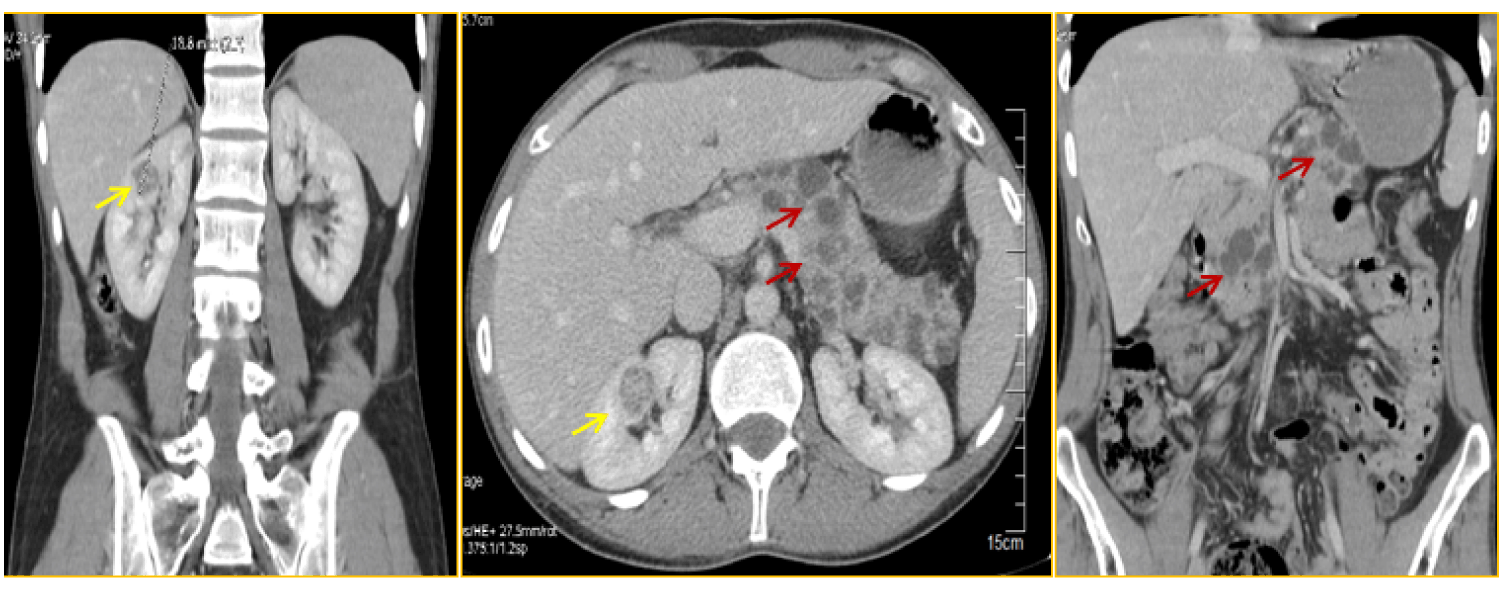
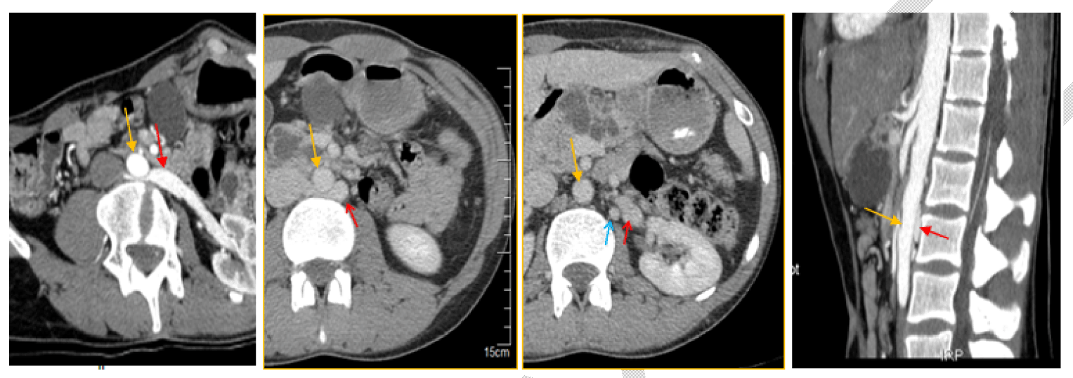
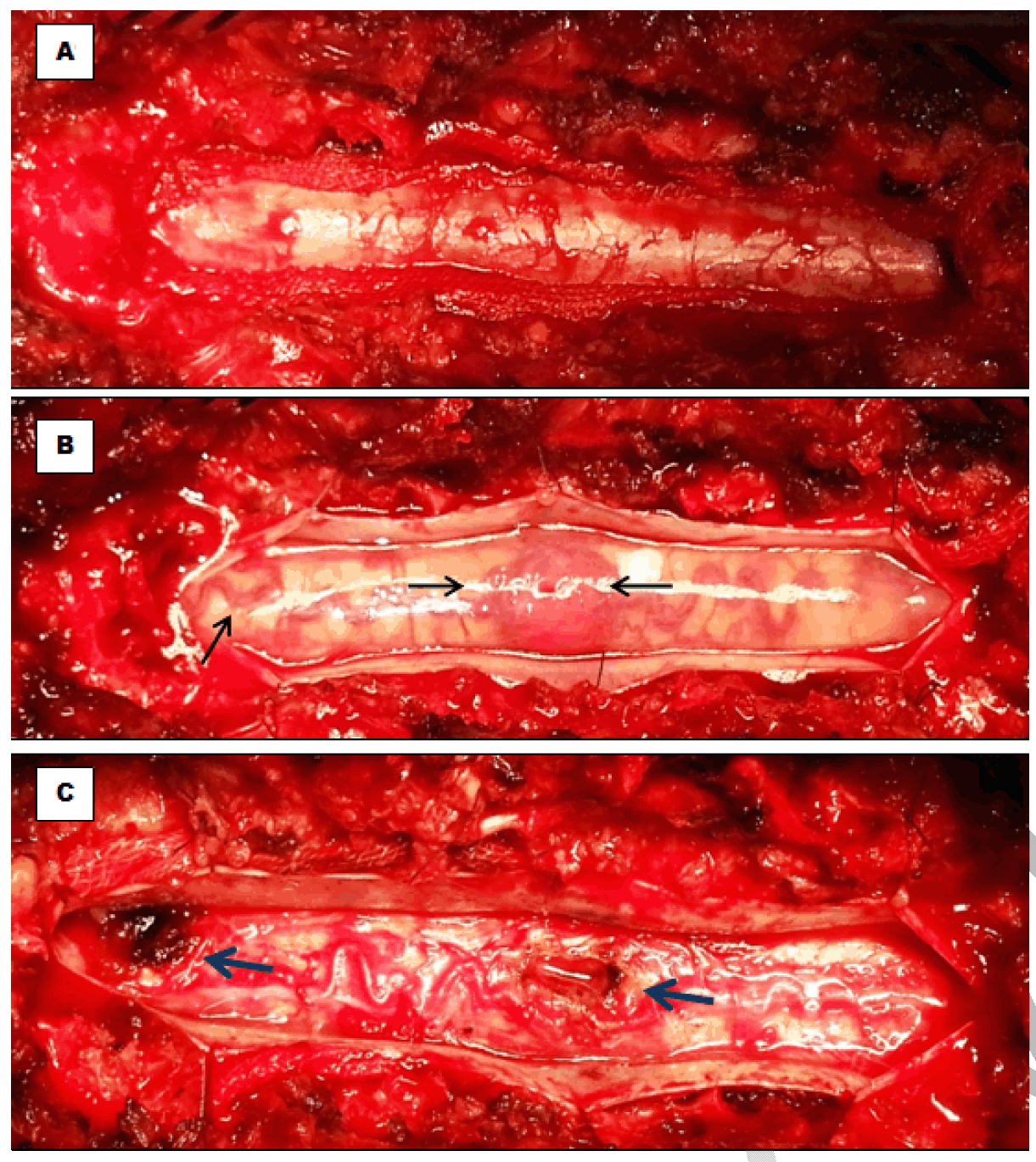
A 27-year-old male was hospitalized at the neurosurgery department because of signs of medullary compression with incomplete tetraplegia especially in the right lower limb, appearing insidiously for six months that did not ameliorate. The patient denied any bladder or bowel dysfunction or problems with handwriting, or fine motor skills. An MRI of the spine showed multiple intramedullary masses. The symptomatic lesions was situated in the terminal cone; the largest mass measured 48×13×1O mm in size. The mass appeared as a cystic lesion with a tissural mural nodule, with hypointense T1 inhomogeneous signal, with increased gadolinium uptake in T1, with perilesional oedema. Just above this mass, there are two other small cystic lesions, up to 1/3 medium of the vertebral body T6; one of the nodules had a subdural extra axial component (Figure 1) and (Figure 2). Based on the characteristics on the MRI, a diagnosis of multiple hemangioblastoma was favored; and thinking of von Hippel–Lindau (VHL) disease, our investigations were expanded: The patient underwent an MRI of the complete neuro-axis including brain and spinal cord showed a vermian hemangioblastoma associated with 2 small right cerebellar hemangioblastomas (Figure 3). The abdominal CT scan revealed 2 small renal masses, less than 2 cm in size; with multiple pancreatic cysts (Figure 4); without liver lesions, or adrenal masses. An ophthalmologic examination demonstrated none of the stigmatisms associated with VHL. Physical examination did not show any café au lait spots. At this first step and on neurological level, a diagnosis of medullary compression as a complication of multiple intramedullary hemangioblastoma in a young male with VHL disease was done. At the second step, and as an incidentally finding, the abdominal computed tomography (CT) scan done for VHL disease investigations, revealed a vascular compression: left renal vein passing posterior to the aorta, entrapped between the aorta and the vertebral column. Left gonadic vein, drained to the second left renal vein, was dilated with multiple pelvic venous collaterals. These aspects were indicative of posterior NCS (Figure 5). Interviewing the patient after discovering the vascular compression (posterior NCS), he complained of intermittent and various symptoms: urinary (intermittent macroscopic hematuria, intermittent left flank pain and chronic pelvic pain aggravated by physical activity) and systemic signs dominated by chronic fatigue and persistent headache. All these symptoms lasted for his childhood; without definitive diagnosis, despite a long investigational history of several imaging examinations and laboratory tests. In addition, high degree of left varicocele was noted at physical examination; laboratory tests revealed anemia at 10.0 mg/dl, microscopic hematuria, and 24 hour urine collection analysis showed elevated proteinuria. Based on this clinical presentation and these radiological and biologic finding, a diagnosis of posterior nutcracker syndrome was confirmed. In summary, we have a young patient with both serious presentation of VHL disease with multiple neurological locations of hemangioblastoma complicated by medullary compression due to spinal hemangioblastoma and right renal carcinomas. Our patient had also missed symptomatic posterior nutcracker syndrome. Prior to surgical intervention, the patient was counseled on the risks entailed in the removal of a spinal cord tumor. After urinary catheter insertion and administration of a steroid-type anti-inflammatory (dexamethasone) and cephalosporin antibiotics, surgical exploration was made through a prone position, and a large median incision. The dissection in the subdural space and especially the dissection of the tumor were performed using an operating microscope. The inspection of the medullary surface showed 3 exteriorised masses red, bloody and very well vascularized (Figure 6). After careful and complete dissection, the tumors were totally ablated (Figure 6), and their vascular pedicle being coagulated and sectioned at the end. Final histopathological examination confirmed the diagnosis of hemangioblastoma. Postoperative course was uneventful; There were no postoperative complications; the drainage tube was removed 48 hours after the operation and precocious mobilization and kinetotherapy was done to prevent deep venous thrombosis. Preoperative deficits were gradually ameliorated after the operation. The MRI scan of the cervical spine performed 8 days after the operation confirmed the total ablation of the 3 tumors and the patient was discharged from the hospital one week later. With 11 months of follow-up, the patient is doing well, and was neurologically cured. Last MRI of the complete neuro-axis including brain and spinal cord showed same aspects of the vermian and the cerebellar hemangioblastoma, without recurrence or appearance of spinal masses.The patient remained neurologically asymptomatic and had returned to his usual life. On the urinary and vascular level, our attitudes were: Concerning the renal masses: tumorectomie was technically impossible; and right nephrectomy was done. Macroscopic examination revealed multiple small tissular masses (more than 3) and histopathological examination confirmed the diagnosis of clear renal cell carcinoma. Concerning the posterior nutcracker syndrome, our attitude was conservative, only the varicocele was treated and medical treatment of anemia was done. Transposition of the left renal vein was proposed in case of severe complication. At the last follow-up, the varicocele improved significantly, but intermittent hematuria persisted. Laboratory tests revealed hemoglobin at 12.0 mg/dl, with persistent microscopic hematuria and proteinuria. The last abdominal CT-scan showed no recurrence of renal mass in the left kidney. | ||||||
|
| ||||||
| ||||||
| ||||||
| ||||||
| ||||||
| ||||||
|
Discussion
| ||||||
|
Hemangioblastoma are rare tumors of the central nervous system characterised by a high vascularization. they account for 1.5–2.5% of spinal cord tumors [1]. They appear spontaneously in most cases; and only 30% of them were associated with von Hippel–Lindau disease and are often multiple [2] [3]. Von Hippel–Lindau disease is a rare autosomal dominant genetic syndrome, characterised by the growth of retinal angiomas, pheochromocytoma, polycythemia, brain and spinal cord hemangioblastoma, renal and pancreatic carcinomas [5]. In our case, hemangioblastoma of the brain and spinal cord, multiple renal carcinomas and pancreatic cysts were found. More than the half of spinal hemangioblastomas are accompanied by a syrinx [5]. In our case syrinx was not noted. The initial symptoms of spinal hemangioblastoma are radicular pain, sensory or motor deficit slowly progressing according to the level of medulla involvement as seen in our case that was complicated by medullary compression. MRI has become the diagnostic tool of choice and is recommended by several authors in the diagnosis of hemangioblastoma; it is also helpful in preoperative planning and the differential diagnosis of spinal cord neoplasms and vascular lesions [6] [7]. On unenhanced T1-weighted images hemangioblastoma appears as an isointense. On T2-weighted images, it appears hyperintense with a homogeneous intense enhancement. Larger hemangioblastoma can appear as a hypointense or mixed hypo-isointense on T1, heterogeneous on T2, and shows an intense heterogeneous enhancement [6]. In addition, cystic components of the hemangioblastoma can be associated with a non-enhancing rim of the cyst. During surgery and because of the risk of massive bleeding, intramedullary hemangioblastoma surgery pose severe problems [3]. Indications for treatment and timing of surgery of hemangioblastoma are still unclear because of the rarity of these tumors and the relatively limited surgical experience. Most of the recently published series recommend microsurgical removal in symptomatic patients or in cases with tumor growth during follow-up [3] [4]. Using the aforementioned principles of microsurgical removal of spinal hemangioblastoma, microsurgical removal is feasible with a low procedure-related morbidity and can be recommended, especially in VHL patients who were symptomatic or with tumor growth during follow-up. Occlusion of the feeding arteries, shrinking of the tumor by bipolar coagulation with low power and its en bloc removal are the key points for a safe microsurgical treatment. It is essential that the ablation must be done en bloc. The most common complications that arise with such lesions are: intramedullary hematoma, epidural hematoma, meningocele or cerebrospinal fluid fistula and arachnoiditis. Postlaminectomy cervical kyphosis is more common with children and it can be prevented by performing laminoplasty or laminotomy [6]. The postoperative course of our present case was not eventful. The follow-up was based on both clinical and MRI examinations to detect tumor recurrence every six months after the operation, or in case of multiple localization to evaluate tumor progress. The neurological follow-up was evaluated according to the classification of McCormick [8]. In our case and on the neurosurgical level, we are faced to two serious problems: The first Problem was the three intramedullary hemangioblastoma complicated by medullary compression: Our attitude was an urgent total ablation. The second problem is non-complicated small vermian and cerebellar hemangioblastoma, that our attitude was conservative, with strict and regular follow-up. At the last follow-up, the patient remained neurologically stable. Our patient had in addition to the neurological presentation, urological presentation of the VHL disease as right multiple carcinomas and had also symptomatic nutcracker syndrome. Posterior nutcracker syndrome results from compression of the left renal vein between the aorta and vertebra, with subsequent development of venous varicosities of the renal pelvis, ureter, and the gonadal vein [9] [10]. The true prevalence of nutcracker syndrome especially the posterior form remains unknown and it might be underdiagnosed and easily delayed [11], as seen in our patient despite several imaging and laboratory investigations. The computed tomography angiography (CTA) is the procedure of choice of the diagnosis with high sensitivity and specificity [10] [11]. The management of the NCS is controversial and the invasive procedures should be considered only when symptoms are severe or persistent, including severe pain, severe hematuria, renal insufficiency, and failure to respond to conservative treatment [9] [10]. Follow-up, conservative approach and surgical therapy are the different treatment options proposed for this syndrome. The available surgical procedure of the PNCS is the transposition of the LRV. Other options were described including intra-vascular or extravascular stents and renal auto transplantation. The indications for laparoscopic surgery continue to augment for the treatment of the nutcracker syndrome [12] [13]. In this case, we are faced to several urological problems: The first we have multiple right renal tumors < 2 cm provided to be clear cell carcinomas in the context of a serious and complicated form of the VHL disease;: conservative attitude with strict and regular follow-up of right renal parenchyma and the left kidney was our proposed procedure of choice but because the lack of the ablative techniques in our country (radiofrequency and cryoablation) and the intraoperative difficulties of multiple tumorectomie, a nephrectomy was made. The second problem was in the left kidney: posterior nutcracker syndrome, our attitude was conservative, only the varicocele was treated and medical treatment of anemia was done; transposition of the LRV was proposed in case of severe complications. | ||||||
|
Conclusion
| ||||||
|
Von Hippel–Lindau (VHL) disease and nutcracker syndrome are rare and severe entities. There association was not described. The complex presentation of the VHL disease must not make some other serious problems easily missed or neglected like renal carcinoma or nutcracker syndrome as seen in our case. | ||||||
|
References
| ||||||
| ||||||

|
[HTML Abstract]
[PDF Full Text]
|
|
Author Contributions
Nadhir Karmani – Substantial contributions to conception and design, Acquisition of data, Analysis and interpretation of data, Drafting the article, Revising it critically for important intellectual content, Final approval of the version to be published Faouzi Mallat – Analysis and interpretation of data, Revising it critically for important intellectual content, Final approval of the version to be published Wissem Hmida – Analysis and interpretation of data, Revising it critically for important intellectual content, Final approval of the version to be published Khaled Ben Ahmed – Analysis and interpretation of data, Revising it critically for important intellectual content, Final approval of the version to be published Oussama Karmani – Analysis and interpretation of data, Revising it critically for important intellectual content, Final approval of the version to be published Sidiya Oueld Chavey – Analysis and interpretation of data, Revising it critically for important intellectual content, Final approval of the version to be published Amel Ben Abdallah – Analysis and interpretation of data, Revising it critically for important intellectual content, Final approval of the version to be published Faouzi Mosbah – Analysis and interpretation of data, Revising it critically for important intellectual content, Final approval of the version to be published Hedi Krifa – Analysis and interpretation of data, Revising it critically for important intellectual content, Final approval of the version to be published |
|
Guarantor of submission
The corresponding author is the guarantor of submission. |
|
Source of support
None |
|
Conflict of interest
Authors declare no conflict of interest. |
|
Copyright
© 2015 Nadhir Karmani et al. This article is distributed under the terms of Creative Commons Attribution License which permits unrestricted use, distribution and reproduction in any medium provided the original author(s) and original publisher are properly credited. Please see the copyright policy on the journal website for more information. |
|
|
|
About The Authors
| |||
| |||
| |||
| |||
| |||
| |||
| |||
| |||
| |||
| |||