| |
|
|
|
Clinical Image
| ||||||
| Bowen's disease involving the dorsal and volar aspects of left thumb: An unusual site diagnostic entity | ||||||
| Shagufta Rather1, Peerzada Sajad2, Iffat Hassan3 | ||||||
|
1MBBS, MD, Consultant, Postgraduate, Department of Dermatology, STD and Leprosy,Government Medical College Srinagar, J&K, India.
2MBBS,MD, Senior Resident, Postgraduate, Department of Dermatology, STD and Leprosy, Government Medical College, Srinagar, J&K, India. 3MBBS, MD, Professor and Head, Postgraduate, Department of Dermatology, STD and Leprosy, Government Medical College, Srinagar, J&K, India. | ||||||
| ||||||
|
[HTML Abstract]
[PDF Full Text]
[Print This Article]
[Similar article in Pumed] [Similar article in Google Scholar] 
|
| How to cite this article |
| Rather S, Sajad P, Hassan I. Bowen's disease involving the dorsal and volar aspects of left thumb: An unusual site diagnostic entity. Int J Case Rep Images 2015;6(7):457–459. |
|
Case Report
| ||||||
|
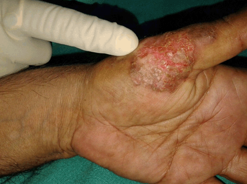
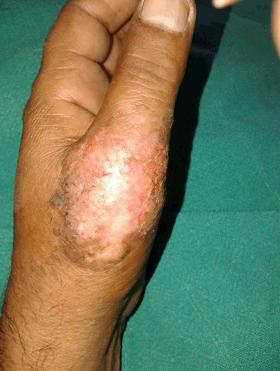
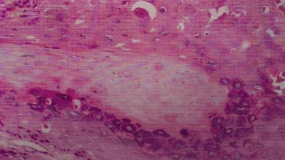
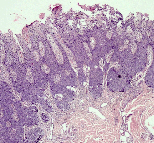
Herein, we report a case of a 40-year-old normotensive, non-diabetic, euthyroid male patient who presented with a non-healing erythematous slightly scaly plaque involving the volar and dorsal aspects of left thumb from last eight months (Figure 1) and (Figure 2). There was history of pruritus and slight burning sensation. There was a history of application of potent topical steroids for few months with no response. A punch biopsy was done which confirmed the diagnosis of eczema psoriasis and Bowen's disease. On histopathology, the patient showed thickened epidermis with full thickness dysplasia of squamous epithelium and atypical keratinocytes with numerous mitotic figures. The basement membrane was intact with no focus of invasion or solar elastosis (Figure 3) and (Figure 4). Thus a diagnosis of Bowen's disease was made. The patient was put on topical 5-fluorouracil and is doing well. | ||||||
| ||||||
| ||||||
| ||||||
|
| ||||||
| ||||||
|
Discussion
| ||||||
|
Bowen's disease (BD) is a form of intra-epidermal or in-situ squamous cell carcinoma with a small potential for invasive malignancy, and commonly involves chronically photo-exposed areas, especially head and neck region. Commonly a persistent, non-elevated erythematous scaly and crusted plaque is seen. Bowen's disease may occur at any age in adults, but is rare before the age of 30 years; most patients are aged over 60. Any site may be affected, although involvement of palms or soles is uncommon. Bowen's disease occurs predominantly in women in whom about 60-85% of patients have lesions on the lower leg, usually in previously or presently sun-exposed areas of skin. Chronic ultraviolet radiation exposure, arsenic exposure, human papillomavirus and immunosuppression are the various aetiological factors [1] [2] [3]. Histopathology is characterized by full-thickness dysplasia of the epidermis, with loss of the normal maturation of its components. Keratinocytes are atypical and disorderly, often described as having a windblown appearance. Basement membrane is intact. Topical 5-fluorouracil, imiquimod, photodynamic therapy, cryotherapy and excision are the various treatment modalities [4]. | ||||||
|
Conclusion
| ||||||
|
The head, neck, and extremities are the most commonly affected anatomic locations in men, while the lower limbs and cheeks are most commonly affected in women. Involvement of palms and soles is an unusual site of occurrence, but it should be considered in the differential diagnosis of non-healing erythematous scaly plaques. | ||||||
|
References
| ||||||
| ||||||
|
[HTML Abstract]
[PDF Full Text]
|
|
Author Contributions
Shagufta Rather – Substantial contributions to conception and design, Acquisition of data, Analysis and interpretation of data, Drafting the article, Revising it critically for important intellectual content, Final approval of the version to be published Peerzada Sajad – Analysis and interpretation of data, Revising it critically for important intellectual content, Final approval of the version to be published Iffat Hassan – Analysis and interpretation of data, Revising it critically for important intellectual content, Final approval of the version to be published |
|
Guarantor of submission
The corresponding author is the guarantor of submission. |
|
Source of support
None |
|
Conflict of interest
Authors declare no conflict of interest. |
|
Copyright
© 2015 Shagufta Rather et al. This article is distributed under the terms of Creative Commons Attribution License which permits unrestricted use, distribution and reproduction in any medium provided the original author(s) and original publisher are properly credited. Please see the copyright policy on the journal website for more information. |
|
|