| |
|
|
|
Case Report
| ||||||
| A rare presentation of calciphylaxis in normal renal function | ||||||
| Parin Rimtepathip1, David Cohen2 | ||||||
|
1MS III, Mercer University School of Medicine.
2MD, Dermatologic Surgery Specialists, Macon, Georgia. | ||||||
| ||||||
|
[HTML Abstract]
[PDF Full Text]
[Print This Article]
[Similar article in Pumed] [Similar article in Google Scholar] 
|
| How to cite this article |
| Rimtepathip P, Cohen D. A rare presentation of calciphylaxis in normal renal function. Int J Case Rep Images 2015;6(6):366–369. |
|
Abstract
|
|
Introduction:
Calciphylaxis is a rare and life- threatening condition in which extensive microvascular calcification in arterioles and occlusion of vessels lead to painful non-healing ulcers with high mortality rate. Calciphylaxis is mainly associated with end stage renal disease or hyperparathyroidism, with rare cases reported in cirrhosis patient.
Case Report: A 55-year-old Caucasian male with significant history of porphyria cutanea tarda and Hepatitis C complicated by cirrhosis with normal renal function presents with history of non-healing ulcers on both of his hands. The diagnosis of calciphylaxis was made by X-ray. Due to rapid progression of the ulcers to tissue necrosis and gangrene with no definite underlying pathology, the patient's hands were eventually amputated due to the inability to withstand pain. Conclusion: Patients presenting with painful ulceration of their fingers with history of cirrhosis and normal renal function should be worked up for calciphylaxis as part of the differential diagnosis, especially with low serum albumin level. Site of calciphylaxis also matters as there is a great difference between mortality rates of proximal versus distal. We postulate the idea of pathophysiological mechanism with further research needed. This case report should alert physicians that calciphylaxis does occurred in patients with cirrhosis and normal renal function. | |
|
Keywords:
Calciphylaxis, Cirrhosis, Porphyria Cutanea Tarda, Hepatitis C
| |
|
Introduction
| ||||||
|
Calciphylaxis is a rare and life-threatening condition in which extensive microvascular calcification due to calcium deposition in arterioles and occlusion of vessels lead to painful, violaceous, mottled skin lesions, which progress to non-healing ulcers, tissue necrosis, gangrene, sepsis, and potentially death [1]. Two years mortality rates from sepsis ranges from 50–80%. Patients with skin involvement over the trunk or proximal extremities have a poorer prognosis and higher mortality rate when compared to distal extremities. The lack of understanding the pathophysiology of the disease and numerous postulation result in unsatisfactory answer to why there is a difference in mortality rate. Biopsy of the calciphylaxis ulcer would reveals calcium deposits lining the vascular intima, while tissue calcification may also be seen on plain radiographs [2]. Risk factors for calciphylaxis are female gender, hyperphosphatemia with elevation of calcium phosphate axis, elevated parathyroid hormone, high alkaline phosphatase, and low serum albumin [3]. Many of the risk factors can be seen in patients on hemodialysis from chronic renal failure especially if the chronic renal failure is the complication of diabetes with 61% had acral gangrene compared to 34% of the non-diabetic calciphylaxis [4]. Calciphylaxis is therefore most common in hyperparathyroidism secondary to chronic renal impairment and rarely occurs in the setting of normal renal function [2]. However, these risk factors are not set in stone or fundamental in diagnosing patients with calciphylaxis. We report an extremely rare case of calciphylaxis in a 55-year-old Caucasian male with significant history of porphyria cutanea tarda and hepatitis C complicated by cirrhosis with normal renal function that presents with non-healing ulcers on both of his hands, with rapid progression to tissue necrosis and gangrene. | ||||||
|
Case Report
| ||||||
|
A 55-year-old Caucasian male with significant history of hepatitis C, porphyria cutanea tarda (PCT), and cirrhosis presents with non-healing ulcers on both hands. The patient was referred to the dermatological center for the evaluation of both hands as a possible complication of PCT. The clinical examination showed a cachectic patient with lesions shown in Figure 1. The patient noticed the new skin lesions on his hands to be different from the lesions of PCT several years ago. Even though PCT caused pain, the new onset of the non healing ulcers were far more painful. The patient described the ulcer as they first appeared as shallow eroding of the skin. Then the ulcers would accompanied by extreme pain and the tissue in the area would started to necroses. Some of the differential diagnoses for the patient's painful ulceration of the fingers were Raynaud syndrome, scleroderma, peripheral vascular disease, and vasculitis. During the course of treatment, patient denied any skin biopsied due to the existence of severe pain. Therefore, we decided to perform X-ray of his left hand. The finding of his left hand (Figure 2) showed mildly increased soft-tissue density and stipple calcification of the vessels. Calciphylaxis was diagnosed. Due to the unknown pathogenesis which makes good clinical treatment extremely difficult, labs were ordered in an attempt to exclude any risk factors. All labs including CBC and biochemistry were normal except the low albumin level of < 3 g/dL. The patient was given analgesic for his pain but increase dosage did not alleviate his condition. Patient's fingers were ultimately amputated due to unbearable pain and unsalvageable tissue necrosis and gangrene. | ||||||
| ||||||
| ||||||
|
Discussion
| ||||||
|
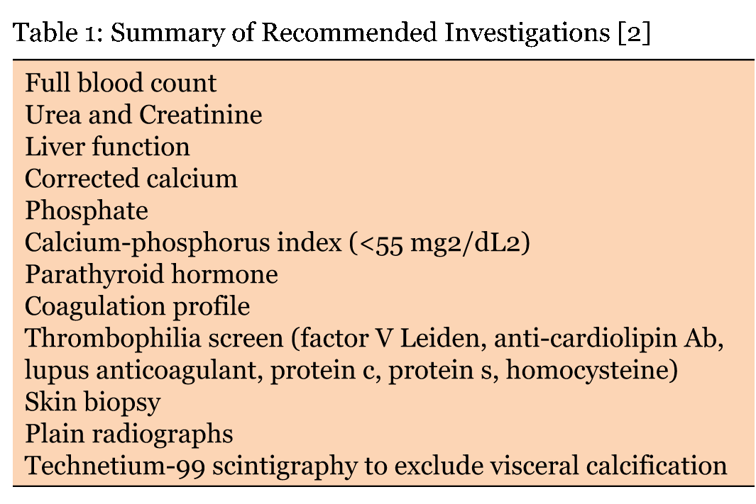
Calciphylaxis (synonym calcific uremic arteriolopathy) results from calcification of the arterioles and subsequent thrombosis which lead to skin ischemia. Calciphylaxis is associated with high morbidity and mortality resulting primarily from infections. According to Mazhar et al., elevated parathyroid hormone levels, elevated serum phosphate and calcium-phosphate products especially in renal failure patients, elevated alkaline phosphatase, high serum levels of iPTH, low serum albumin, female, Caucasian origin, ESRD, medications such as warfarin, prednisone, calcitriol, and calcitriol salts are associated with risk factors at the time of diagnosis of calciphylaxis. Since most calciphylaxis typically occurs in patients with end-stage renal disease undergoing dialysis or patients who have secondary hyperparathyroidism, patients without these underlying risk factors are often misdiagnosed at presentation [3]. An important postulated pathophysiologic mechanism is proposed by Danziger and Demer about the increase risk for calciphylaxis in patient with hepatic disease, like our presenting patient. A possible reason is that vitamin K is reduced in liver disease, which is required for post-translation gamma-carboxylation of matrix gamma-carboxyglutamic acid protein, fetuin, or growth arrest-specific gene 6. These are calcifications inhibitors produced by vascular smooth muscle cells. Warfarin, which inhibits vitamin K-dependent carboxylation of these calcification inhibitors, is thought to encourage vascular calcification in this way [5] [6]. Another propose mechanism by Goli, Shah, Byrd, and Roy, is the deficiencies in protein C and S from cirrhosis as the cause of calciphylaxis as low levels of these anticoagulant factors may be an important etiologic factor [7]. Low serum albumin level is one of the main interests as a risk factor for our patient since all lab values were normal except the albumin level. Bleyer et al. reported a 17-fold increase in the risk of developing calciphylaxis with each decrease in albumin by 1 g/dL (OR= 16.9, 95% CI, 5.25 to 54.5) [8]. This finding is later supported by Coates et al., who reported a loss of 10 % body weight over six months preceding the diagnosis of calciphylaxis in 7 out of 16 patients in their series [9]. As mentioned earlier, two years mortality rates from infections and eventually sepsis for these patients might be due to underlying poor would healing as a result of low albumin. Correcting albumin level in patients with calciphylaxis with no other risk factors might enhance their survival rates by enhancing the wound healing process, which would lead to less infection and less mortality rate. However, further research concerning the possible role of albumin level should be pursued. Suyin and I.H. Coulson proposed a summary of recommended investigations in Table 1 [2]. Why do patients with skin involvement over the trunk or proximal extremities have a poorer prognosis and higher mortality rate when compared to distal extremities which occurs below the knee? Is the fact that our patient beat the two years mortality rates of 50–80% because only his distal extremity was involved? The lesions of calciphylaxis typically develop suddenly and progress rapidly. However, does this concept apply to distal calciphylaxis with better prognosis? According to Mazhar et al., eleven patients with calciphylaxis and seven controls died during the follow-up period. Seven out of ten patients who died of complications related to calciphylaxis had proximal lesions and died of infections [3]. Proximal lesions have been observed in many studies to be one of the worst prognoses, but none of the articles postulated the underlying pathophysiologic mechanisms. A raising question surrounding this area: Is it because proximal type of calciphylaxis involves major arteries while distal type involves arterioles? In humans, vascular calcification is an active process and is not sufficient to produce skin necrosis. Vascular calcification and thrombosis are both required to produce lesions of calciphylaxis [10]. According to Rayz, V. L., et al., in addition to biochemical factors, hemodynamic factors that are governed by luminal geometry and blood flow rates likely play an important role in the thrombus formation and deposition process [11]. Arteries and arterioles have different luminal geometry and blood flow rates, therefore, calcification and thrombus formation can possibly formed much faster and much more aggressive in the proximal calciphylaxis. Also, the proximal arteries increase the chance of infections developing into sepsis because they are closer to many important organs. The idea behind this theory stimulates further research concerning the difference between mortality rates in proximal versus distal calciphylaxis. | ||||||
| ||||||
|
| ||||||
|
Conclusion
| ||||||
|
In conclusion, patients presenting with painful ulceration of their fingers with significant history of porphyria cutanea tarda and Hepatitis C with cirrhosis with normal renal function should be worked up for calciphylaxis as part of the differential diagnosis. Calciphylaxis is a very rare disease with approximately 160 case reports worldwide. This case report should alert physicians that calciphylaxis does occurred in patients with cirrhosis and normal renal function. | ||||||
|
References
| ||||||
| ||||||
|
[HTML Abstract]
[PDF Full Text]
|
|
Author Contributions
Parin Rimtepathip – Substantial contributions to conception and design, Drafting the article, Final approval of the version to be published David Cohen – Substantial contributions to conception and design, Drafting the article, Final approval of the version to be published |
|
Guarantor of submission
The corresponding author is the guarantor of submission. |
|
Source of support
None |
|
Conflict of interest
Authors declare no conflict of interest. |
|
Copyright
© 2015 Parin Rimtepathip et al. This article is distributed under the terms of Creative Commons Attribution License which permits unrestricted use, distribution and reproduction in any medium provided the original author(s) and original publisher are properly credited. Please see the copyright policy on the journal website for more information. |
|
|