| |
|
|
|
Case Report
| ||||||
| A rare cause of quadriplegia: Bilateral medial medullary syndrome presenting with "heart appearance sign" | ||||||
| Suryanarayana Sharma P. M.1, Mahendra J. V.2, Rohan R. Mahale2, Acharya P. T.3, Madhusudhan B. K.1, Srinivasa R.4 | ||||||
|
1D. M. Neurology Resident.Department of Neurology, M. S. Ramaiah Medical College, Bangalore, Karnataka, India.
2D. M. Neurology, Assistant Professor. Department of Neurology, M. S. Ramaiah Medical College, Bangalore, Karnataka, India. 3D. M. Neurology, Sr. Professor, Department of Neurology, M. S. Ramaiah Medical College, Bangalore, Karnataka, India. 4D. M. Neurology, Sr. Professor & Head, Department of Neurology, M. S. Ramaiah Medical College, Bangalore, Karnataka, India. | ||||||
| ||||||
|
[HTML Abstract]
[PDF Full Text]
[Print This Article]
[Similar article in Pumed] [Similar article in Google Scholar] 
|
| How to cite this article |
| Sharma SPM, Mahendra JV, Mahale RR, Acharya PT, Madhusudhan BK, Srinivasa R. A rare cause of quadriplegia: Bilateral medial medullary syndrome presenting with "heart appearance sign". Int J Case Rep Images 2015;6(6):338–342. |
|
Abstract
|
|
Introduction:
Bilateral medial medullary infarct (MMI) is a very rare form of cerebrovascular disease presenting with quadriplegia, tongue weakness and posterior column sensory deficit. Initial reports of bilateral MMI were on autopsy. Only 38 magnetic resonance imaging (MRI) proven cases of bilateral MMI have been published in English literature till March 2011.
Case Report: In the present case, patient presented with progressive quadriplegia of three days duration with respiratory involvement, MRI scan of brain revealed diffusion restriction in bilateral paramedian medulla appearing as characteristic heart appearance sign diagnostic of bilateral MMI. Conclusion: High index of suspicion is required to make early diagnosis in this rare stroke subtype. Optimal respiratory management may significantly improve the clinical outcome. | |
|
Keywords:
Bilateral medial medullary infarct (MMI), Quadriplegia, Heart appearance sign
| |
|
Introduction
| ||||||
|
Medial medullary infarct (MMI) was initially described in late 19th century by Spiller [1]. Subsequent description has been credited to Dejerine [2]. Medial medullary infarction is uncommon; accounting for less than 1% cases of all brain infarctions [3]. Bilateral MMI is even rarer. Previously, the diagnosis of bilateral MMI was made at autopsy. Presently, with increasing use of the magnetic resonance imaging (MRI) in acute ischemic strokes, increasing number of bilateral MMI cases have been identified. Clinical presentation, stroke mechanism and outcome in patients with bilateral MMI is variable. Only 38 MRI proven cases of bilateral MMI have been published in English literature till March 2011 [4]. Herein, we report a patient with bilateral medial medullary infarction with a typical heart shaped sign on brain MRI. | ||||||
|
Case Report
| ||||||
|
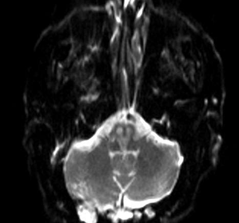
A 74-year-old male with diabetes was referred to emergency department of our hospital with giddiness, dyspnea and left hemiparesis of three days duration. He was intubated in local hospital and was referred to us for further management. Computed tomography (CT) scan of brain done outside did not reveal any acute infarct/bleed. On examination, the patient was drowsy, but obeying simple commands. His blood pressure was 190/100 mmHg and heart rate was 96/min. He had bilateral horizontal nystagmus with torsional component in all directions. His pupils were equal, reactive and eye movement was full. Gag reflex was impaired. Tongue movement could not be assessed. He had left hemiparesis (3/5 on MRC scale). He had generalized hyperreflexia with bilateral Babinski sign. Sensory system could not be evaluated. He had an ulcer over dorsum of right foot with amputated 4th and 5th toes. Other systems are unremarkable. Hematological investigations did not show any significant abnormality. He had deranged renal parameters are creatinine 1.8, random blood sugar 283, HbA1C 8.7, total cholesterol 198, triglyceride 264.3, low-density lipoprotein 115.3 and his urine analysis revealed 15–20 pus cells with 1+ proteinuria. MRI scan of brain diffusion weighted (DW) imaging (1.5 T) revealed heart shaped hyperintensity areas in the bilateral ventral medulla with apparent diffusion coefficient (ADC) reversal as shown in (Figure 1). Similar findings were observed in the same region on T2 and fluid attenuated inversion recovery sequence (FLAIR) (Figure 2) and (Figure 3). Old infarcts in right occipital and parietal region were noted. Based on these findings, patient was diagnosed as having an acute bilateral medial medullary infarction (Figure 4) and he was treated with dual anti-platelets, enoxaparin, atorvastatin, ventilatory support, antibiotics and chest physiotherapy and DVT prophylaxis. Echo was normal. However, weakness progressed to quadriplegia on day-5 of hospitalization. He developed aspiration pneumonitis. He remained quadriplegic during his subsequent stay for 34 days in the hospital. Endotracheal culture revealed Pseudomonas and Acinetobacter, antibiotics were escalated to meropenem and colistin. He died on day-34 of hospitalization due to pneumonia and sepsis. | ||||||
| ||||||
| ||||||
| ||||||
| ||||||
|
Discussion
| ||||||
|
Medial medullary syndrome is a rare stroke subtype characterized by ipsilateral hypoglossal nerve palsy, contralateral hemiparesis sparing face and impairment of contralateral deep sensation [5]. It results from infarction of paramedian region of medulla oblongata due to occlusion of vertebral/anterior spinal artery or their small branches. Davison described a case of bilateral MMI in 1937 [6]. Bilateral MMI is even rarer stroke subtype and only 12 anatomically proven cases have been reported in English literature till date [7]. Subsequently, with advances in imaging technology with widespread use of MRI in diagnosis of acute ischemic strokes worldwide, additional 38 cases have been reported in the last 20 years. Bilateral medial medullary syndrome is characterized by classical triad of quadriplegia, tongue weakness and posterior column sensory deficits [8] . Katoh and Kawamoto classified bilateral MMI into type I, with an infarction area from medullary pyramid to pontine medial longitudinal fasciculus and type II with infarction confined to bilateral medullary pyramids [9]. Our patient was classified as having type I disease which has a worse prognosis.The vascular events likely to be associated with bilateral MMI are occlusion of vertebral artery or anterior spinal artery and its intrinsic penetrating branches. The infarcted area usually includes the pyramidal tracts, medial lemniscus, medial longitudinal fasciculus, hypoglossal nucleus or hypoglossal nerve fibres and medullary reticular formation bilaterally [10]. Before the advent of MRI scan, it was often confused with Guillain-Barre syndrome as it presented predominantly with quadriplegia. Limb weakness is a constant finding in this syndrome (85.3%) as in our case where patient initially presented with hemiparesis, subsequently progressing to quadriplegia with dysarthria and dysphagia [11]. Tongue weakness was not evident clinically as he was intubated elsewhere. Respiratory disturbance occurs in significant number of cases (29.4%) requiring ventilator support [10]. Computed tomography scan of brain is not a sensitive tool for posterior fossa, especially for medullary infarcts as in our case, CT scan of brain was normal even after three days of onset of symptoms. DWI sequence of MRI shows the characteristic heart appearance sign due to infarction of anteromedial and anterolateral territory of medulla [12]. Acute to subacute MMI can be differentiated accurately only by MRI. In acute MMI, there will be diffusion restriction with ADC reversal and no abnormality on T2/FLAIR sequences. Subacute MMI shows T2 shine through phenomena- no ADC reversal with hyperintense signal changes on T2/FLAIR. According to vascular supply, medulla is divided into anteromedial, anterolateral, lateral and posterior territory. Blood supply to these areas is predominantly by vertebral and anterior spinal arteries [13]. It is often difficult to identify to occluded vessel on MRA due to vastly complex network often formed by these vessels. Misdiagnosis/delay in diagnosis in this syndrome is common as patient presenting with areflexic quadriplegia sparing face may be misdiagnosed as Guillain-Barre syndrome [14], myasthenia gravis, brain stem encephalitis, inflammatory myopathy, periodic paralysis and paraneoplastic syndrome as other possible differential diagnosis. Bilateral MMI should be suspected in patients presenting with acute onset quadriparesis, tongue weakness and facial sparing. Pongmoragot et al. [4] have done a systematic review of bilateral MMI and have identified 38 MRI proven cases and concluded rostral medullary infarct (V-shaped) as the most common MRI finding in these cases. Aspiration pneumonia is the major cause of death in up to 66% of cases in different series [4]. Urosepsis and pulmonary thromboembolism contribute to other causes of mortality. In our case, patient succumbed to respiratory infection. Recognizing the severity of the respiratory symptoms in these patients is critical to avoid recurrent aspiration pneumonia and to improve the clinical outcome. Intravenous/intra-arterial thrombolysis using recombinant tissue plasminogen activator (r-TPA) may be beneficial in cases with bilateral MMI. Pfefferkorn et al. have demonstrated that a combination of intravenous thrombolysis with consecutive in endovascular mechanical thrombectomy may be a option in this difficult clinical situation [15]. | ||||||
|
Conclusion
| ||||||
|
In summary, patient presenting with rapid onset quadriplegia with sensory loss and bulbar weakness should raise the suspicion of bilateral medial medullary infarct (MMI). Advances in imaging technology like diffusion weighted MRI has greatly improved the yield of early diagnosis which is a key factor in predicting the outcome. Bilateral MMI which was previously considered fatal can be effectively treated if respiratory management is optimally performed during acute period. We report this case because of rarity of its occurrence. | ||||||
|
Acknowledgements
| ||||||
|
We are thankful to Anas A. Majeed and Sudhindra Aroor for their help and support. | ||||||
|
References
| ||||||
| ||||||
|
[HTML Abstract]
[PDF Full Text]
|
|
Author Contributions
Suryanarayana Sharma P. M. – Substantial contributions to conception and design, Acquisition of data, Analysis and interpretation of data, Drafting the article, Revising it critically for important intellectual content, Final approval of the version to be published Rohan R. Mahale – Substantial contributions to conception and design, Analysis and interpretation of data, Drafting the article, Revising it critically for important intellectual content, Final approval of the version to be published Mahendra J. V. – Analysis and interpretation of data, Drafting the article, Revising it critically for important intellectual content, Final approval of the version to be published Madhusudhan B. K. – Analysis and interpretation of data, Revising it critically for important intellectual content, Final approval of the version to be published Acharya P. T. – Analysis and interpretation of data, Revising it critically for important intellectual content, Final approval of the version to be published Srinivasa R. – Substantial contributions to conception and design, Revising it critically for important intellectual content, Final approval of the version to be published |
|
Guarantor of submission
The corresponding author is the guarantor of submission. |
|
Source of support
None |
|
Conflict of interest
Authors declare no conflict of interest. |
|
Copyright
© 2015 Suryanarayana Sharma et al. This article is distributed under the terms of Creative Commons Attribution License which permits unrestricted use, distribution and reproduction in any medium provided the original author(s) and original publisher are properly credited. Please see the copyright policy on the journal website for more information. |
|
|