| |
|
|
|
Case Report
| ||||||
| Successful bail-out stenting of severe stenosis of the left main trunk coronary artery using guiding catheter exchange with the anchor balloon technique | ||||||
| Daizaburo Yanagi1, Takeshi Serikawa2, Masanori Okabe3, Yusuke Yamamoto4 | ||||||
|
1MD, PhD, Chief Resident Physician, Department of Cardiology, Cardiovascular and Aortic Center of Saiseikai Fukuoka General Hospital, Fukuoka, Japan.
2MD, Ph.D, Manager of the Catheterization Laboratory, Department of Cardiology, Cardiovascular and Aortic Center of Saiseikai Fukuoka General Hospital, Fukuoka, Japan. 3MD, PhD, Assistant Director, Department of Cardiology, Cardiovascular and Aortic Center of Saiseikai Fukuoka General Hospital, Fukuoka, Japan. 4MD, PhD, Director, Department of Cardiology, Cardiovascular and Aortic Center of Saiseikai Fukuoka General Hospital, Fukuoka, Japan. | ||||||
| ||||||
|
[HTML Abstract]
[PDF Full Text]
[Print This Article]
[Similar article in Pumed] [Similar article in Google Scholar] 
|
| How to cite this article |
| Yanagi D, Serikawa T, Okabe M, Yamamoto Y. Successful bail-out stenting of severe stenosis of the left main trunk coronary artery using guiding catheter exchange with the anchor balloon technique. Int J Case Rep Images 2015;6(6):376–380. |
|
Abstract
|
|
Introduction:
Trans radial intervention (TRI) is less invasive. However, percutaneous coronary intervention (PCI) operators may be concerned that trans femoral approach (TFI) is better than TRI according to the state of the patients, for example the patients with acute coronary syndrome (ACS) under the shock state, with severe winding subclavian artery and with the spasming radial artery.
Case Report: We herein report a case of an unstable angina and acute heart failure. Coronary angiography (CAG) revealed evidence of 90% ostial stenosis of the left main trunk (LMT). But we were unable to engage a 6 Fr guiding catheter (GC) because of severe tortuosity of the left subclavian artery. Therefore, we attempted intracoronary passage of a 4 Fr JL3.5 catheter exchange the 4 Fr diagnostic catheter with a 6 Fr GC using an extension wire. However, before entering the left coronary artery, the guidewire coiled around the catheter, which prolapsed; therefore, the 6 Fr GC could not be engaged. We carefully inserted a 3.0-mm semi-compliant balloon up to the LMT lesion without GC support and were able to engage the GC by the anchor balloon technique. The process took approximately 5 s and the patient's hemodynamic state were not affected. TRI or a downsizing stenting system is essential for patients in whom the approach site is limited in size. Conclusion: The use of an extension wire after insertion of the diagnostic catheter and the anchor balloon technique has been successful in limited cases when insertion of GC is difficult. | |
|
Keywords:
Trans radial intervention, Acute coronary syndrome, Left main trunk, Anchor balloon technique, Guiding catheter exchange
| |
|
Introduction
| ||||||
|
Although the transradial intervention (TRI) is increasingly used globally for coronary angiography and interventions, performing percutaneous coronary intervention (PCI) in arteries with complex anatomy remains a clinical problem. In particular tortuosity within a subclavian artery is frequently encountered and can hamper delivery of guiding catheter to coronary artery. The management of these conditions remains controversial, with only a few reports in literature. | ||||||
|
Case Report
| ||||||
|
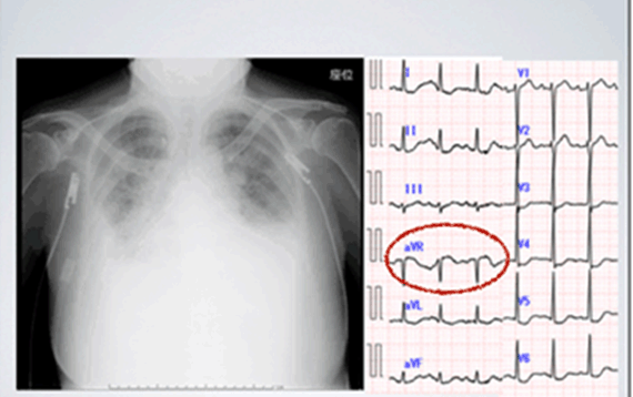
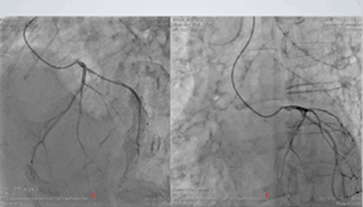
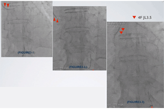
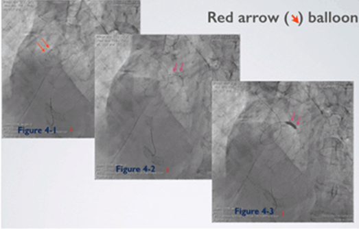
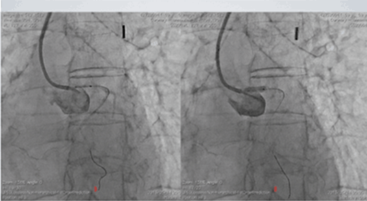
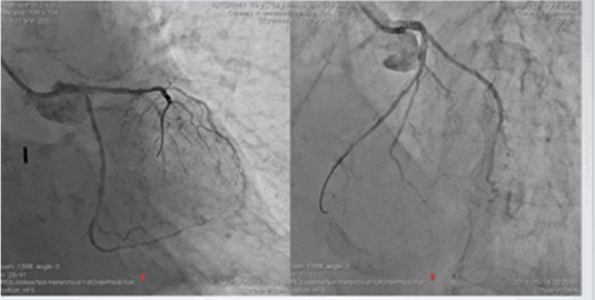
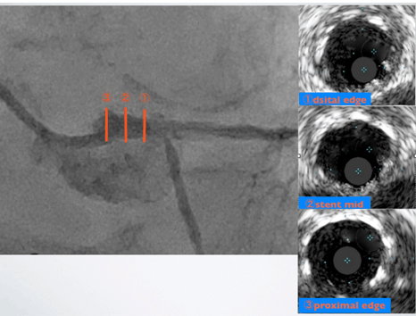
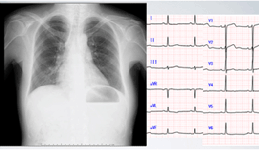
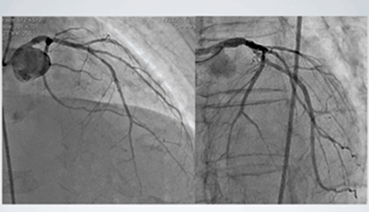
A female in her 60s complaining of severe chest pain and dyspnea was admitted to our hospital with a diagnosis of unstable angina and acute heart failure. Chest radiography revealed pulmonary congestion, echocardiography showed evidence of markedly decreased ventricular wall motion in all ventricular walls except the inferior wall, and electrocardiography demonstrated both marked ST-segment depression in precordial leads and ST elevation in the aVR lead (Figure 1). Risk factors for coronary artery disease, including diabetes mellitus and dyslipidemia, were noted. Access to the same region was not possible because the patient underwent right femoral artery bypass surgery five years earlier. After the insertion of an intra-aortic balloon pump (IABP) into the left femoral artery, we inserted a 4 French (Fr) sheath into the right radial artery. At this time, coronary angiography revealed evidence of 90% ostial stenosis of the left main trunk (LMT) (Figure 2). However, because of spasm in the right radial artery, we changed to the left radial artery. Using a long sheath, we attempted to continue percutaneous coronary intervention (PCI), but were unable to engage a 6 Fr guiding catheter (GC) because of severe tortuosity of the left subclavian artery. Therefore, we attempted intracoronary passage of a 4 Fr JL3.5 GC to exchange the 4 Fr diagnostic catheter with a 6 Fr GC using an extension wire (Figure 3-1, 3-2, and 3-3). However, before entering the left coronary artery, the guidewire coiled around the catheter, which subsequently prolapsed; therefore, the 6 Fr GC could not be engaged. We carefully inserted a 3.0-mm semi-compliant balloon up to the LMT lesion without GC support and were able to engage the GC by dilating the balloon and using the anchor balloon technique (Figure 4-1, 4-2, 4-3). The process took approximately 5 s, and hemodynamics were not affected. The procedure was concluded after placement of an XIENCE V® Everolimus Eluting Coronary stent (Abbott Laboratories, Abbott Park, IL, USA), and stent apposition was confirmed by intravascular ultrasound (IVUS; (Figure 5) (Figure 6) (Figure 7)). Fluoroscopy time was 40 min, and the radiation exposure dose was 1.2 Gy. A total of 220 ml of radiocontrast medium was used and the total procedure time was 80 min. The patient was subsequently admitted to the coronary care unit, where heart function rapidly improved (Figure 8). The patient was subsequently given an ambulatory discharge on hospitalization day 14. Her postoperative course was uneventful. An angiography performed at the 2-month follow-up showed no recurrence of stenosis (Figure 9). | ||||||
| ||||||
| ||||||
|
| ||||||
|
| ||||||
| ||||||
| ||||||
| ||||||
| ||||||
|
| ||||||
| ||||||
|
Discussion
| ||||||
|
The patient was admitted with post-acute coronary syndrome (ACS) with complicating cardiogenic shock and underwent right femoral artery bypass surgery five years earlier. Transradial intervention (TRI) was performed after insertion of an IABP because of limited vascular access. We attempted to insert a diagnostic catheter with a 0.014-inch guidewire before replacing this with a GC using an extension wire because insertion of the GC was initially difficult and insertion of a diagnostic catheter was possible. However, because of the severe tortuosity of the left subclavian artery, the guidewire prolapsed during the procedure, making insertion impossible. A balloon was inserted without the use of a GC, and the anchor balloon technique was implemented. Yoshimachi et al. [1] reported performing IVUS and inserting a balloon when performing PCI using the King's cloth technique without the use of a GC, although a 0.035-inch guidewire or microcatheter might have been useful in this patient. We carefully inserted a semi-compliant balloon up to the LMT, even though it was fixed to a prolapsed wire. A risk of negative effects on vital signs was present; however, inflation time was maintained within approximately 5 s. The anchor balloon technique was also simultaneously used to engage the GC. The balloon was dilated for a short time, and the GC was inserted without any negative effects on the hemodynamic state of the patient because passage of the balloon was difficult due to severe stenosis of the LMT. Prognoses of TRI and transfemoral coronary intervention (TFI) are not very different [2] [3] [4] . With regard to complications in ACS [5], ST segment elevation myocardial infarction [6][7] [8] [9] [10] or IABP support is associated with a favorable prognosis for TRI [11]. Transradial PCI may be considered for severely obese patients [12] and women [13] who are at a higher risk of bleeding complications. The limitations for transradial PCI were few, and this made it particularly suitable for our patient in whom the approach site was limited in size. Maneuvering of the GC might have been easier by downsizing to a 5 Fr GC [14] or inserting a 0.035-inch guidewire during the procedure. TRI or a downsizing stenting system is essential for patients in whom the approach site is limited. The use of an extension wire after the insertion of the diagnostic catheter and the anchor balloon technique has been successful in limited cases when insertion of a GC is difficult. | ||||||
|
Conclusion
| ||||||
|
The guiding catheter (GC) insertion is difficult during transradial intervention for patients with acute coronary syndrome (ACS) and stenosis of the left main trunk and in whom the approach site is limited in size. The GC insertion and percutaneous coronary intervention can be achieved when the diagnostic catheter is exchanged with a GC using a 0.014-inch wire as well as an extension wire, after which the balloon anchor technique should be used. | ||||||
|
References
| ||||||
| ||||||
|
[HTML Abstract]
[PDF Full Text]
|
|
Author Contributions
Daizaburo Yanagi – Substantial contributions to conception and design, Acquisition of data, Drafting the article, Revising it critically for important intellectual content, Final approval of the version to be published Masanori Okabe – Substantial contributions to conception and design, Revising it critically for important intellectual content, Final approval of the version to be published Takeshi Serikawa – Substantial contributions to conception and design, Acquisition of data, Drafting the article, Revising it critically for important intellectual content, Final approval of the version to be published Yusuke Yamamoto – Substantial contributions to conception and design, Acquisition of data, Drafting the article, Revising it critically for important intellectual content, Final approval of the version to be published |
|
Guarantor of submission
The corresponding author is the guarantor of submission. |
|
Source of support
None |
|
Conflict of interest
Authors declare no conflict of interest. |
|
Copyright
© 2015 Daizaburo Yanagi et al. This article is distributed under the terms of Creative Commons Attribution License which permits unrestricted use, distribution and reproduction in any medium provided the original author(s) and original publisher are properly credited. Please see the copyright policy on the journal website for more information. |
|
|