| |
|
|
|
Case Series
| ||||||
| A novel approach of human embryonic stem cells therapy in treatment of Friedreich's ataxia | ||||||
| Geeta Shroff | ||||||
|
MBBS, Director, Nutech Mediworld, New Delhi, India.
| ||||||
| ||||||
|
[HTML Abstract]
[PDF Full Text]
[Print This Article]
[Similar article in Pumed] [Similar article in Google Scholar] 
|
| How to cite this article |
| Shroff G. A novel approach of human embryonic stem cells therapy in treatment of Friedreich's ataxia. Int J Case Rep Images 2015;6(5):261–266. |
|
Abstract
|
|
Introduction:
Friedreich's ataxia (FRDA) is an autosomal recessive inherited disease that damages nervous system and impairs muscle coordination. FRDA usually begins in childhood and is caused by expanded GAA triplet repeat within the first intron of the frataxin (FXN) gene leading to reduced level of mitochondrial protein frataxin. There is no effective treatment for FRDA. If stem cells are transplanted near the affected cells under oxidative stress in FRDA patients, they can produce tropic factors, thereby increasing the survival of the cells. In FRDA, the mechanism to remove the reactive oxygen species (ROS) is impaired leading to oxidative stress and cell death. Stem cells may have ability to protect cells susceptible to oxidative stress that occurs in FRDA. In our previous studies we have shown the improvement in the patients' condition who were suffering from cerebral palsy and cortical visual impairment after human embryonic stem cells (hESCs) therapy.
Case Series: Herein, I report three cases of FRDA patients who were treated with hESCs therapy. All the patients were suffering from problems like difficulty in walking, standing or climbing stairs and muscle weakness. After undergoing hESCS therapy, improvement in condition of all the patients was observed. Conclusion: The hESCs therapy was effective in treating patients with FRDA. Further research is required to understand the mechanism of action of hESCs. | |
|
Keywords:
Friedreich's ataxia, GAA triplet repeat human embryonic stem cells, Neurodegenerative diseases
| |
|
Introduction
| ||||||
|
An autosomal recessive inherited disease Friedreich's ataxia (FRDA) is the most common form of hereditary ataxia that damages nervous system and impairs muscle coordination (ataxia). FRDA usually begins in childhood and is characterized by movement problems such as gait ataxia, or walking difficulty that worsens over time. FRDA is a rare disorder [1] caused by expanded GAA triplet repeat within the first intron of the frataxin (FXN) gene leading to reduced level of frataxin, a mitochondrial protein. [2] [3] FXN protein is involved in regulation of iron homeostasis, biosynthesis of iron-sulfur clusters, energy conversion and stimulation of oxidative phosphorylation. FXN prevents highly redox-reactive metal from generating oxidative stress [4] [5] [6] . Lack of FXN causes iron overloading and increase free-radical production [7] that triggers a series of metabolic derangements [4]. The underlying molecular mechanisms for instability in GAA repeat are currently unknown [8]. There is no effective treatment for FRDA [1]. Only few treatments with free-radical scavengers or antioxidants are available that counteract oxidative stress in FRDA. But there is no proof of the positive results in the neurological aspects of the disorder. Thus, alternative approaches must be developed to treat FRDA [9]. Human embryonic stem cells (hESCs) have unlimited proliferative capacity and potential to differentiate into all types of somatic cells [10] [11]. According to Jones et al., if stem cells are transplanted near the affected cells that are under oxidative stress, they can produce tropic factors, thereby increasing the survival of the cells in FRDA patients [9]. In our previous studies we have shown the improvement in patient's condition who were suffering from cerebral palsy and cortical visual impairment after hESCs therapy [12] [13]. Herein, I report three cases of FRDA patients who were treated with hESCs therapy. Two of these patients (Case I and II) were siblings. In this study, animal free and chromosomally stable hESCs (NTECH 2000n/nn) were used. The cell lines are cultured and maintained as per our proprietary in-house patented technology in a GMP (Good manufacturing practices), GLP (Good Laboratory Practice) and GTP (Good Tissue Practices) certified Nutech Mediworld laboratory (Patent-WO 2007/141657A PCT/1B 2007 Published 13 December 2007). The evidence for the use of hESCs at Nutech Mediworld has been submitted and accepted at House of Lords, Regenerative Medicine, Science and Technology Committee [8]. Prior to start of the treatment all three patients provided a written informed consent. A thorough examination of the patients was done by the doctors and the rehabilitation team during the treatment. Video recordings were also made. The treatment consisted of three phases in which 0.25 mL hESCs were administered through intramuscular route twice daily and 1 mL of hESCs were administered through intravenous route twice every 7th day for 12 weeks initially and then four weeks thereafter. The details of the technique have been elaborated elsewhere [12] . | ||||||
|
Case Series
| ||||||
|
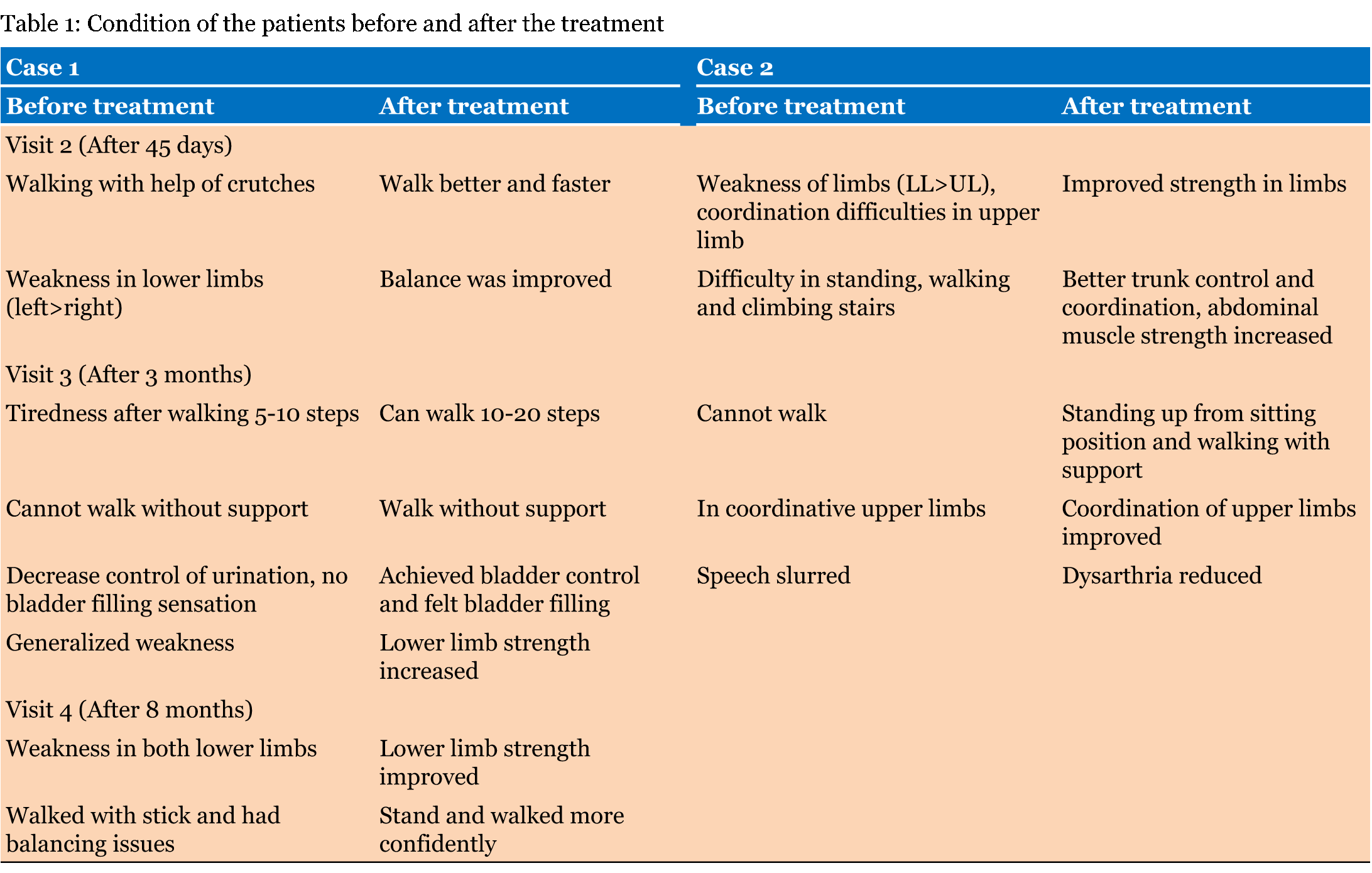
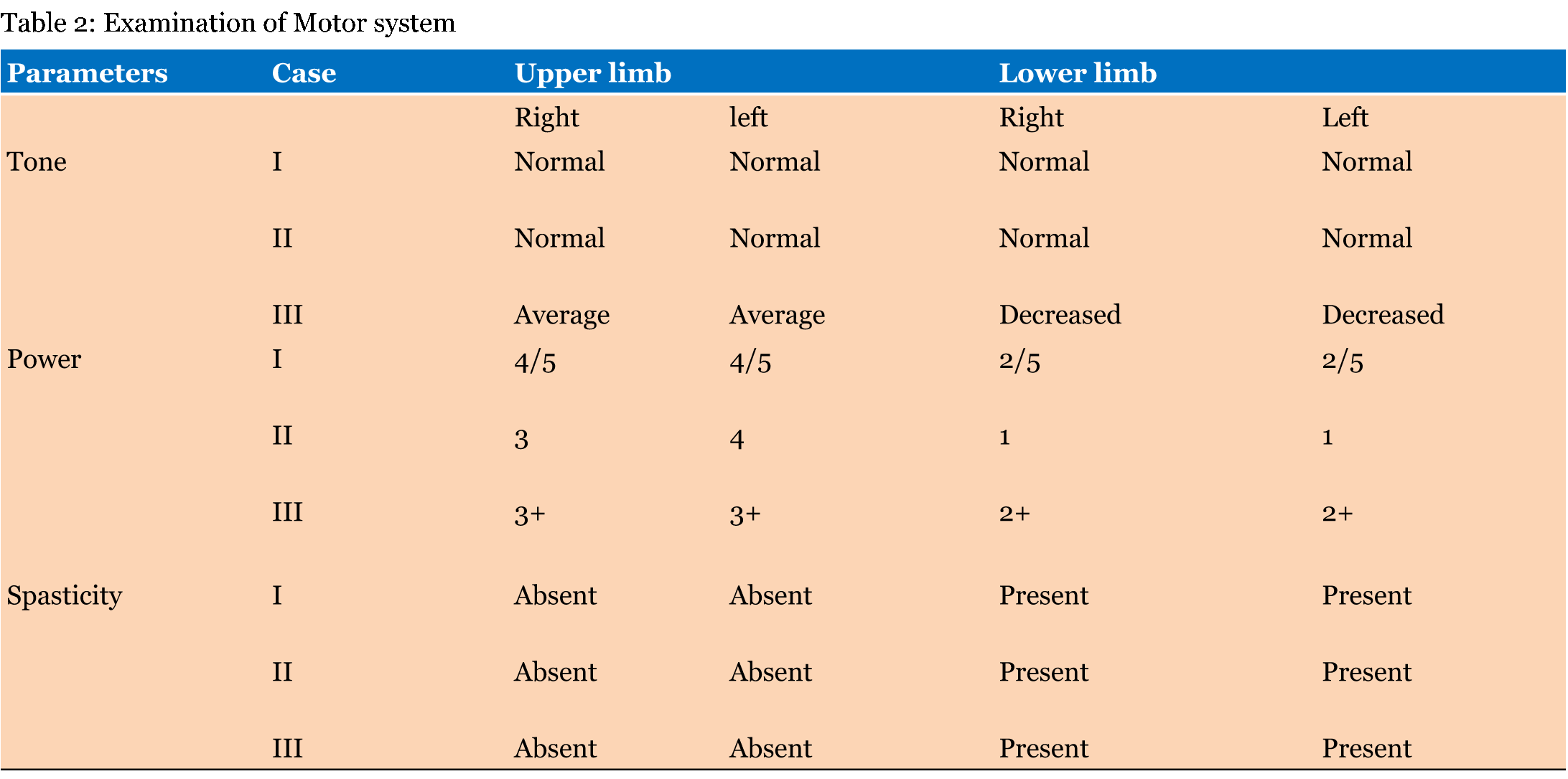
Case 1 and Case 2: A 25-year-old female (Case 1) and a 28-year-old male (Case 2) were admitted at Nutech Mediworld on July 2011 with chief complaints of difficulty in walking, standing, and climbing stairs, weakness in lower limbs and slowness of speech. They both were siblings. There was a history of five siblings death. They were born of a consanguineous marriage and father was diabetic. Both patients were born normal with a normal birth weight. Case 1: Patient was well till 16 years of age, when she developed weakness of lower limbs which gradually increased with time. Her central nervous system (CNS) coordination was affected. The flaccidity in the upper limb was absent. She was wheelchair bound and needed help for her daily activities. Case 2: Patient was apparently well until 12 years of age when he developed progressive weakness in all limbs and trunk with frequent falls. The weakness in lower limbs, difficulty in walking and climbing stairs deteriorated gradually with time followed by generalized weakness of body. He was bound to wheelchair for last eight years. There was atrophy of upper limb and weakness of hand muscles. The speech was affected from last two years. He also had complaints of edema in right foot. At our facility, both were given hESCs therapy as a primary treatment. In Case 1, after the first treatment, overall stamina, lower limb strength and knee flexion was improved. She could stand without support for 20 seconds and stand and walk with the help of a stick. After a gap of 45 days, patient was admitted second time with the complaints of weakness in lower limbs and sustained burns in legs while taking shower. On examination, difficulty in initiating knee flexion in prone position was observed, but she could stand without support or with help of stick for few seconds. Again treatment was given. After the treatment balance was improved and she was able to walk faster. A gap of three months was given. She was again admitted for third time with complaints of tiredness after walking 5–10 steps, decrease control of urination especially in morning with no bladder filling sensation and generalized weakness. She could not walk or stand without support. Human embryonic stem cells treatment was given again. After the treatment, she could walk 10–20 steps without support, strength in lower limbs was increased and bladder filled sensation was improved. At fourth visit, the patient presented with complaints of generalized weakness of whole body, balancing issues while walking, and difficulty in bending both knees. After the treatment, the patient reported further improvement in her condition. Her lower limb muscle strength was improved. She could stand and walk more confidently without the fear of falling down. She could perceive fullness of urinary bladder and her bladder control was improved. Her exercise endurance during physiotherapy increased and could do static cycling more effectively. The status of the patient before and after hESC therapy at visit 2, 3 and 4 is given in Table 1. In Case 2, electrophysiology studies showed that lower limb was suggestive of severe motor sensory asymmetric peripheral neuropathy. After clinical examination, a brisk knee jerk, absence of ankle reflexes, pitting pedal edema, fasciculation in tongue, bilateral hypothenar wasting and dysdiadochokinesia was reported. Examination details of CNS and motor system is given in Table 2. The patient was given hESC therapy as a primary treatment four times over a period of 18 months (including gap phases). After the treatment, overall stamina and endurance was better, strength in upper limbs was increased, he could flex his knees, and toes, walk in walkers with calipers, spasticity in legs was reduced and sitting balance was also improved. After 45 days, on visit 1, he was admitted with chief complains of weakness of limbs. hESC therapy was given again to this patient. The status of the patient before and after hESC therapy at visit 2, 3 is given in Table 1. At the end of the treatment, overall stamina and coordination was improved. The strength in limbs, trunk control, waking gait with walker, sitting, standing and walking with support was improved. Case 3: A 35-year-old male was admitted at Nutech Mediworld on February 2006 with chief complaints of progressive weakness of the body over the last 20 years. The patient had a history of choking sensation with liquids and cardiomyopathy (left ventricular ejection fraction, LVEF- 15–20%) for which he was taking medicines. His elder sister had same disorder and she died due to heart failure in 2004. The patient was not able to stand, walk or sit. He used wheel chair and was unable to do his day to day activities. He managed to have food and dress by himself. His coordination and balance was lost and had slurred speech and hearing loss. A mild difficulty in swallowing with aspiration of liquids infrequently was also reported. The patient was given hESCs therapy as a primary treatment. The examination details of patient's central nervous system and motor system is given in Table 2. After the treatment, spine curvature was improved, neck was more erect, leg movement was significantly increased, lower limbs spasticity was reduced and endurance was better. The patient was able to walk 5–7 steps forward and backward with full calipers. | ||||||
| ||||||
|
| ||||||
| ||||||
|
Discussion
| ||||||
|
In this study I observed that hESCs therapy was effective in treating patients with FRDA. Insufficiency of the protein frataxin in FRDA patients leads to spinocerebellar neurodegeneration along with associated movement disorders. FRDA patient also have increased risk of diabetes and cardiomyopathy, which are the leading cause of death [14]. In this cases, I observed that the patients were suffering from various problems like difficulty in walking, standing or climbing stairs and muscle weakness. After undergoing hESCs therapy, a marked improvement in the condition of all patients was noticed. At the end of the treatment, endurance of all patients was increased. They were able to stand and walk confidently. Other changes includes improved urinary bladder control in Case I, improved overall stamina and coordination in Case II and improved spine curvature in Case III. Our finding that the hESCs are capable of treating FRDA is supported by our previous studies, where hESCs have successfully treated the patients with cerebral palsy [12]. Human embryonic stem cells might have the potential to treat patients with neurodegenerative diseases such as cerebral palsy, Friedreich's ataxia, etc. Currently, there is no cure for FRDA. However, associated symptoms and complications can be treated [1] . Neurons and cardiomyocytes are the two affected cell types in FRDA [8]. As human stem cells have ability to migrate and home to the injury site [15], we could assume that hESCs might have also migrated to the affected brain and differentiated into neurons and cardiomyocytes. The size of the hESCs used in our study was < 1 µm, so it can be assumed that hESCs have permeated through the parenchyma via blood brain barrier. Though neurodegenerative diseases like Parkinson's disease, multiple sclerosis, multiple system atrophy, Alzheimer disease, FRDA and Huntington's disease have different etiology, but all these disease share a common trait involving mitochondrial dysfunction that lead to iron accumulation and ultimately cell death [2] [16] [17]. Several studies have shown the clinical benefits of hESCs in treating neurodegenerative diseases such as Parkinson's disease and multiple sclerosis [18] [19] [20]. In current cases, I expect that hESCs have similar clinical benefits in reducing the clinical symptoms associated with FRDA. An efficient antioxidant system in our body removes the oxidative stress. In FA, the mechanism to remove the reactive oxygen species (ROS) is impaired leading to oxidative stress and cell death [7] [9]. Stem cells may have ability to protect cells susceptible to oxidative stress that occurs in many neurodegenerative diseases like FRDA [9]. Jones et al. cultured FRDA cells undergoing oxidative stress in healthy human adipose stem cell conditioned medium. Increased cell survival and frataxin expression and, unregulated oxidative-stress-related genes were observed. This result was due the presence of trophic factors such brain-derived neurotrophic factor (BDNF) expressed by the adipose stem cells in the conditioned medium [21]. Jones et al. in their another study isolated mouse bone marrow mesenchymal stem cell-conditioned medium and cultured dorsal root ganglia neurons isolated from an FRDA mouse model. The results showed that the conditioned medium increased the neurons cell survival, decreased apoptosis when exposed to oxidative stress. The transcription of certain oxidative stress-related genes was also activated. Thus, autologous stem cells transplantation in FRDA patients may protect the affected neurons [9]. Stem cells such as mesenchymal stem cell, ESCs, induced pluripotent stem cells are capable of producing trophic factors [22]. So it is possible to consider that hESCs might have produced trophic factors at the affected part, thereby increasing the frataxin expression and preventing the cells degeneration. However, studies supporting this assumption are lacking. We can infer from our study that hESCs can lead to mild to moderate and short term improvement of few weeks in the patients with FRDA. In this study, the three patients did not come back on time after the therapy. FRDA is aprogressive disease that requires regular injections of hESCs every three months in first year, every 4–8 months in second year and every 6–8 months in third years and thereafter yearly. | ||||||
|
Conclusion
| ||||||
|
This case series provides a novel direction in which human embryonic stem cells (hESCs) therapy may be used in treatment of Friedreich's ataxia (FRDA). I can postulate that hESCs have ability to modulate disease frequency and severity. I would continue to monitor the condition of the patients before retreatment is required. Further research is required to shed further light on understanding the mechanism of action of hESCs. | ||||||
|
List of Abbreviations
| ||||||
|
FRDA- Friedreich's ataxia | ||||||
|
References
| ||||||
| ||||||
|
[HTML Abstract]
[PDF Full Text]
|
|
Author Contributions:
Geeta Shroff – Substantial contributions to conception and design, Acquisition of data, Analysis and interpretation of data, Drafting the article, Revising it critically for important intellectual content, Final approval of the version to be published |
|
Guarantor of submission
The corresponding author is the guarantor of submission. |
|
Source of support
None |
|
Conflict of interest
Authors declare no conflict of interest. |
|
Copyright
© 2015 Geeta Shroff. This article is distributed under the terms of Creative Commons Attribution License which permits unrestricted use, distribution and reproduction in any medium provided the original author(s) and original publisher are properly credited. Please see the copyright policy on the journal website for more information. |
|
|
|
About The Author
| |||
| |||