Case Report
|
| |
|
Internal herniation beneath mesodiverticular band producing ambiguous picture in intestinal obstruction: A management dilemma
|
|
Rakesh Chauhan1,
Rajesh Chaudhary2,
R. Dayashankar2
|
1MBBS, MS, Assistant Professor Surgery Dr. RP Govt. Medical College Kangra at Tanda, HP, India.
2MBBS, Junior Resident, Dr. RP Govt. Medical College Kangra at Tanda, Himachal Pradesh, India.
|
doi:10.5348/ijcri-201532-CR-10493
|
Address correspondence to:
Dr. Rakesh Chauhan
MS, Assistant Professor Surgery, Dr. RP Govt. Medical College Kangra at Tanda
Himachal Pradesh
India
Email: drrakeshchauhan66@gmail.com
|
Access full text article on other devices
 |
|
Access PDF of article on other devices
 |
|
|
[HTML Abstract]
[PDF Full Text]
[Print This Article]
[Similar article in Pumed]
[Similar article in Google Scholar]

|
Introduction
|
|
Intestinal obstruction because of entrapment of small gut loop between mesodiverticular band and mesentery is a rare cause of obstruction due to Meckel's diverticulum complication. It cannot be diagnosed preoperatively and because of the loosely entrapped loop obstruction is not complete. So it produces difficult situation for the surgeon to take decision in favor of surgery [1]. We report a case of 13-year-old boy presenting with intestinal obstruction due to mesodiverticular band which presented an ambiguous picture difficult to take decision for surgery.
|
Case Report
|
|
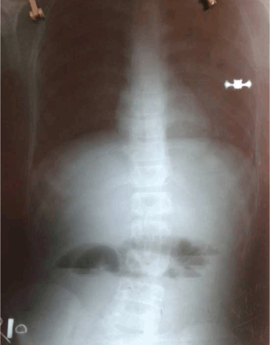
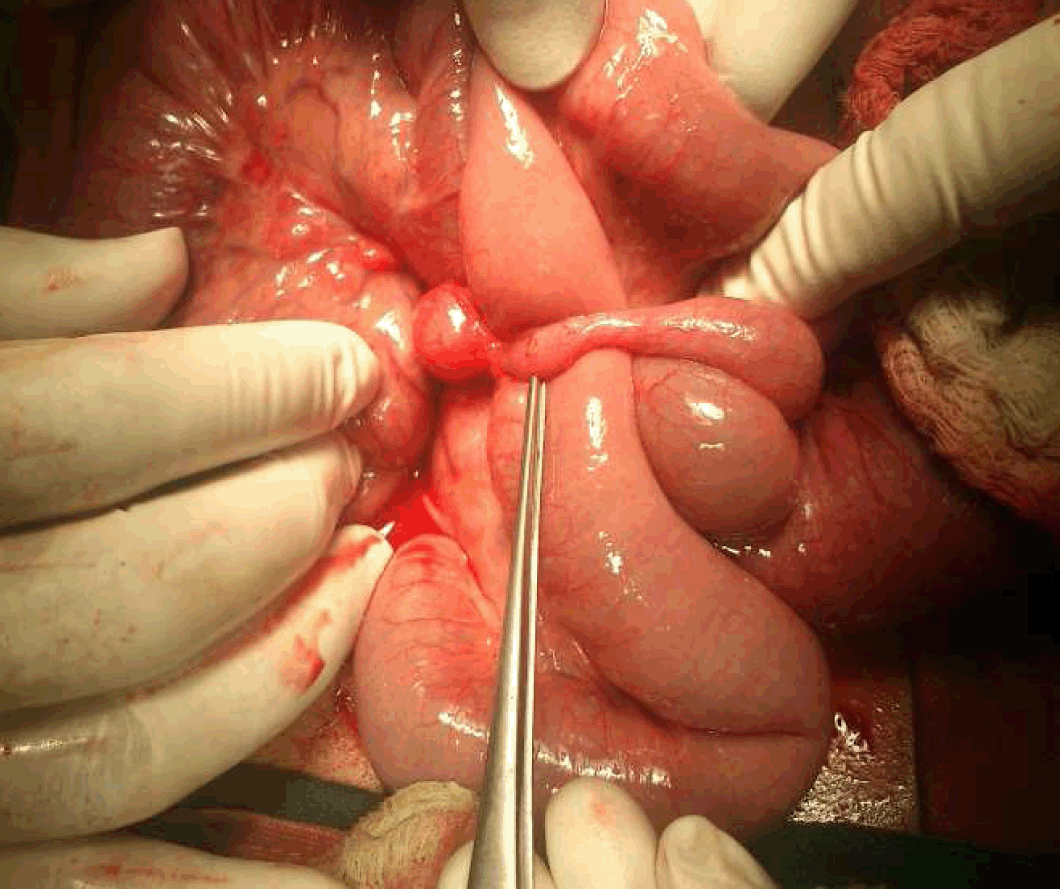
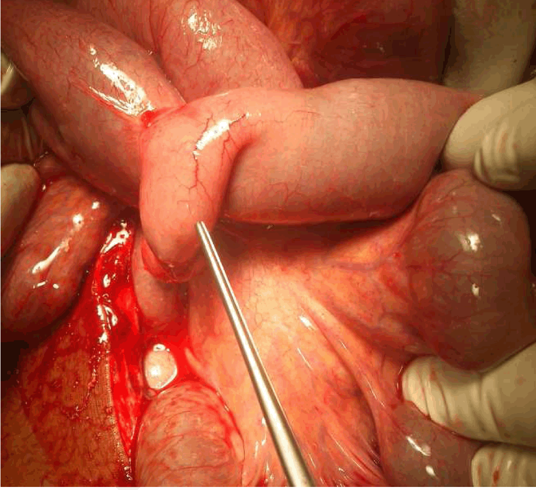
A 13-year-old boy from poor socio-economic family background presented to the emergency outpatient department in the night with a history of colicky pain in umbilical region associated with bilious vomiting and constipation for the last two days. There was a history of ingestion of some dry fruits and half cooked food before the appearance of symptoms. There was no history suggestive of worm infestation. On examination, the patient was afebrile and pulse was 80/min. Blood pressure was normal and there was no dehydration. Abdomen was mildly distended and soft, but bowel sounds were not present. Hernia sites were normal and on digital rectal examination (DRE) there was slight ballooning with hard fecal matter impacted. Biochemistry and hematology was normal especially the TLC which was 5700/cc. Urine output was adequate. Ryle's tube aspirate was about 1 L. Chest X-ray was normal and standing abdominal X-ray showed multiple air fluid levels in the small gut and there was no gas in the large gut (Figure 1). Peritoneal aspiration showed straw colored fluid. Diagnosis of acute intestinal obstruction was made with possibility of ileocecal tuberculosis or bolus obstruction. So conservative trial was given with proctoclysis enema. Patient passed multiple hard stools with passage of the flatus. With this pain subsided altogether and distention was decreased. Next day air fluid levels persisted in the abdominal X-ray. However, patient was pain free and Ryle's tube aspirate was decreased to 200 mL/d. Patient gave the history of passage of flatus in between. Sluggish bowel sounds were also heard. On second day of admission the patient was again pain free, feeling hungry, pulse was 78/min, abdomen was soft, bowel sounds were very sluggish and rectum was collapsed but air fluid levels persisted in the X-ray (Figure 2). Therefore, decision for laparotomy was taken. Patient was taken up for surgery with prior anesthetic clearance with one unit of whole blood arranged. On exploration mesodiverticular band extending from the tip of Meckel's diverticulum to the right side of the mesentery was found forming a tunnel of about 2–3 cm through which small gut loops were loosely herniated without forming complete obstruction or constriction band in its wall (Figure 3). Proximal segment was dilated and there was about 200 cm3 straw colored fluid in the cavity. Diverticulum was about 7 cm with wide base (Figure 4). Excision of band and diverticulum with ileo-ileal anastomosis was done. Patient recovered well without any complication and discharged on eighth day of surgery. Specimen was sent for histopathological examination. Histopathological examination report confirms Meckel's diverticulum with a fibrous band attached to its tip.
|
|
|
|
|
|
|
|
|
|
|
Discussion
|
|
Meckel's diverticulum, a true diverticulum, is the most common congenital abnormality of the gut. First reported in 1598 by Hildanus, it was described in anatomical detail by Johann Meckel in 1809 whose name it carries [2]. It is a remnant of intestinal end of omphalomesenteric duct which normally gets obliterated during eighth week of gestation. It is present in 2% of the population, however, about 96% of the cases are asymptomatic in which it is incidentally detected on surgery, radiographic imaging, endoscopically for other purposes or on autopsy [3]. Only in 4% of the cases symptoms arise as a result of complications related to diverticulum. More than 50% of the symptomatic patients are less than two years of age [4]. The complications related to the Meckel's diverticulum include bleeding, intestinal obstruction, infection, perforation, neoplasms. The most common symptom of complicated Meckel's diverticulum is intestinal obstruction. The reasons for obstruction are volvulus around the band between diverticulum and umbilicus, intussusception of the inverted diverticulum acting as a lead point, stricture, entrapment of gut loops under the mesodiverticular band and litter's hernia [5]. Mesodiverticular band is a rare intraoperative finding leading to entrapment of the gut loop between it and mesentery. Mesodiverticular band cannot be diagnosed preoperatively by any investigation and is an incidental finding during surgery only. It is a flimsy mesentery carrying independent blood supply to the diverticulum through persistent vitelline vessels and runs between the tip of diverticulum and the mesentery. Through the space between the two (mesentery and Meckel's diverticulum carrying band) internal herniation of the small gut loops take place [6]. Investigations for Meckel's diverticulum depends upon the complication it produces. For the bleeding which is the most common complication in the children sodium 99Tc pertechnetate scan is investigation of the choice with accuracy of more than 90% [7]. In adults accuracy of scan is less than 50% because of less prevalence of the gastric mucosa. In adults, accuracy can be increased by pentagastrin, cimetidine or glucagon [8]. Angiography can also be used to detect the bleeding. Obstruction as a result of the intussusception can be detected with the help of the ultrasonography or computed tomography scan or barium enema [7] [8]. But in most of the cases of obstruction, cause is confirmed on laparotomy. This patient also presented to the emergency department with a history of colicky pain abdomen, vomiting and not passing stool and flatus for the last two days. Vitals were normal but standing X-ray of abdomen showed multiple air fluid levels in small gut. Abdomen was mildly distended but soft, however, shifting dullness was present. Paracentesis showed straw colored fluid. RT aspiration showed bilious fluid about one liter on presentation. There was no history of contact with tuberculosis patient or intake of ATT but ileocecal tuberculosis was kept one of the possibilities. With conservative treatment patient settled clinically but not radiologically and on exploratory laparotomy internal herniation of small gut through a mesodiverticular band was found. This picture leading to the incomplete obstruction makes the decision difficult if to carry on conservative treatment or do emergency laparotomy. But since the air fluid levels were not disappearing despite the improvement in the clinical status we decided in the favor of laparotomy and found mesodiverticular band. Emptied sigmoid colon with enema reduced the pressure and allowed luminal contents to pass through herniated gut leading to relief of symptoms but loop was still entrapped under the mesodiverticular band not allowing to disappearance of the air fluid levels. It projected ambiguous picture leading to difficult situation for the surgeon to take decision for surgery.
|
Conclusion
|
|
Mesodiverticular band is a rare cause of intestinal obstruction due to loose entrapment of small gut loops in a tunnel produced by it and mesentery. So it does not produce complete obstruction. On conservative treatment patient gives an improving clinical picture but radiologically obstruction is not settled. Therefore, surgeon faces a dilemma weather to operate or to continue manage the patient conservatively. We faced this challenge and decided to operate the patient.
|
References
|
-
Park JJ, Wolff BG, Tollefson MK, Walsh EE, Larson DR. Meckel diverticulum: The Mayo Clinic experience with 1476 patients (1950-2002). Ann Surg 2005 Mar;241(3):529–33.
[CrossRef]
[Pubmed]

-
Opitz JM, Schultka R, Gobbel L. Meckel on developmental pathology. Am J Med Genet A 2006 Jan 15;140(2):115–28.
[CrossRef]
[Pubmed]
-
Anderson DJ. Carcinoid tumor in Meckel's diverticulum: Laparoscopic treatment and review of the literature. J Am Osteopath Assoc 2000 Jul;100(7):432–4.
[Pubmed]
-
Ruscher KA, Fisher JN, Hughes CD, et al. National trends in the surgical management of Meckel's diverticulum. J Pediatr Surg 2011 May;46(5):893–6.
[CrossRef]
[Pubmed]
-
Elsayes KM, Menias CO, Harvin HJ, Francis IR. Imaging manifestations of Meckel's diverticulum. AJR Am J Roentgenol 2007 Jul;189(1):81–8.
[CrossRef]
[Pubmed]
-
Chieh-Wen L, Internal Herniation of Ileum Caused by Band of sMeckel's Diverticulum. J Med Sci 2011;31(2):79–80.
-
Connolly SA, Drubach LA, Connolly LP. Meckel's diverticulitis: Diagnosis with computed tomography and Tc-99m pertechnetate scintigraphy. Clin Nucl Med 2004 Dec;29(12):823–4.
[CrossRef]
[Pubmed]
-
Rerksuppaphol S, Hutson JM, Oliver MR. Ranitidine-enhanced 99mtechnetium pertechnetate imaging in children improves the sensitivity of identifying heterotopic gastric mucosa in Meckel's diverticulum. Pediatr Surg Int 2004 May;20(5):323–5.
[Pubmed]
|
[HTML Abstract]
[PDF Full Text]
|
|
Author Contributions
Rakesh Chauhan – Substantial contributions to conception and design, Acquisition of data, Analysis and interpretation of data, drafting the article, Critical revision of the article and final approval of the version to be published
Rajesh Chaudhary – Substantial contributions to conception and design, Acquisition of data, Analysis and interpretation of data, drafting the article, Critical revision of the article and final approval of the version to be published
Dayashankar – Substantial contributions to conception and design, Acquisition of data, Analysis and interpretation of data, drafting the article, Critical revision of the article and final approval of the version to be published
|
Guarantor of submission
The corresponding author is the guarantor of submission.
|
Source of support
None
|
Conflict of interest
Authors declare no conflict of interest.
|
Copyright
©
2015 Rakesh Chauhan et al. This article is distributed under the terms of Creative Commons Attribution License which permits unrestricted use, distribution and reproduction in any medium provided the original author(s) and original publisher are properly credited. Please see the copyright policy on the journal website for more information.
|
|
About The Authors
|
|
|

| |
Rakesh Chauhan is Assistant Professor Department of Surgery at Dr. Rajendera Prasad Medical College, Kangra at Tanda, Himachal Pradesh, India. He earned undergraduate (MBBS) and postgraduate (MS General Surgery) degrees from Indira Gandhi Medical College, Shimla, Himachal Pradesh, India. His research work include general surgery, gastrointestinal surgery, oncosurgery and hospital administration.
|
|
|
|

| |
Rajesh Chaudhary is Junior Resident in Department of Surgery at Dr. Rajendera Prasad Medical College, Kangra at Tanda, Himachal Pradesh, India. He earned undergraduate degree from Indira Gandhi Medical College, Shimla, Himachal Pradesh, India.
|
|
|
|
|
| |
R. Dayashankar is Junior Resident in Department of Surgery at Dr. Rajendera Prasad Medical College, Kangra at Tanda, Himachal Pradesh, India.
|
|
|
|