|

|
 |
|
Case Report
| ||||||
| Short left main coronary artery causing dynamic left ventricular outflow tract obstruction and new onset left bundle branch block | ||||||
| Geoffrey Chibuzor Nwuruku1, Godsent Chichebem Isiguzo2, Joel Tamayo Brooks3, Ernest Madu4 | ||||||
|
1MD, Registered Cardiologist and Family Physician, DOCS VIP Clinic, Enugu, Nigeria.
2MBBS, FWACP, Consultant Cardiologist, DOCS VIP Clinic, Enugu, Nigeria. 3MD, Interventional Cardiologist, Heart institute of Caribbean (HIC), Kingston, Jamaica. 4MD, FACP, FACC, FRCP Edin, Professor of Cardiology, Heart institute of Caribbean (HIC), Kingston, Jamaica, and Director, DOCS VIP Clinic Enugu, Nigeria. | ||||||
| ||||||
|
[HTML Abstract]
[PDF Full Text]
[Print This Article]
[Similar article in Pumed] [Similar article in Google Scholar] 
|


| How to cite this article |
| Nwuruku GC, Isiguzo GC, Brooks JT, Madu E. Short left main coronary artery causing dynamic left ventricular outflow tract obstruction and new onset left bundle branch block. Int J Case Rep Images 2015;6(3):156–160. |
|
Abstract
|
|
Introduction:
Left bundle branch block can be seen in conditions like aortic stenosis, extensive coronary artery disease, primary disease of the cardiac electrical conduction system, dilated cardiomyopathy, Lyme disease; it is also associated with short left main coronary artery and dynamic left ventricular outflow tract obstruction. While the former is caused by the shearing force on septal branches of the left anterior descending artery, the latter is related to either Venturi effect in hypertrophic cardiomyopathy or effect of systolic anterior motion caused by abnormal geometric relationship of papillary muscle and the mitral apparatus.
Case Report: A 50-year-old male, former smoker with a history of dyslipidemia, presented with shortness of breath and exertional chest pain. After clinic review, he was thought to have stable angina, electrocardiogram, and echocardiography were normal and lifestyle modification was advised. Patient had some improvement, but represented seven months later with worsening of symptoms. Repeat electrocardiograph showed a new onset left bundle branch block, with short left main coronary artery on coronary angiogram and dynamic left ventricular outflow tract obstruction in stress echocardiography, with a gradient of 40 mmHg. Conclusion: Short left main coronary artery is a rare cause of left bundle branch block, and it should be considered when evaluating patients with new onset left bundle branch block without hypertrophic cardiomyopathy. | |
|
Keywords:
Erectile dysfunction, Hypertrophic cardiomyopathy, Left bundle branch block (LBBB), Left main coronary artery (LMCA), Left ventricular outflow tract (LVOT), Lyme disease, Microvascular dysfunction (MVD), Septal dyssynchrony, Short left main coronary
| |
|
Introduction
| ||||||
|
Although left bundle branch block (LBBB) is a common finding in conditions such as aortic stenosis, extensive coronary artery disease, primary disease of the cardiac electrical conduction system, dilated cardiomyopathy, Lyme disease; it can also be associated with short left main coronary artery (LMCA) and a dynamic left ventricular outflow tract (LVOT) obstruction [1]. While the former is caused by the shearing force on the septal branches of the left anterior descending artery, the latter is related to these major possibilities among others: the Venturi effect in hypertrophic cardiomyopathy (HCM) as well as systolic anterior motion of the mitral valve (SAM) generated largely by drag effect, that is hydrodynamic pushing force of flow directly on the leaflets, and the SAM caused by abnormal geometric relationship of papillary muscle and the mitral apparatus. This abnormal geometry is in part as a result of non-coordinated contraction (mechanical dyssynchrony) of interventricular septum and left ventricular posterior or posterolateral walls which lead to the displacement of the papillary muscles and the chordae tendineae (chordae tendineae SAM). | ||||||
|
Case Report
| ||||||
|
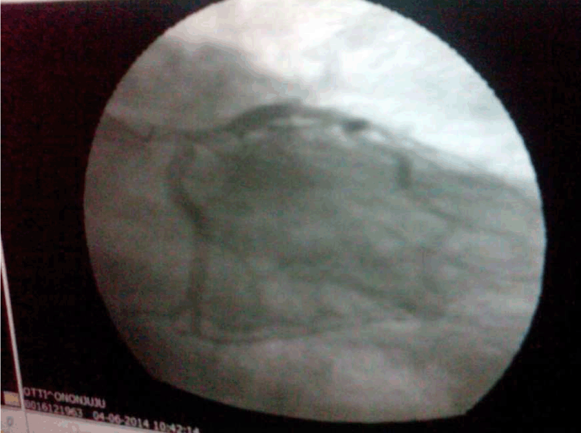
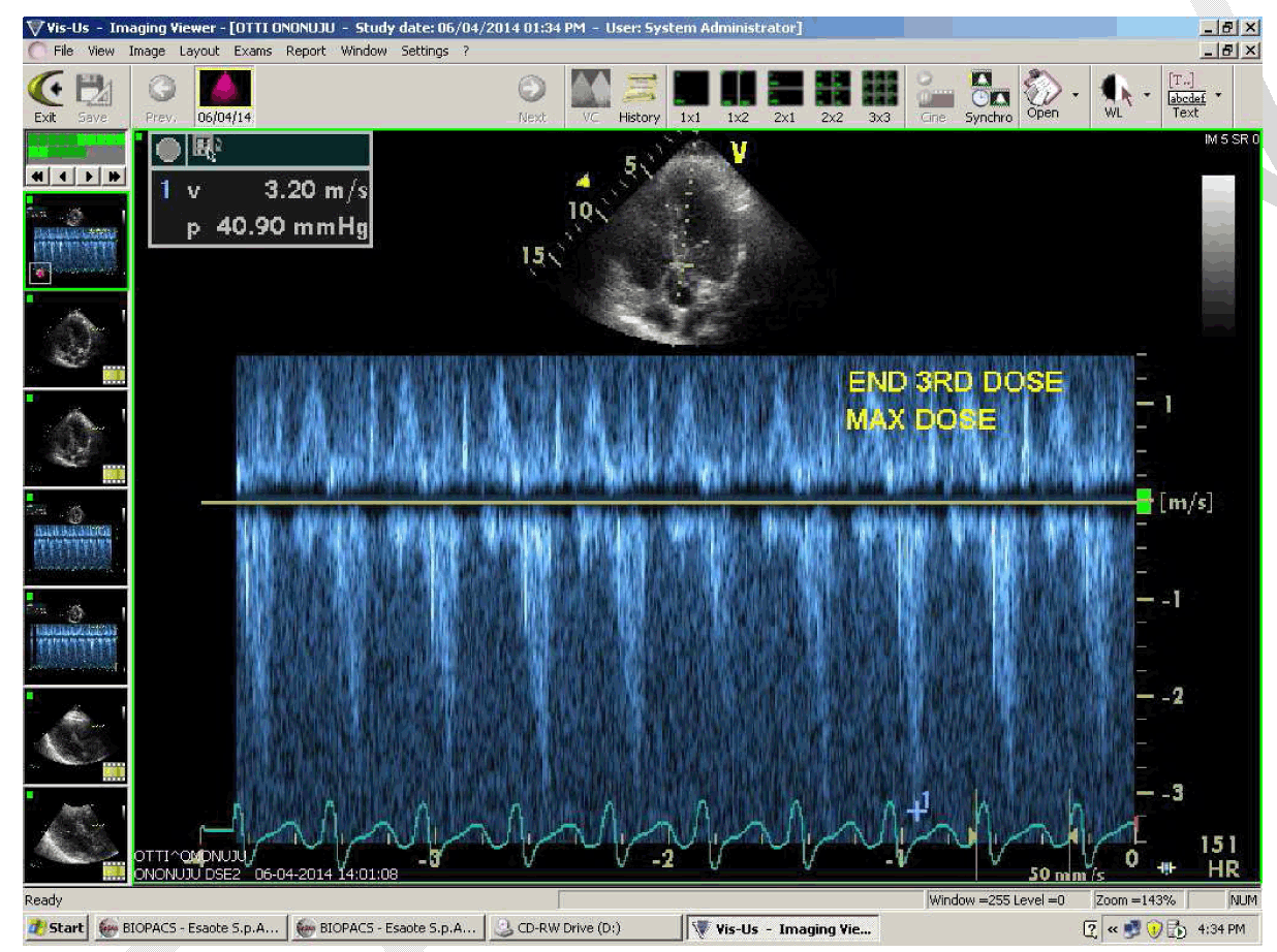
A 50-year-old male with a history of dyslipidemia and erectile dysfunction, stopped smoking 10 years ago, not known to be hypertensive or have diabetes mellitus and no significant family history. He presented with shortness of breath and exertional chest pain at variable threshold. Physical examination was negative while blood workup showed total cholesterol (223 mg/dL), LDL (165 mg/dL) and the remainder of the blood test results were normal. Various other tests conducted including ECG (Figure 1A), ECHO/Doppler and Stress ECHO were not significant while 24-hour Holter monitoring revealed sinus arrhythmias with a solitary ventricular ectopic beat and rare supraventricular ectopic beat with a couplet. A treatment plan that included lifestyle modification was instituted for stable angina. The patient noted improvement but less than a year later the same symptoms recurred and even became more severe. He presented to the clinic again and a repeat ECG revealed a new onset BBB (Figure 1B) that was not seen in the previous rest ECG before stress test (Figure 1A). He was suspected to have a progressive ischemia, and repeat echocardiography showed septal dyssynchrony with good left ventricular systolic function. A coronary angiogram was then done, showing a short left main coronary artery ( (Figure 2), while a stress ECHO revealed a significant dynamic LVOT obstruction (chordae tendineae SAM) with a gradient of 40.9 mmHg (Figure 3) at the end of a third dose with symptoms that prompted the termination of the test. He was discharged on beta blocker (bisoprolol 5 mg daily) management and has reported great improvement in his functional capacity. | ||||||
| ||||||
| ||||||
| ||||||
|
Discussion
| ||||||
|
This is a case of a 50-year-old male patient with a new onset LBBB associated with systolic anterior motion of the mitral valve (chordae tendineae SAM). This finding triggered a LVOT obstruction with a gradient of 40.9 mmHg on a stress echo. The LBBB was spontaneous and thought to have resulted from progressive ischemia as previous ECG and resting ECG before stress test did not reveal this. The echocardiographic findings were related to shortness of breath and angina (on a mild activity) noticed on the patient that was also reproduced during stress test. Studies have shown longstanding LVOT obstruction (basal gradient, ≥30 mmHg) as a strong determinant of HCM-related progressive heart failure symptoms and cardiovascular death but however, showed only a weak relationship evident between outflow obstruction and specifically the risk for sudden cardiac death (usually in patients without significant heart failure symptoms) [2]. We did not find any study that demonstrated this relationship in a patient with SAM caused purely by abnormal geometry of the mitral valve apparatus. A study by Rodriguez Rodrigo et al. demonstrates microvascular dysfunction (MVD) as the cause of deterioration of left ventricular function in patients with isolated LBBB [3]. The study only considered the functional influence of LBBB on the coronary arteries; in contrast our report shows the prognostic influence of the coronary arteries anatomy in the pathogenesis of a spontaneous LBBB of which MVD plays a vital role. Short LMCA has been shown to be a prelude to development of coronary artery disease, and a cause of LBBB, with the later known as a marker of slowly progressing ischemic or non-ischemic cardiac disease and increase cardiovascular disease related death risk, and lower survival rate [4], hence the need for an exhaustive evaluation. In the index patient, finding of new onset LBBB, without any apparent trigger associated with progressive symptoms was a source of concern and lead to our further evaluation with a resting echocardiogram which revealed a septal dyssynchrony while a stress echo showed SAM of the mitral valve with LVOT obstruction demonstrated by a gradient of 40.9 mmHg. He was thereafter studied on coronary angiogram and a short LMCA was observed with no significant coronary artery obstruction. There are divergent results on the role of short LMCA in the pathogenesis of LBBB, most of which are not recent. A study by Lewis et al. in 1969 found that the length of the left main coronary artery (LMCA) was less than 6 mm in all but one of 12 patients with LBBB and was longer than 7 mm in a control group of 25 patients [1]. This data suggest that the cardiac diseases associated with LBBB were not etiologically important or that they were important only when the LMCA was short. We posited that the association between a short LMCA and LBBB could be explained by greater shearing forces imposed during systole in the short arteries (LMCA). This, in turn might compromise flow through the early septal branches of the left coronary system and thus produce ischemia and fibrosis of the left bundle branch. Ischemia as a possible pathogenetic mechanism producing fibrosis, when it affects conducting system, has been found to be the histologic abnormalities in patients with LBBB [5]. This could explain the new onset conducting system abnormality noticed in this patient after one year of a normal ECG. In a study based on pathological observations by Gazetopoulos et al. suggested that short LMCA should be considered as a congenital factor predisposing to the development of atherosclerotic coronary artery disease, confirming their earlier finding that length of the LMCA is an anatomic factor that may influence the rate of development of atheromatosis on its branches [6]. But in contrast to the findings, De Mots et al. showed that the length of LMCA in their 13 patients with LBBB was not significantly different compared to the 78 patients in the control group [7]. The LBBB leads to electrical conduction disorder which by the way of mechanical dyssynchrony causes abnormalities in the movement of the mitral apparatus including anterior and inward or central displacement of the papillary muscle and leaflet elongation that can ultimately lead to SAM and left ventricular outflow tract obstruction in patients without HCM. Several explanations have been adduced for the occurrence of SAM in patients with hypertrophic cardiomyopathy (HCM), notable among which are Venturi effect and anatomic difference in position of papillary muscles and valve leaflets [8] [9]. More recent studies have also attributed the dominant force in the anterior displaced mitral valve leaflets as drag force that is in proportion to velocity of flow in LVOT and to the angle between the anterior mitral leaflet and the direction of flow in the LVOT [10]. Many other factors may play roles in causing patients without HCM to present with LVOT obstruction as seen in the index. These include primary abnormalities of the mitral valve apparatus, like anterior and inward or central displacement of the papillary muscles, leaflet elongation, geometric disarray of the left ventricle caused by LBBB. Short LMCA may also cause progressive microvascular disease, and its clinical consequence depends on the level of obstruction at the LVOT, dyssynchrony impact of LBBB and dynamic state of the patient. | ||||||
|
Conclusion
| ||||||
|
Though there are divergent reports and paucity of recent studies regarding the role of a short left main coronary artery in causing left bundle branch block (LBBB), it has been shown that isolated LBBB with microvascular dysfunction is associated with worse outcome. Therefore, there is need for physicians to evaluate for causes of new onset LBBB in their patients as early discovery and prompt intervention may alter the cause of potential cardiovascular disease mortality and morbidity as seen in the index patient. | ||||||
|
References
| ||||||
| ||||||


|
[HTML Abstract]
[PDF Full Text]
|
|
Author Contributions
Geoffery Chibuzor Nwuruku – Substantial contribution to conception design, Acquisition of data, Drafting of article, Revising it critically for important intellectual content, Final approval of version to be published Godsent Chichebem Isiguzo – Substantial contribution to conception design, Analysis and interpretation of data, Drafting of article, Revising it critically for important intellectual content, Final approval of version to be published Joel Tamayo Isiguzo – Substantial contribution to conception design, Acquisition of data, Revising critically for important intellectual content, Final approval of version to be published Madu Ernest – Substantial contribution to conception design, Acquisition of data, Revising it critically for important intellectual content, Final approval of version to be published |
|
Guarantor of submission
The corresponding author is the guarantor of submission. |
|
Source of support
None |
|
Conflict of interest
Authors declare no conflict of interest. |
|
Copyright
© 2015 Geoffery Chibuzor Nwuruku et al. This article is distributed under the terms of Creative Commons Attribution License which permits unrestricted use, distribution and reproduction in any medium provided the original author(s) and original publisher are properly credited. Please see the copyright policy on the journal website for more information. |
|
|
|
About The Authors
| |||
| |||
| |||
| |||
| |||