| |
|
|
|
Case Report
| ||||||
| ST-elevation myocardial infarction secondary to paradoxical coronary emboli in a patient with massive pulmonary embolism and essential thrombocytosis: A case report | ||||||
| Fahad S. Almehmadi1, Albayda M. Mehdar2, Kumar Sridhar3, Patrick Teefy4 | ||||||
|
1MBBS, FRCPC, Adult Cardiology Resident, Department of Medicine, University of Toronto, Toronto, Ontario, Canada, Adult Cardiology Scholar at King Saud Bin Abdulaziz University for Health Sciences, Jeddah, Saudi Arabia.
2MBBS, Internal Medicine Resident, Department of Medicine, University of Toronto, Toronto, Ontario, Canada. 3MD, FRCPC, Associate Professor, Department of Medicine, University of Western Ontario, London, Ontario, Canada. 4MD, FRCPC, Associate Cardiology Professor, Department of Medicine, University of Western Ontario, London, Ontario, Canada. | ||||||
| ||||||
|
[HTML Abstract]
[PDF Full Text]
[Print This Article]
[Similar article in Pumed] [Similar article in Google Scholar] 
|
| How to cite this article |
| Almehmadi FS, Mehdar AM, Sridhar K, Teefy P. ST-elevation myocardial infarction secondary to paradoxical coronary emboli in a patient with massive pulmonary embolism and essential thrombocytosis: A case report. Int J Case Rep Images 2015;6(3):149–155. |
|
Abstract
|
|
Introduction:
Essential thrombocytosis (ET) is a myeloproliferative disorder with higher incidence of thrombotic events. To our knowledge, we present the first case of ST-segment elevation myocardial infarction (STEMI) secondary to paradoxical right coronary artery (RCA) embolus through a patent foramen ovale (PFO) in a patient with essential thrombocytosis and pulmonary embolus.
Case Report: A 67-year old female with a history of ET presented to the emergency room with dyspnea. Physical examination revealed an elevated JVP, an S1Q3T3 pattern on her presenting ECG, and an elevated D-dimer. V/Q scan showed a high probability for pulmonary embolism as well as unusual evidence of right-to-left cardiac shunting. After starting low molecular weight heparin, she developed new-onset chest pain and her ECG showed ST-elevation in the inferior leads. Emergency left and right heart catheterization showed an acutely occluded RCA with heavy thrombus burden. This was managed successfully with thrombus aspiration only. Massive bilateral pulmonary embolism was seen on thoracic computed tomography (CT) scan, which was managed by systemic thrombolysis. A Transesophageal echocardiogram was performed, which confirmed a patent PFO with right-to-left shunting. The patient was treated medically with dual antiplatelets, anticoagulation with heparin and hydroxyurea. Given the degree of thrombotic burden PFO closure was not performed and the patient was managed conservatively with lifelong anticoagulation. The patient has been followed closely, and three years post-event, she has done remarkably well on warfarin with no evidence of further thromboembolism. Conclusion: We describe the first case of paradoxical coronary artery embolism through a PFO in a patient with ET and massive PE. Our patient was managed conservatively on oral anticoagulation without further thromboembolic events at three years post-event. | |
|
Keywords:
Essential thrombocytosis, Myocardial infarction, Paradoxical embolism, Patent foramen ovale, Pulmonary embolism
| |
|
Introduction
| ||||||
|
Essential thrombocytosis (ET) is a myeloproliferative disorder characterized by pathological clonal proliferation of megakaryocytes and thrombocythemia [1]. Essential thrombocytosis can be complicated by both thrombotic and/or hemorrhagic sequelae. De Novo coronary thrombus is rare, but has been described in literature [2] [3] [4] [5] [6] [7] [8] [9] [10] [11]. We present a patient with ET and an unusual presentation of inferior myocardial infarction secondary to paradoxical embolus through a patent foramen ovale (PFO) in the setting of acute pulmonary embolism. | ||||||
|
Case Report
| ||||||
|
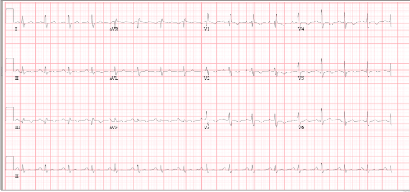
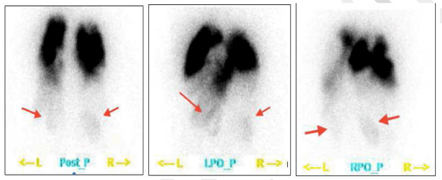
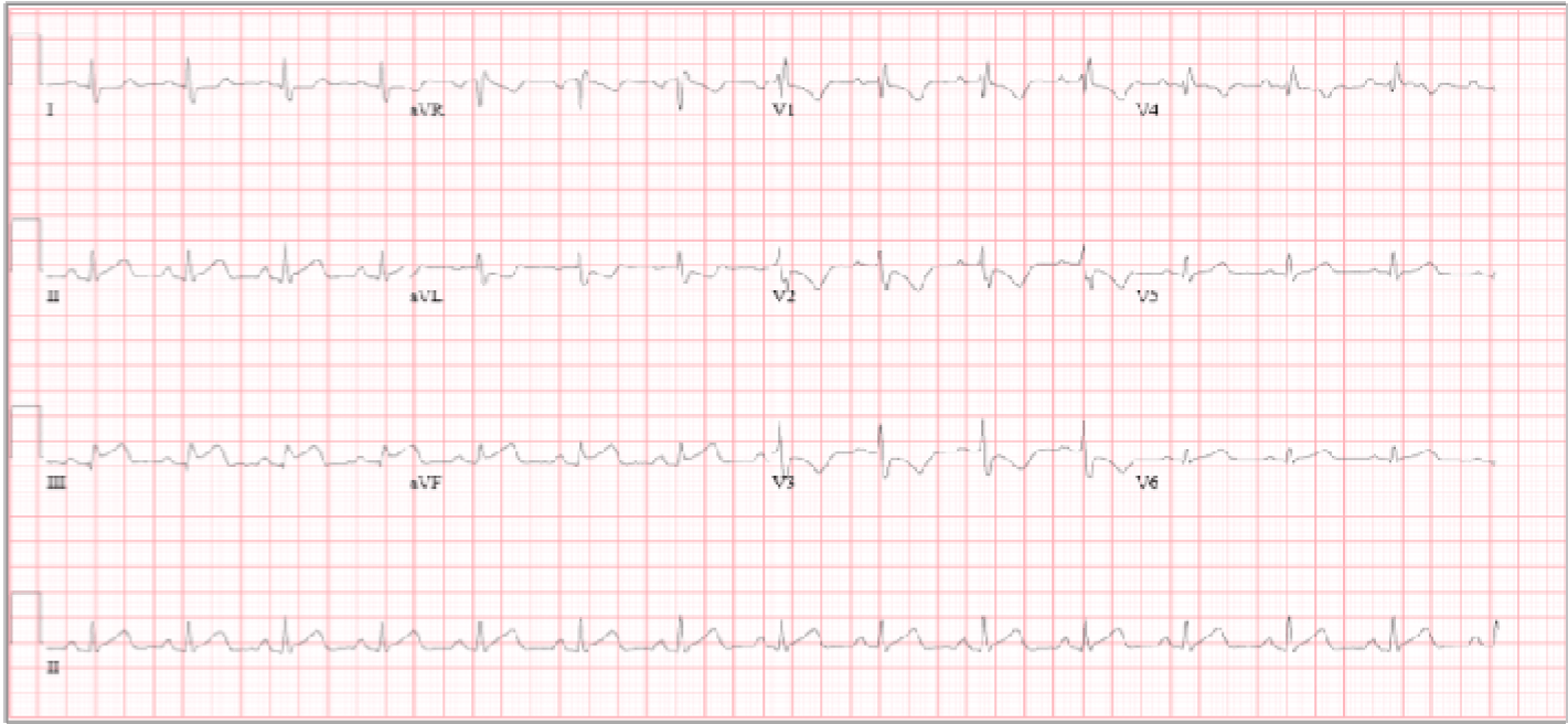
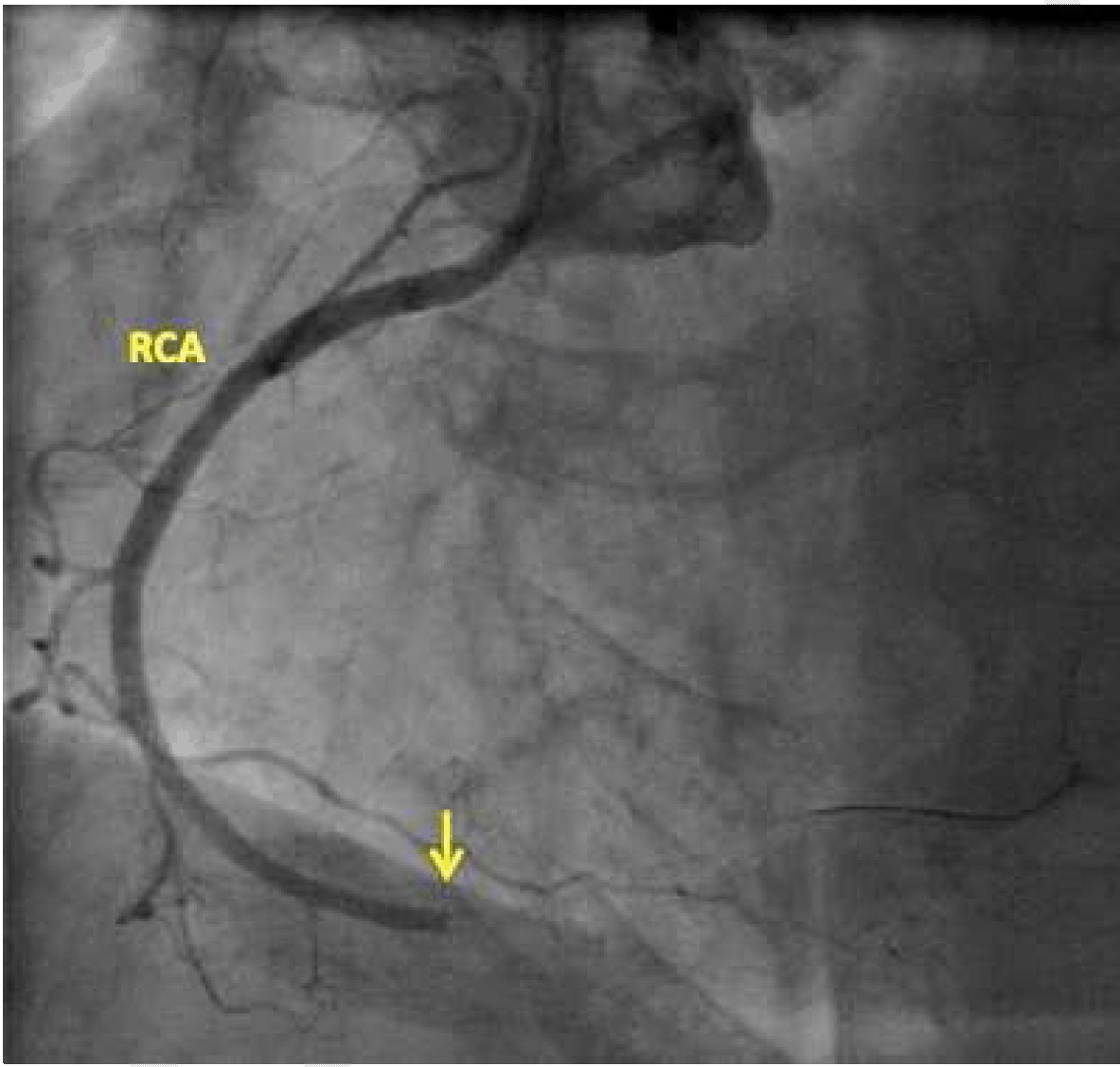
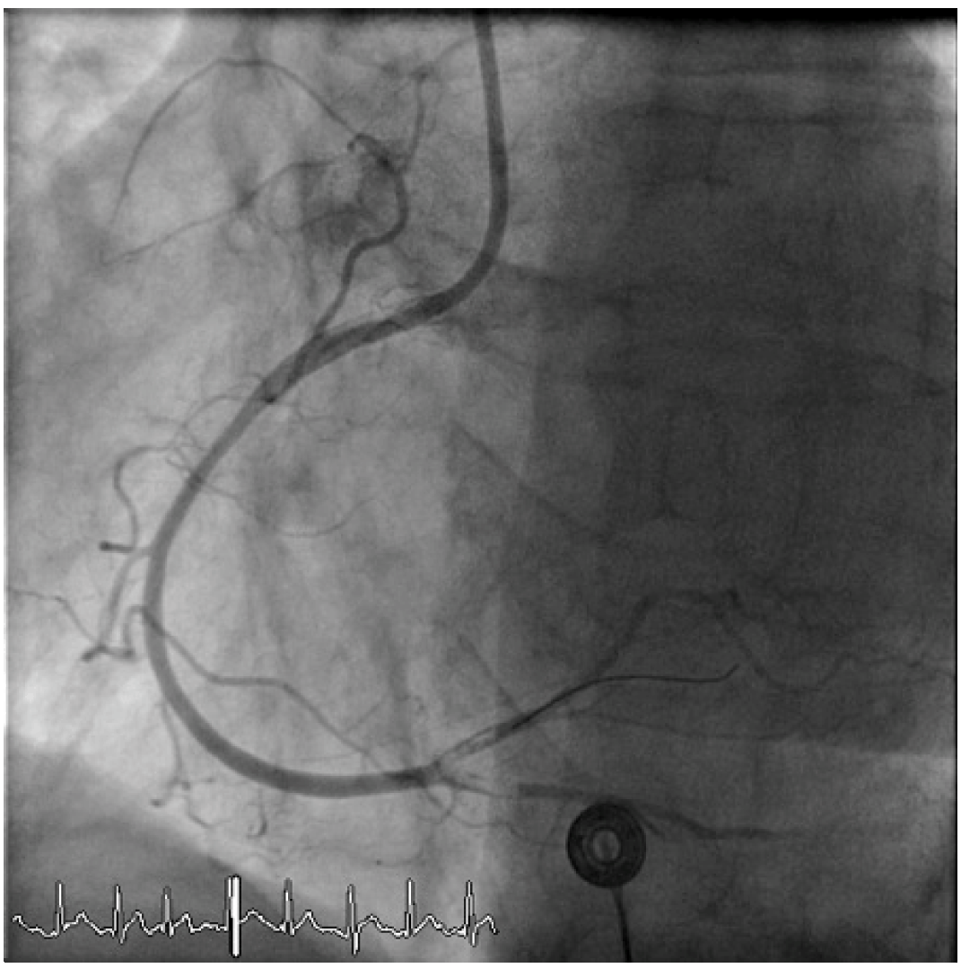
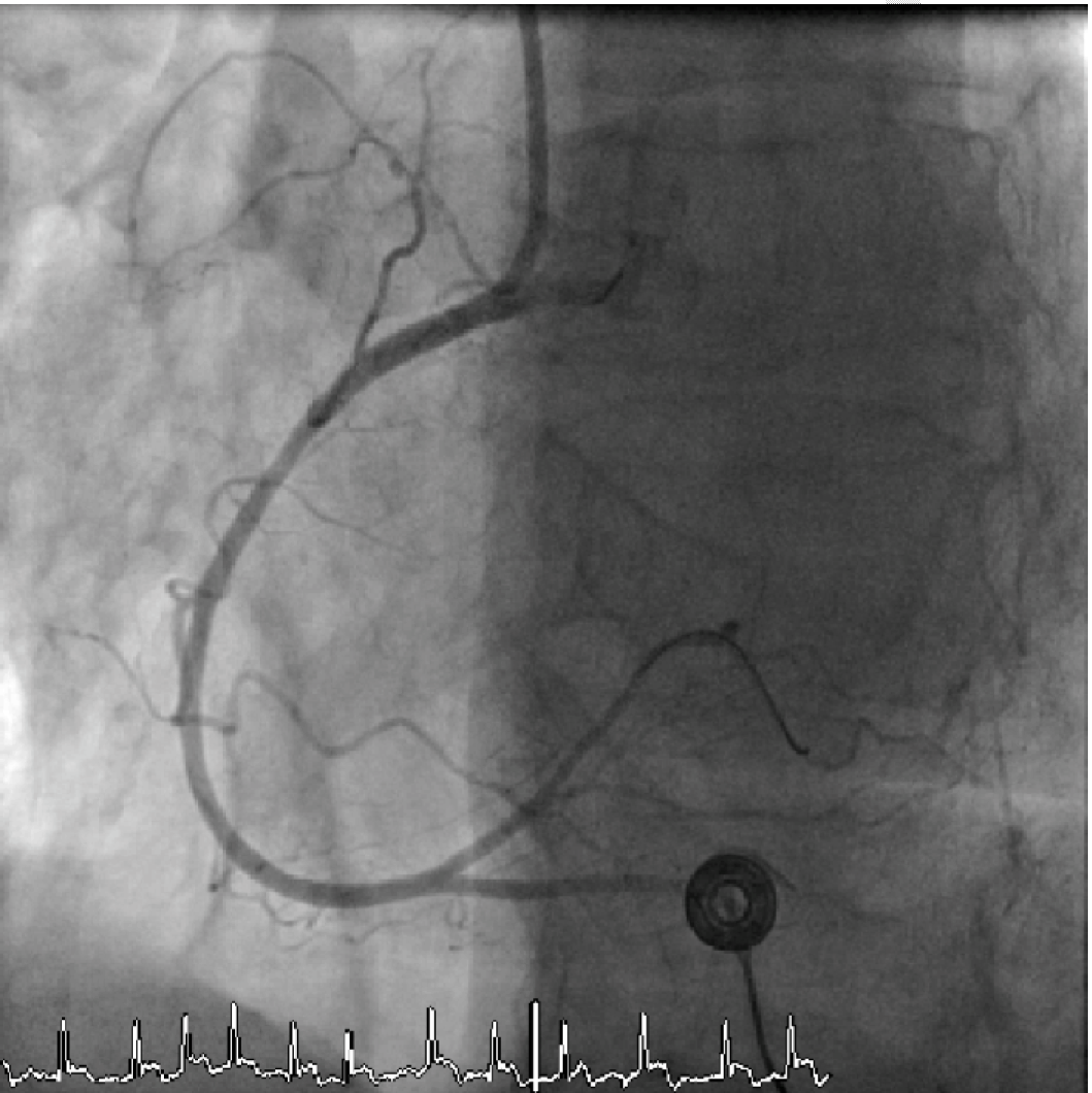
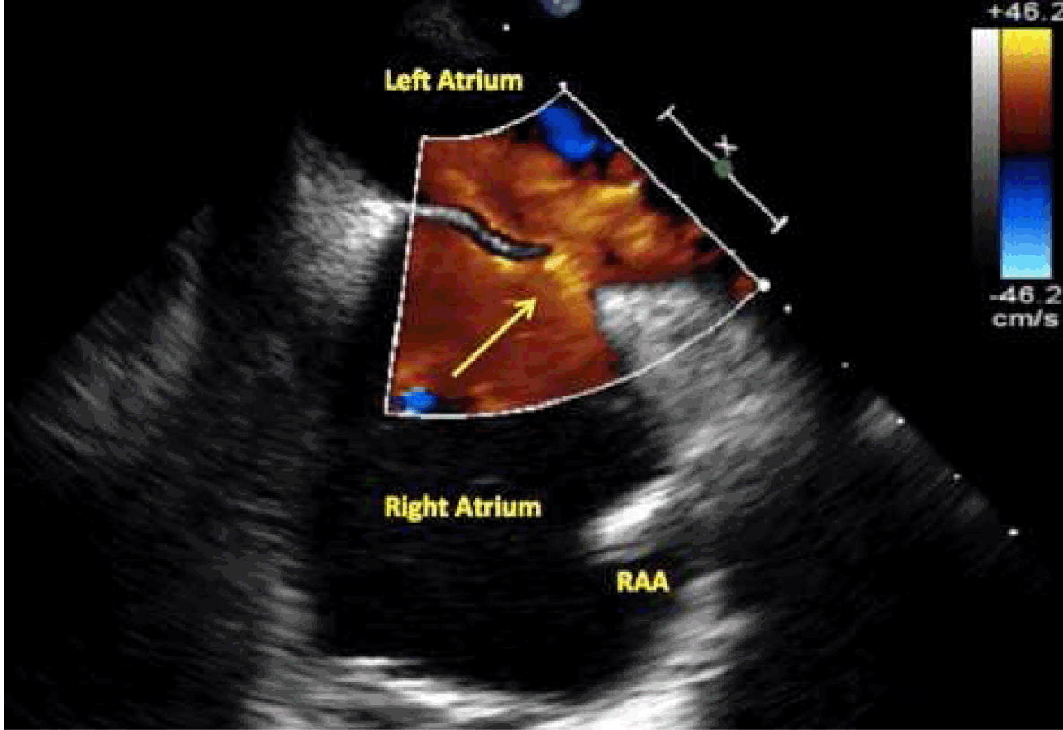
A 67-year-old female presented to the emergency department with rapidly progressing dyspnea and epigastric pain. Her past medical history was significant for ET, hypertension, atrial fibrillation, and osteoporosis. Her ET was diagnosed six years ago based on persistent thrombocythemia (1100×109/dL). Her medications included aspirin 81 mg and hydroxyurea 1000/500 mg on alternate days. On presentation, she was tachycardic, tachypneic and hypoxic. Physical examination revealed an elevated JVP and bilateral lower limb edema. Initial blood work showed a white blood cell count 7.1×10⁹/dL, hemoglobin 14.9 g/dL, platelet count 412×109/dL, D-dimer >4000, creatine kinase 59, and troponin-T 0.03. Presenting ECG showed a typical S1Q3T3 pattern and symmetric T-wave inversion in anterior precordial leads (Figure 1). Her chest X-ray was unremarkable. Acute pulmonary embolus was suspected and a V/Q scan revealed high probability of pulmonary embolism. V/Q scan unexpectedly revealed uptake of radiotracer in the kidneys, suspicious for a right-to-left cardiac shunt. (Figure 2) Transthoracic echocardiogram showed severe right ventricular dilation with septal shift and left-to-right intra-atrial shunting, suggestive of a PFO. The patient was started on subcutaneous low molecular weight heparin for a presumed pulmonary embolism. She remained clinically stable for eight hours, and then suddenly developed worsening chest pain, hypotension, and worsening hypoxia. Repeat ECG revealed ST-segment elevation in leads II, III and aVF consistent with STEMI (Figure 3). She was immediately intubated, started on dopamine and taken for cardiac catheterization. Left and right heart catheterization showed normal left main, left anterior descending, and left circumflex arteries, and a completely occluded right coronary artery (RCA) (Figure 4). Thrombectomy to the RCA was performed with a 6 F Export® thrombectomy catheter with concomitant administration of intra-coronary abciximab. TIMI 3 flow in RCA was established though distal embolization to the posterior descending and posterior-lateral arteries was apparent (Figure 5) that subsequently cleared when additional unfractionated heparin was bloused (Figure 6). Right heart catheterization showed an elevated RV pressure of 70/18 mmHg. Oxygen saturation analysis of the right-sided chambers did not reveal a "step-up" to suggest a large left-to-right shunt. Moreover, trans-septal puncture and left sided chamber oxygen saturation measurements also did not reveal a significant step down form femoral artery's readings. The patient was started on an abciximab infusion for 12 hours and admitted to the intensive care unit. She continued to deteriorate hemodynamically and recombinant tissue plasminogen activator (tPA) was given. Transesophageal echocardiography showed a markedly dilated and hypokinetic right ventricle, a redundant and hypermobile inter-atrial septum with an 8-mm connection suggestive of a PFO (Figure 7). Bubble study showed intense and immediate opacification of the left atrium, confirming right-to-left shunting (Figure 8). There was no evidence of left atrial or left ventricular thrombi. The computed tomography (CT) pulmonary angiography confirmed the diagnosis of massive bilateral pulmonary emboli. The patient did well post coronary thrombectomy and systemic thrombolysis with no further thrombotic sequelae or hemorrhagic complications. She was started on unfractionated heparin and transferred to the cardiology ward one week later. She was transitioned to warfarin with a target INR between 2 and 3, and also maintained on hydroxyurea 1000 mg/500 mg on alternate days. She continued to improve clinically and was eventually discharged home. The decision was not to close her PFO and resume therapeutic anticoagulation. The patient was done well on medical therapy, symptom free at three-year follow-up | ||||||
| ||||||
| ||||||
|
| ||||||
| ||||||
|
| ||||||
|
| ||||||
| ||||||
| ||||||
|
Discussion
| ||||||
|
Essential thrombocytosis (ET) is a rare myeloproliferative disorder characterized by megakaryocytic lineage expansion leading to sustained thrombocythemia and increased risk of bleeding and thrombosis. Essential thrombocytosis affects 0.5–2.5/100,000 patients with higher predisposition in females [1]. Thrombosis can involve any vascular bed and is associated with a higher morbidity and mortality than bleeding, which is typically limited to the skin and mucus membranes [1]. Deep venous thrombosis and pulmonary embolism are the most common thrombotic manifestation [12]. The incidence of ET related thrombotic events is 15–26.4% and in western studies [3]. Factors predisposing to thrombosis are age >60, previous thrombotic event, higher cardiac risk factors, and leukocytosis >15,000 [1] [13]. Platelet counts do not predict thrombotic events, as 10–20% of severe thrombotic complications occur in patients with platelets counts of <60x109/L [14] [15]. Our case also represents an example of life-threatening thrombosis with normal platelets counts. Typical ECG changes in setting of pulmonary embolism include sinus tachycardia, S1Q3/S1Q3T3 pattern, right axis deviation, transient complete or incomplete right bundle branch block, and T-wave inversions in the right pericardial lead. All of these findings, except sinus tachycardia were seen on our first ECG. The patients ECG also had symmetrical T-Wave inversion in leads V1-V4, which is a marker for worse prognosis in hospitalized patients with pulmonary embolism [16]. ST-segment elevation has been described in the setting of pulmonary embolism [17] [18]. It is presumed to be secondary to pressure overload and subendocardial ischemia involving the right ventricle, but paradoxical embolism with subsequent coronary occlusion has also been postulated [17] [19]. STEMI is a rare but well described complication of ET both as an initial manifestation and during follow-up. Approximately, 20 cases of STEMI occurring in the setting of ET have been described. Left main and left anterior descending (LAD) occlusions represent the majority of cases [8] [11]and only five cases have right coronary artery (RCA) involvement [5] [6] [7] [20][21], and no isolated circumflex involvement has been described. To our knowledge, our case is the first published report of RCA occlusion secondary to paradoxical coronary embolism in a patient with ET and massive bilateral pulmonary embolism. As per criteria suggested by Johnson et al., a paradoxical embolism (PDE) can be presumed when there is evidence of arterial embolization in the absence of a source in the left heart, or when there is a venous source of embolism and an abnormal communication between the venous and the arterial circulation is demonstrated [22]. All three criteria are applicable to our case [22]. Most cases of paradoxical embolism are associated with PFO, while ASD, pulmonary AV malformation, or VSD have been described less frequently [23]. Paradoxical embolism to the coronary tree is rare and constitutes 5–10% of all paradoxical emboli [22]. Paradoxical emboli are further supported if pressure gradients support right-to-left shunting. Pulmonary embolism is associated with 60% of the cases of paradoxical embolism in patients with a PFO and right-to-left shunting [23]. We decided to treat the coronary emboli with manual aspiration and intracoronary abciximab without stenting given the absence of residual disease post-aspiration. Manual thrombectomy has been shown to be beneficial in patients with STEMI in the setting of primary PCI [24]. Murhtay et al. described a successful aspiration of LAD emboli using Export® catheter with eptifibatide infusion in treating PDE resulting from an underlying DVT [22] Kujime et al. described successful use of Thrombuster® aspiration catheter in treating paradoxical RCA clot [25]. Wilson et al. successfully used Export® catheter in conjunction with heparin and eptifibatide in treating an occluded obtuse marginal coronary embolus [26]. Glycoprotein IIb/IIIa infusion post-catheterization has been used in conjunction with thrombectomy to treat high thrombus burden in ET related coronary emboli. Murthay et al. described the use of eptifibatide infusion for 18 hours post-procedure in a similar case [22]. Michaels et al. described the utility of abciximab as an effective sole therapy for decreasing the thrombus burden. In a 50-year-old male with ET and STEMI, angiogram showed extensive clot to left coronary system and total occlusion of RCA. He was successfully treated with thrombectomy to the RCA alone with abciximab infusion for 24 hours [6]. We decided not to close the patient's PFO. The thrombotic potential of the closure device may have negated any potential benefit from closure. Three relatively recent randomized controlled trails, CLOSURE I [27], PC [28], and PROSPECT [29], failed to prove any benefit of percutaneous closure over medical therapy in patients with paradoxical embolism secondary to PFO. Also a recent meta-analysis by Ntaios et al. showed the same results [30]. Our patient is unique, as her paradoxical embolic event was a STEMI. She has remained stable with rigorous maintenance of therapeutic anticoagulation with warfarin. | ||||||
|
Conclusion
| ||||||
|
Essential thrombocytosis is a disorder of dysfunctional platelet overproduction leading a high risk of catastrophic thrombotic events even with normal platelets count. This case shows a rare presentation of paradoxical coronary embolism through a patent foramen ovale in patients with massive pulmonary embolism. Coronary embolism itself is rare, and should always be considered in venous thromboembolism patients with cardiac deterioration. There is no clear treatment consensus in such patients, but combination therapy of thrombectomy and a GPIIb/IIIa inhibitor in the acute setting with chronic anticoagulation was successfully employed in our case. Patent foramen ovale closure may be considered to prevent further embolism, however, our patient did well conservatively, with anticoagulation only. | ||||||
|
References
| ||||||
| ||||||
|
[HTML Abstract]
[PDF Full Text]
|
|
Author Contributions
Fahad S. Almehmadi – Conception and design, Drafting the article, Critical revision of the article, Final approval of the version to be published Albayda M. Mehdar – Conception and design, Drafting the article, Critical revision of the article, Final approval of the version to be published Kumar Sridhar – Analysis and interpretation of data, Drafting the article, Critical revision of the article, Final approval of the version to be published Patrick Teefy – Acquisition of data, Analysis and interpretation of data, Drafting the article, Critical revision of the article, Final approval of the version to be published |
|
Guarantor of submission
The corresponding author is the guarantor of submission. |
|
Source of support
None |
|
Conflict of interest
Authors declare no conflict of interest. |
|
Copyright
© 2015 Fahad S. Almehmadi et al. This article is distributed under the terms of Creative Commons Attribution License which permits unrestricted use, distribution and reproduction in any medium provided the original author(s) and original publisher are properly credited. Please see the copyright policy on the journal website for more information. |
|
|
|
About The Authors
| |||
| |||
| |||
| |||
| |||