| |
|
|
|
Case Report
| ||||||
| Epigastric pain: Incarceration or rotation? | ||||||
| Ana Franky Carvalho1, Ana João Rodrigues2, Pedro Leão3 | ||||||
|
1MD, PhD, Serviço de Cirurgia Geral - Hospital de Braga, Life and Health Sciences Research Institute (ICVS), School of Health Sciences, University of Minho, Braga, ICVS/3B's - PT Government Associate Laboratory, Braga/Guimarães, Portugal.
2PhD, Life and Health Sciences Research Institute (ICVS), School of Health Sciences, University of Minho, Braga, ICVS/3B's - PT Government Associate Laboratory, Braga/Guimarães, Portugal. 3MD, Ph.D, Serviço de Cirurgia Geral - Hospital de Braga, Life and Health Sciences Research Institute (ICVS), School of Health Sciences, University of Minho, Braga, ICVS/3B's - PT Government Associate Laboratory, Braga/Guimarães, Portugal. | ||||||
| ||||||
|
[HTML Abstract]
[PDF Full Text]
[Print This Article]
[Similar article in Pumed] [Similar article in Google Scholar] 
|
| How to cite this article |
| Carvalho AF, Rodrigues AJ, Leão P. Epigastric pain: Incarceration or rotation? Int J Case Rep Images 2015;6(3):145–148. |
|
Abstract
|
|
Introduction:
Acute intrathoracic gastric volvulus occurs when the stomach has a twist mesenteroaxial/organoaxial or chest cavity resulting in a dilatation or rupture of the diaphragmatic hiatus or diaphragmatic hernia. The purpose of this work is to show a interesting case of gastric volvulus in a patient with several comorbidities.
Case Report: A 77-year-old female with past history of hiatal hernia and mental disease associated with diabetes and atrial fibrillation. Patient went to the emergency department due to vomiting associated with blood. Analytical parameters (WBC, HGB, PCR, metabolic panel and liver function), showed no significant alterations. Thoracic X-ray revealed an enlarged mediastinum due to herniation of the stomach. A computed tomography (CT) scan confirmed intrathoracic localization of the gastric antrum with twist. Patient's symptoms were relieved by nasogastric intubation and analgesia. After six months, the patient is still asymptomatic. Conclusion: In general, the treatment of an acute gastric volvulus requires an emergent surgical repair. In patients who are not surgical candidates (with comorbidities or an inability to tolerate anesthesia), endoscopic reduction should be attempted. Chronic gastric volvulus may be treated non-emergently, and surgical treatment is increasingly being performed using a laparoscopic approach. In this case, it is a chronic form that was solved with the placement of the nasogastric tube. A nasogastric decompression is an option in the chronic form of hiatal hernia associated to gastric volvulus in patients with serious comorbidities. | |
|
Keywords:
Epigastric pain, Gastric volvulus, Hiatal hernia, Mesenteroaxial, Nasogastric tube
| |
|
Introduction
| ||||||
|
Gastric volvulus (GV) is an abnormal rotation of the stomach. The primary etiology is laxity or agenesis of the gastric ligaments. It may also develop adhesions, hiatal hernia and other causes [1], It was first described by Berti in 1866 [2]. According to the axis of rotation, it is classified into organoaxial:
The acute GV, represented by sudden abdominal pain, vomiting and triad Borchardt (epigastric distention, inability to pass the gastric tube and ineffectual efforts to vomit), is prone to tissue ischemia, necrosis and gastric perforation, and is considered an emergency surgery [2] [3] [4]. The chronic form of presentation is asymptomatic or oligosymptomatic, and may be responsible for uncharacteristic symptoms of abdominal discomfort and heart burn [4] [5] and often requires a new procedure to treat the condition, especially if the patient presents several comorbidities. Here, we present a different concept in GV treatment. | ||||||
|
Case Report
| ||||||
|
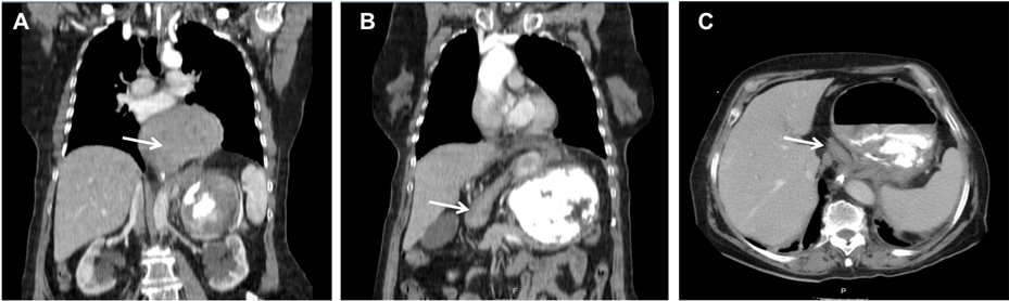
A 77-year-old female with past history of diabetes, mental disease and hiatal hernia complained of epigastric pain and abdominal cramps, more evident after meals and relieved by vomiting. Patient went to the emergency department due to vomiting associated with blood and halitosis. Analytical parameters (WBC, HGB, PCR, metabolic panel and liver function), showed no significant alterations. Thoracic X-ray revealed an enlarged mediastinum due to herniation of the stomach. The stomach bubble (gastric fundus) could be seen in its usual position suggesting herniation of the gastric antrum (Figure 1). A computed tomography (CT) scan confirmed intrathoracic localization of the gastric antrum (Figure 2). The diagnosis is a hiatal hernia with gastric mesenterioaxial rotation type, as depicted in Figure 2, where the arrow indicates the duodenum near the hernia position. An upper gastrointestinal endoscopy showed gastric stasis. The exam was interrupted due to patient intolerance, suggestive of gastric torsion. The patient's symptoms were relieved by nasogastric intubation and analgesia. After six months of follow-up the patient still asymptomatic. | ||||||
| ||||||
|
| ||||||
|
Discussion
| ||||||
|
Gastric volvulus is a complete obstruction of the gastric lumen by rotation, that can occur at any age, with equal frequency in men and women [6]. In 25% of patients, gastric rotation is primary or idiopathic and occurs because there is a lengthening of ligaments [5] [6] [7]. In 75% of patients, GV is associated with a pathological factor namely: hiatus hernia, diaphragmatic hernia resulting from trauma, herniation of the left diaphragm phrenic nerve injury, chronic pyloric obstruction with dilatation of the stomach or prior gastroesophageal surgery [6] [7]. In the presented case, the most probable etiology is laxity ligaments and hiatal hernia, although we cannot exclude other causes. The most commonly used classification was proposed by Singleton and describes three types of GV, according to the rotational axis: Type 1 or organoaxial is the most common (59%) and the rotation occurs about a line drawn from the pylorus to the esophagus gastric junction. Type 2 or mesenteroaxial occurs in about 29% of patients and rotation turns on an axis that connects the greater curvature and the hepatic hilum. As we can see in the given images of CT scan; Type 3 is a rare form (3%), which combines both types 1 and 2 [1] [2] [3] [4] . Supplementary examination in these patients is important for diagnosis. In the analytical control, there may be a hyperamylasemia and elevated levels of lactate dehydrogenase and alkaline phosphatase. Gastrointestinal contrast studies, barium or gastrografin, have high sensitivity and specificity. The endoscopy usually shows a high deformation with gastric pylorus and difficult access, and in the most advanced stage of the disease, mucosal ulcerations. The CT scan has important diagnostic value in GV, as for example in this case, where it was performed in the acute phase and provided a rapid diagnostic. Moreover, this examination can detect the presence of pneumatosis or pneumoperitoneum in case of necrosis and perforation [6]. Recent data suggest that routine elective repair of completely asymptomatic paraesophageal hernias may not be indicated. Surgical treatment of gastric voluvus includes reduction of the stomach and limited gastric resection in cases of gastric necrosis. The laparoscopic approach can be used in most of the cases, but conversion to open access should be considered for complex problems or for the safety of the patient [8] [9]. Large hiatal hernias with or without gastric volvulus can be repaired either transabdominally (open or laparoscopic) or via thoracotomy. However to date, there is no randomized trials directly comparing open transthoracic versus open transabdominal repair. In the surgical approach, we need to take to consideration the following four hallmarks: Hernia sac excision Sac dissection during paraesophageal hernia repair is thought to release the tethering of the esophagus, facilitating reduction of the hernia and the decrease of early recurrence, as well as protecting the esophagus from iatrogenic damage. [10] Reinforced repair Primary sutured crural repair has been the main option for many years, but follow-up has suggested very high recurrence rates (>42%) after laparoscopic paraesophageal hernia repair [11]. Several case series suggests benefit with mesh, however, there are a few which question the use of meshed repair [12]. Fundoplication The majority of reports in the recent literature describe the performance of a fundoplication as a step of the repair. In a case-controlled study, surgeons found increased dysphagia with fundoplication, and of reflux symptoms in the group without fundoplication, thus routine fundoplication should be avoided [13]. Gastropexy One of the first studies using anterior gastropexy to reduce the recurrence rate after laparoscopic hiatal hernia repair with gastric volvulus showed no recurrences up to two years of follow-up evaluation [14]. This finding has been supported by a recent study showing that the addition of an anterior gastropexy significantly reduced recurrent hernias. However, other reports found no significant difference in recurrence rate [15]. In this case, the placement of a nasogastric tube has the function of gastric decompression, and solved the gastric volvulus. A nasogastric decompression is possible only because the cardia mesenteroaxial VG is open. The endoscopic "des rotation" have satisfactory results but it is a temporary solution, being the definitive treatment the surgical approach. Urgent surgery in acute cases is fundamental and its delay increases mortality [4]. Contraindications for surgical treatment involve conditions or comorbidities in which the patient cannot tolerate general anesthesia. The surgeon should also use clinical judgment and make sure the patient conditions are optimal before the operation. | ||||||
|
Conclusion
| ||||||
|
A nasogastric decompression is an option in chronic form of hiatal hernia associated to gastric volvulus. | ||||||
|
References
| ||||||
| ||||||
|
[HTML Abstract]
[PDF Full Text]
|
|
Author Contributions
Ana Franky Carvalho – Substantial contributions to conception and design, Acquisition of data, Analysis and interpretation of data, Drafting the article, Revising it critically for important intellectual content, Final approval of the version to be published Ana João Rodrigues – Acquisition of data, Analysis and interpretation of data, Revising it critically for important intellectual content, Final approval of the version to be published Pedro Leão – Acquisition of data, Analysis and interpretation of data, Revising it critically for important intellectual content, Final approval of the version to be published |
|
Guarantor of submission
The corresponding author is the guarantor of submission. |
|
Source of support
None |
|
Conflict of interest
Authors declare no conflict of interest. |
|
Copyright
© 2015 Ana Franky Carvalho et al. This article is distributed under the terms of Creative Commons Attribution License which permits unrestricted use, distribution and reproduction in any medium provided the original author(s) and original publisher are properly credited. Please see the copyright policy on the journal website for more information. |
|
|