| |
|
|
|
Case Report
| ||||||
| Ureteroscopy assisted retrograde nephrostomy (UARN) in patient with lower pole renal stone | ||||||
| Takashi Kawahara1, Hiroki Ito2, Hideyuki Terao1, Hiroji Uemura3, Yoshinobu Kubota3, Junichi Matsuzaki1 | ||||||
|
1Department of Urology, Ohguchi Higashi General Hospital, 2-19-1, Irie, Kanagawa-ku, Yokohama City, Kanagawa, Japan.
2Department of Urology, Ohguchi Higashi General Hospital, Department of Urology, Yokohama City University Graduate School of Medicine, Japan. 3Department of Urology, Yokohama City University Graduate School of Medicine, Japan. | ||||||
| ||||||
|
[HTML Abstract]
[PDF Full Text]
[Print This Article]
[Similar article in Pumed] [Similar article in Google Scholar] 
|
| How to cite this article |
| Kawahara T, Ito H, Terao H, Uemura H, Kubota Y, Matsuzaki J. Ureteroscopy assisted retrograde nephrostomy in patient with lower pole renal stone. Int J Case Rep Images 2015;6(3):142–144. |
|
Abstract
|
|
Introduction:
We previously reported a new technique, ureteroscopy-assisted retrograde nephrostomy (UARN) for percutaneous nephrolithotomy (PCNL). This case report describes a new approach for treating renal stones in the lower calyx using the UARN technique.
Case Report: A 68-year-old female was referred to our department for the treatment of a right renal stone. We performed PCNL using the technique for UARN. Conclusion: We herein report a new technique using UARN for lower pole renal stones. This method may represent a new treatment option for renal stones in the lower calyx. | |
|
Keywords:
Nephro access sheath (NAS), Percutaneous nephrolithotomy (PCNL), Renal stones, Ureteral access sheath, Ureteroscopy, Ureteroscopy-assisted retrograde nephrostomy (UARN)
| |
|
Introduction
|
|
For large renal stones in the lower calyx, percutaneous nephrolithotomy (PCNL) has become the preferred method of treatment in patients with a large or complex stone burden. We previously reported a new technique, ureteroscopy-assisted retrograde nephrostomy (UARN) for PCNL, which allows for continuous visualization from puncture to the insertion of the nephro access sheath (NAS), resulting in a higher stone-free rate and fewer complications. Although the ureteroscope can usually be used to reach the target puncture spot, the URS with a puncture wire cannot reach the lower calyx because it is too stiff. This report describes a new approach for treating renal stones in the lower calyx using the UARN technique. |
|
Case Report
|
|
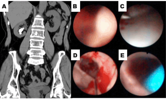
A 68-year-old female was referred to our department for the treatment of a right renal stone. In April 2012, the patient was admitted to our department for PCNL of a right lower pole renal stone. A preoperative kidney ureter bladder (KUB) film and computed tomography (CT) scan showed a maximum stone diameter of 23 mm, with no evidence of hydronephrosis (Figure 1A). We did not consider that ureteroscopic lithotripsy would provide a stone-free outcome because the stone's diameter was more than 20 mm. Therefore, we performed UARN, following the method described in our previous manuscript [1]. Briefly, we confirmed that performing puncture from the lower pole was difficult. However, puncturing from the middle calyx was considered to be associated with a higher risk of not being able to reach the target stone with the nephroscope because the entrance to the lower calyx was occupied by the NAS (Figure 1B). Despite generating hydronephrosis using a high irrigation pressure supplied by the URS, performing percutaneous puncture under ultrasonography with fluoroscopic guidance was not successful, as the lower calyx was not sufficiently dilated for puncture. We first punctured the site from the middle calyx using the UARN technique, as described in our previous report [2]. Briefly, after the puncture wire was easily passed through the renal capsule and the skin at the posterior axillary line was tented, a 12-Fr safety catheter was inserted under visualization with the URS; the outer 12-Fr catheter was left in place to drain the irrigation flow. After conducting ureteroscopic lithotripsy using a holmium:yttrium aluminum garnet (Ho:YAG) laser to create stone fragments, the calculus fragments were washed out from the lower calyx into the renal collecting system using the irrigation flow (Figure 1C-E). Then, a 24-Fr NAS (X-Force® Nephrostomy Balloon Dilation Catheter, BARD, Murray Hill, NJ, USA) was inserted, and the stone fragments were retracted using a nephroscope. Finally, a ureteral stent was inserted at the conclusion of the PCNL procedure and subsequently removed two weeks after the operation. Postoperative nephrostomy was not performed. A stone-free status was confirmed on computed tomography (CT) scan; the stone composition included calcium oxalate monohydrate. |
|
|
|
Discussion
|
|
Treating renal stones in the lower calyx is challenging, even when shockwave lithotripsy can be used to successfully create stone fragments, due to the anatomical structure in this area [3]. URS is a candidate for a new therapeutic approach for treating renal stones in the lower calyx [4] [5]. The American Urological Association (AUA) and European Association Urology (EAU) guidelines both recommend PCNL as the first-line therapy for renal stones larger than 2 cm [4] [5]. We previously described a new technique for UARN [1]. Using UARN, it is possible to continuously visualize the region from the initial puncture to the insertion of the NAS. The most important limitation of UARN is that it cannot be used to puncture from the lower calyx. Notably, the URS with the puncture wire is too stiff, making it impossible to reach the lower calyx. Therefore, in most cases without a horseshoe kidney, the target calyx is the middle calyx on the dorsal side [6]. However, when the target stone is located in the lower calyx, inserting the NAS may block the entrance to the lower calyx, which can also make it difficult or impossible to advance the nephroscope to the target stone [7]. In this case, before dilating the balloon, we inserted a 12-Fr catheter and created stone fragments using Ho:YAG laser lithotripsy. After washing out the stone fragments from the lower calyx via the irrigation flow, balloon dilation and NAS insertion was performed and the stone fragments were easily removed through the NAS. This procedure allowed for the use of a higher irrigation flow with less intrarenal pressure as a result of the drainage provided by the 12-Fr catheter. This method may help decrease the risk of postoperative urinary tract infections. The most critical issue associated with PCNL is the proper selection of the puncture site to minimize the risk of hemorrhage, the most common major complication of this procedure [1] [8]. US-guided puncture to the renal collecting system with subsequent placement of a drainage tube under fluoroscopic guidance is the standard modality for performing PCNL. However, applying US-guided nephrostomy is difficult without dilating the renal collecting system, even if an occlusion balloon catheter is used to create hydronephrosis [9]. The limitation of this procedure is that it is only effective for relatively small stones after initial fragmentation using a Ho:YAG laser. |
|
Conclusion
|
|
We herein reported a new technique using ureteroscopy-assisted retrograde nephrostomy (UARN) for lower pole renal stones. This method may represent a new treatment option for renal stones in the lower calyx. |
|
References
|
|
|
[HTML Abstract]
[PDF Full Text]
|
|
Author Contributions
Takashi Kawahara – Substantial contributions to conception and design, Acquisition of data, Analysis and interpretation of data, Drafting the article, Revising it critically for important intellectual content, Final approval of the version to be published Hiroki Ito – Analysis and interpretation of data, Revising it critically for important intellectual content, Final approval of the version to be published Hideyuki Terao – Analysis and interpretation of data, Revising it critically for important intellectual content, Final approval of the version to be published Hiroji Uemura – Analysis and interpretation of data, Revising it critically for important intellectual content, Final approval of the version to be published Yoshinobu Kubota – Analysis and interpretation of data, Revising it critically for important intellectual content, Final approval of the version to be published Junichi Matsuzaki – Analysis and interpretation of data, Revising it critically for important intellectual content, Final approval of the version to be published |
|
Guarantor of submission
The corresponding author is the guarantor of submission. |
|
Source of support
None |
|
Conflict of interest
Authors declare no conflict of interest. |
|
Copyright
© 2015 Takashi Kawahara et al. This article is distributed under the terms of Creative Commons Attribution License which permits unrestricted use, distribution and reproduction in any medium provided the original author(s) and original publisher are properly credited. Please see the copyright policy on the journal website for more information. |
|
|