  |

|
 |
|
Clinical Image
| ||||||
| The lung riddle: A Blesovsky's syndrome case | ||||||
| Aviral Vij1, Anshu Singh2 | ||||||
|
1MBBS, MD, Internal Medicine Resident at John H. Stroger Hospital of Cook County, Chicago, IL.
2MBBS, MD, Attending Physician and Firm Chief at John H. Stroger Hospital of Cook County, Chicago, IL. | ||||||
| ||||||
|
[HTML Abstract]
[PDF Full Text]
[Print This Article]
[Similar article in Pumed] [Similar article in Google Scholar] 
|


| How to cite this article |
| Vij A, Singh A. The lung riddle: A Blesovsky's syndrome case. Int J Case Rep Images 2015;6(3):187–188. |
|
Case Report
|
|
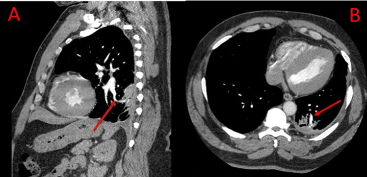
A 49-year-old male presented with worsening shortness of breath and reduction in exercise tolerance over eight months. Review of systems was negative. Past history was significant for untreated hypertension, a 40 pack-year smoking history and work in construction, where he was exposed to asbestos and silica for over 20 years without use of masks. Vital signs were relevant for blood pressure of 159/94 mmHg. Physical examination revealed reduced breath sounds in the left infra axillary area. Rest of the examination was unremarkable. Chest X-ray showed a small, well-circumscribed pleural-based density in the left lower zone. A subsequent computed tomography (CT) scan of the chest showed a rounded, well-defined, 14x17 mm pleural based mass with pleural thickening in the posterior segment of the left lower lung which was identified as 'rounded atelectasis'. At sixth month follow-up, the CT scan findings remained unchanged and his symptoms had improved with treatment of underlying chronic obstructive pulmonary disease with salmeterol and tiotropium inhalers. |
|
|
|
Discussion
|
|
Rounded atelectasis (RA) is a benign process, incidentally found on follow-up X-ray of patients with known occupational exposure or on routine testing. It has been associated with several etiologies [1] like exposure to mineral dust of asbestos (most common cause) and silica, exudative pleural effusions like tuberculosis and para-pneumonic effusions, infections like Legionella pneumonia and histoplasmosis and also heart failure. Rounded atelectasis is a radiological diagnosis and characteristic findings on CT scan include [2] :
As in our patient, RA can be diagnosed with characteristic CT scan. Atypical radiographic presentations may require additional testing such as MRI scan, PET scan or invasive procedures such as VATS or thoracotomy. Absence of lymphadenopathy with minimal to no pleural effusion and metabolically inactive mass seen on PET scan can help differentiate Rounded atelectasis from the most common differential which is malignancy. RA by itself is benign and need not be followed, however, such patients often are exposed to asbestos and should be followed-up considering the malignant potential of asbestosis. Awareness of this radiographic entity can help avoid invasive procedures in patients with typical CT scan findings. There is no specific treatment for rounded atelectasis and management is usually symptom guided. Surgical resection of the mass is rarely indicated, when there is significant compromise of lung function or if there is high suspicion of malignancy which could not be ruled out by other diagnostic modalities. |
|
Conclusion
|
|
Rounded atelectasis is a rare but increasingly recognized entity due to enhanced screening especially in patients with exposure to asbestosis. Radiographic features are often typical but in the presence of atypical features or in equivocal cases, malignancy must be ruled out. |
|
References
|
|

|
[HTML Abstract]
[PDF Full Text]
|
|
Author Contributions
Aviral Vij – Substantial contributions to conception and design, Acquisition of data, Drafting the article, Revising it critically for important intellectual content, Final approval of the version to be published Anshu Singh – Substantial contributions to conception and design, Acquisition of data, Drafting the article, Final approval of the version to be published. |
|
Guarantor of submission
The corresponding author is the guarantor of submission. |
|
Source of support
None |
|
Conflict of interest
Authors declare no conflict of interest. |
|
Copyright
© 2015 Aviral Vij et al. This article is distributed under the terms of Creative Commons Attribution License which permits unrestricted use, distribution and reproduction in any medium provided the original author(s) and original publisher are properly credited. Please see the copyright policy on the journal website for more information. |
|
|