|

|
 |
|
Case Report
| ||||||
| Septic cavernous sinus thrombosis secondary to a mycotic pseudoaneurysm of a cubital arteriovenous fistula | ||||||
| Ahmed Mohamed Elhassan Elfaki Osman1, Saif Eldin Mohammed Ali Ibrahim1 | ||||||
|
1MBBS, Ibn Sina Specialized Hospital - Senior House-officer, Unit of Vascular and Endovascular Surgery, Ibn Sina Specialized Hospital, Khartoum, Sudan.
2MBBS, MD, MRCS (ENG); D.MAS; F.MAS; F. Vasc/Endovasc (MAL), Ibn Sina Specialized Hospital - Head, Unit of Vascular and Endovascular Surgery, Ibn Sina Specialized Hospital, Khartoum, Sudan. | ||||||
| ||||||
|
[HTML Abstract]
[PDF Full Text]
[Print This Article]
[Similar article in Pumed] [Similar article in Google Scholar] 
|


| How to cite this article |
| Osman AMEE, Ibrahim SMA. Septic cavernous sinus thrombosis secondary to a mycotic pseudoaneurysm of a cubital arteriovenous fistula. Int J Case Rep Images 2015;6(2):103–107. |
|
Abstract
|
|
Introduction:
The cavernous sinuses are part of the dural sinuses. In 1831, Bright described cavernous sinus thrombosis (CST) as a complication of epidural and subdural infections. Cavernous sinus thrombosis usually results as a complication of paranasal sinus infection or infections of the face, in an area called the 'danger triangle', trauma, bacteremia or ear infections.
Case Report: A 35-year-old female presented with one month history of high grade fever and progressively increasing periorbital swelling. This presentation was preceded by failure and infection of her arteriovenous fistula (AVF) three weeks beforehand, which was resistant to medical therapy. On examination, she was febrile and had periorbital swelling with bilateral closure of the eyes. The left jugular vein was distended. At the site of the AVF, there was a pulsatile swelling which was also discharging pus. Following aneurysmectomy and AVF ligation, an angiogram of the head and neck showed a long segment occlusion on the left internal jugular vein extending to the left brachiocephalic vein plus a cavernous sinus thrombus. A diagnosis of a septic CST was made and management was conservative. We report this case because, to the best of our knowledge, no literature was found describing a mycotic AVF complicated by a jugular and brachiocephalic vein thrombosis causing a septic CST. Conclusion: In patients presenting with a mycotic AVF, the risk of developing a septic CST should be kept in mind. The goal of intervention should be to control the source of infection and prevent complications. | |
|
Keywords:
Aneurysm, Cavernous sinus thrombosis (CST), Mycotic arteriovenous fistula, Septic cavernous sinus thrombosis
| |
|
Introduction
| ||||||
|
The cavernous sinuses are part of a group of sinuses which, together, form the dural sinuses (composed of sagittal, lateral and cavernous sinuses). In 1831, Bright described cavernous sinus thrombosis as a complication of epidural and subdural infections. Cavernous sinus thrombosis (CST) is the most important thrombosis compared to other intracranial thrombosis, because of its complex relationship, both neurovascular and anatomic [1]. It usually results as a complication of paranasal sinus infection [1] or infections of the face, in an area called the "danger triangle" which lies between the corners of the mouth to the bridge of the nose including the nose itself and the maxilla) [2]. Other causes may include trauma, bacteremia or ear infections [1]. | ||||||
|
Case Report
| ||||||
|
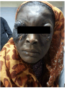
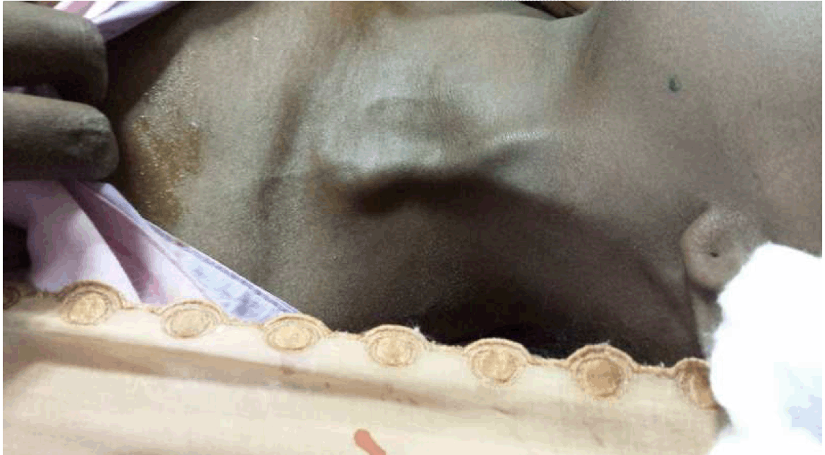
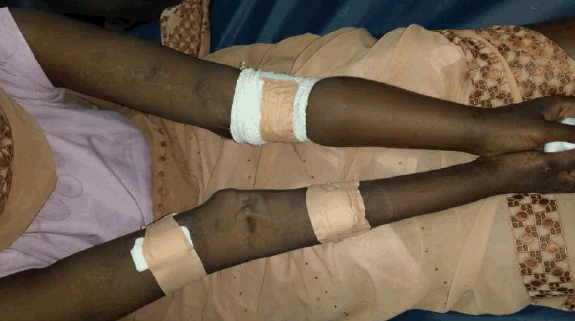
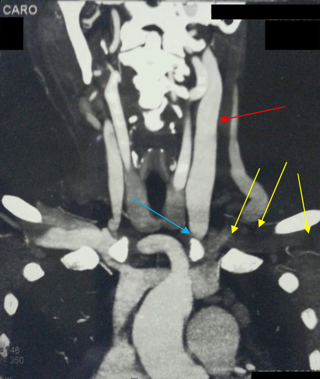
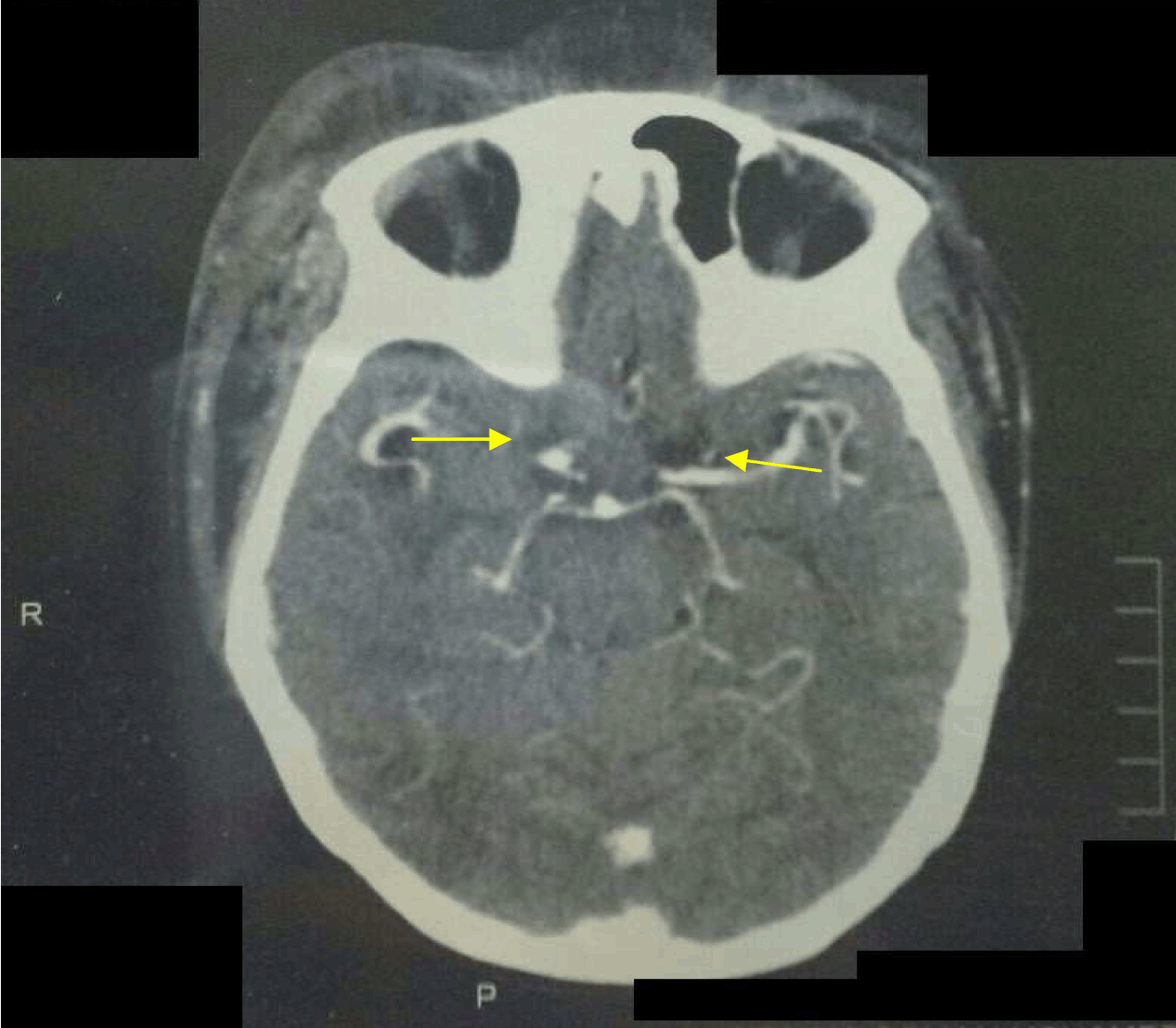
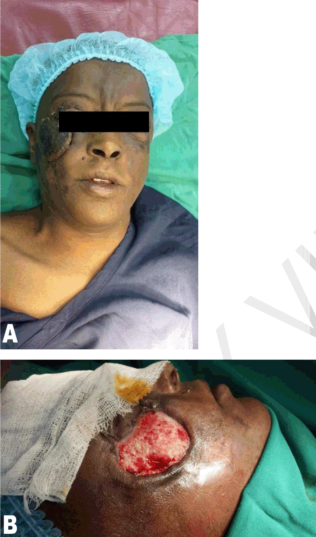
A 35-year-old female, who is on regular hemodialysis (HD) via an arm arteriovenous fistula (AVF), originally presented to the medical department complaining of high grade fever and failure of her AVF associated with a swelling and pus discharge. A diagnosis of a mycotic AVF was made and she received intravenous (IV) antibiotics for three weeks with no improvement. During that time, the patient complained of a progressively increasing periorbital swelling which became associated with complete ptosis of the right eye and partially on the left eye. The swelling was also accompanied by skin discoloration and pus discharge (Figure 1). She was referred to us for further management of her condition. On general examination, the patient looked ill and pallor was noted on the palms. Both the radial and ulnar pulses were intact bilaterally with a pulse rate of 112 beats per minute. Her blood pressure was 105/60 mmHg and her temperature was 39.1°C (102.38°F). There was a generalized swelling on the face, more prominent periorbitally, with complete ptosis of the right eye and an incomplete ptosis of the left eye (Figure 1). The periorbital swelling was firm, ulcerated and discharging pus bilaterally. No neurological deficit was noted. The left jugular vein was prominent and distended along its course (Figure 2). Upper limbs were asymmetrical (Figure 3), the left upper limb was swollen. On the left cubital fossa, there was a pulsatile, firm swelling which was discharging pus. Blood samples and a swab from the AVF were taken for laboratory investigations which revealed signs of infection. With the source of infection being the mycotic AVF pseudoaneurysm, the initial step of management included aneurysmectomy and AVF ligation to control the systemic infection. Postoperatively intravenous antibiotics and analgesia were administered. The fever subsequently subsided. Next, computed tomography angiogram of the head and neck showed a long segment occlusion on the left internal jugular vein extending to left brachiocephalic and subclavian veins (Figure 4) and evidence of a cavernous sinus thrombus (Figure 5); a diagnosis of a septic cavernous sinus thrombosis was made. The treatment included anticoagulant therapy, IV 3rd generation cephalosporin and periorbital eye dressing with honey and water. After the periorbital swelling and local infection subsided, she was discharged on oral anticoagulant therapy and daily eye dressing. She was later booked for debridement of a necrotic skin infection just below the right eye (Figure 6). After complete resolution of her condition, she now presents with ectropion of the right lower eye lid. | ||||||
| ||||||
| ||||||
| ||||||
| ||||||
| ||||||
| ||||||
|
Discussion
| ||||||
|
With regards to the anatomy, the cavernous sinuses are irregularly shaped and trabeculated cavities which, together with other sinuses, are collectively known as the dural sinuses. The cavernous sinuses lie at the base of the skull on either side of the sella turcica and of all the dural sinuses, they are the most centrally located in the skull. Relations to the cavernous sinuses include the sphenoid sinus, which lies inferomedially from the cavernous sinus and the optic chiasm, located anterior to the cavernous sinuses. Structures that pass through the cavernous sinuses include the internal carotid artery with its surrounding sympathetic plexuses. Attached to the lateral wall of the sinus are the oculomotor, trochlear and abducens nerves (cranial nerves III, IV and VI, respectively) and embedded in the wall are the ophthalmic and maxillary divisions of the trigeminal nerve. This complex and intimate relationship of vital structures within and around the cavernous sinuses accounts for the characteristic presentation of CST. Venous blood drains from the superior and inferior ophthalmic veins to the facial veins which drain into the cavernous sinuses. The cavernous sinuses also receive venous blood from the sphenoid and middle cerebral veins. Both cavernous sinuses then drain into the inferior petrosal sinuses which drain, via the superior petrosal sinuses, into the inferior jugular veins and sigmoid sinuses. Depending on the dominant pressure gradients, blood can flow in either direction due to the fact that this complex structure of cerebral veins contains no valves and infections usually extend from one cavernous sinus to the other because multiple connections exist between them [1]. In a study conducted by Stolic R regarding complications of AV fistulas, it was found that infections were very rare and respond well to antibiotic treatment lasting 4–6 weeks and that AVF ligature is only indicated if it is a source of recurrent septic pulmonary emboli [3]. In this case, a source of jugular vein occlusion and septic CST. Lucian et al., undertook a study in which they concluded that complications of autogenous arteriovenous fistulas are divided into two categories-acute and chronic. The acute complications included thrombosis, bleeding and hematoma formation, while the chronic complications were thrombosis, anastomotic pseudoaneurysms, venous aneurysm, venous pseudoaneurysm, skin necrosis, hand ischemia, hyperdynamic syndrome, hand edema, lymphorrhea and infection. Regarding anastomotic pseudoaneurysms and infections as a chronic complication, their rate was 0.8% and 0.1%, respectively. The formation of the pseudoaneurysm is almost always preceded by a septic process, either intraoperatively or due to an infected hemodialysis catheter or wrongful AVF cannulation techniques [4]. A case reported by Watsona and Russoa describes arterialized flow within a cavernous sinus due to an upper extremity arteriovenous dialysis fistula. The arterialized flow was due to occlusion of the left brachiocephalic vein resulting retrograde flow of blood via the left internal jugular vein. Despite it being a rare presentation, common causes for left brachiocephalic vein occlusion include venous catheter placement and malignancies [5]. In a retrospective study of 100,942 patients in a three- year period, conducted by Oymak et al., the prevalence of brachiocephalic veins and the superior vena cava (SVC) occlusion was rare. Only 33 patients (0.03%) were diagnosed with brachiocephalic or SVC thrombosis. They evaluated the causes and found that malignancies, chronic diseases, central venous lines, peripheral venous lines and thrombophilia came in at 42%, 39%, 27% and 38%, respectively. The manifestations included arm, head and neck swellings in 97% of the patients, which coincide with the findings in the patient being reported, and pulmonary embolism in 36% of the patients. No accounts of cavernous sinus thrombosis were observed [6]. Otten et al. performed a study of nine patients with brachiocephalic vein occlusion and found that three of these patients had AV fistulas as their means of dialysis with no history of central lines. They concluded that a sufficient number of patients develop brachiocephalic vein occlusion to consider a diagnosis, especially in patients with malignancies or central venous lines [7]. The list of complications provided in an article by Mueller regarding internal jugular vein thrombosis included pulmonary embolism, subclavian vein and superior sagittal sinus thrombosis, superior vena cava syndrome, pseudotumor cerebri, laryngeal and lower airway edema and infected thrombophlebitis, but again, no mention was given regarding CST as a complication [8]. Boedeker et al. described, in general, the complications of internal jugular vein thrombosis, which included pulmonary embolism, sepsis with septic emboli to different organs and tissues and thrombus propagation intracranially [9] . | ||||||
|
Conclusion
| ||||||
|
Cavernous sinus thrombosis (CST) can result as a complication of infections within the danger triangle of the face or paranasal sinuses, trauma, bacteremia or ear infections. Brachiocephalic and jugular vein thrombosis propagation is another important cause. Most importantly, in patients presenting with a mycotic arteriovenous fistula (AVF), the risk of developing a septic CST as a complication should be kept in mind and intervention, either conservative or surgical, should be based upon the risks versus benefits of either form of management and prevention of further complications. | ||||||
|
References
| ||||||
| ||||||

|
[HTML Abstract]
[PDF Full Text]
|
|
Author Contributions
Ahmed Mohamed Elhassan Elfaki Osman – Acquisition of data, Drafting the article, Final approval of the version to be published Saif-Eldin Mohammed Ali Ibrahim – Substantial contributions to conception and design, Revising it critically for important intellectual content, Final approval of the version to be published |
|
Guarantor of submission
The corresponding author is the guarantor of submission. |
|
Source of support
None |
|
Conflict of interest
Authors declare no conflict of interest. |
|
Copyright
© 2015 Ahmed Mohamed Elhassan Elfaki Osman et al. This article is distributed under the terms of Creative Commons Attribution License which permits unrestricted use, distribution and reproduction in any medium provided the original author(s) and original publisher are properly credited. Please see the copyright policy on the journal website for more information. |
|
|
|
About The Authors
| |||
| |||
| |||