| |
|
|
|
Case Report
| ||||||
| An unusual case of gastric gangrene, diaphragmatic gangrene and autosplenectomy due to mucormycosis in a diabetic patient | ||||||
| Amrit Manik Nasta1, Kushal Bairoliya2, Shashi Ranjan2 | ||||||
|
1MS, Senior Registrar in Department of General Surgery at KEM Hospital, Mumbai, Maharashtra, India.
2MS, Junior Registrar in Department of General Surgery at KEM Hospital, Mumbai, Maharashtra, India. | ||||||
| ||||||
|
[HTML Abstract]
[PDF Full Text]
[Print This Article]
[Similar article in Pumed] [Similar article in Google Scholar] 
|
| How to cite this article |
| Nasta AM, Bairoliya K, Ranjan S. An unusual case of gastric gangrene, diaphragmatic gangrene and auto-splenectomy due to mucormycosis in a diabetic patient. Int J Case Rep Images 2015;6(2):95–98. |
|
Abstract
|
|
Introduction:
Stomach has a rich vascular supply and undergoes gangrene in rare circumstances. Some of the known causes include gastric volvulus, diaphragmatic herniation, caustic injury and infectious necrotising gastritis. Mucormycosis, a life-threatening infection caused by fungi of the subphylum Mucoromycotina, order Mucorales, usually affects the para-nasal sinuses, orbit, brain and lung.
Case Report: We report an unusual presentation of gastric mucormycosis in a 50-year-old diabetic male, leading to perforative peritonitis with diaphragmatic gangrene and auto-splenectomy. Conclusion: Clinical suspicion of mucormycosis in immunosuppressed to aid diagnosis and adequate treatment in the form of antifungal agents and surgery may help improve outcome. | |
|
Keywords:
Autosplenectomy, Diaphragmatic gangrene, Gangrene of stomach, Gastric Mucormycosis
| |
|
Introduction
| ||||||
|
Mucormycosis of stomach is a rare cause of gangrene and perforation of gastric wall [1]. Fungi of the order Mucorales are causative agents of Mucormycosis, commonly of genus Rhizopus and Mucor. Risk factors for the development of invasive mucormycosis include diabetes, immunosuppressive states, corticosteroid use, organ or stem cell transplantation, and increased levels of available serum iron [2]. Common sites of infection are the paranasal sinuses, orbit, brain and lung. Gastrointestinal mucormycosis, in the past, was usually seen in premature neonates with widespread dissemination of the disease [3]. We report an unusual presentation of gastric mucormycosis leading to gangrene and perforation, associated with gangrene of diaphragm and autosplenectomy, in a diabetic male. | ||||||
|
Case Report
| ||||||
|
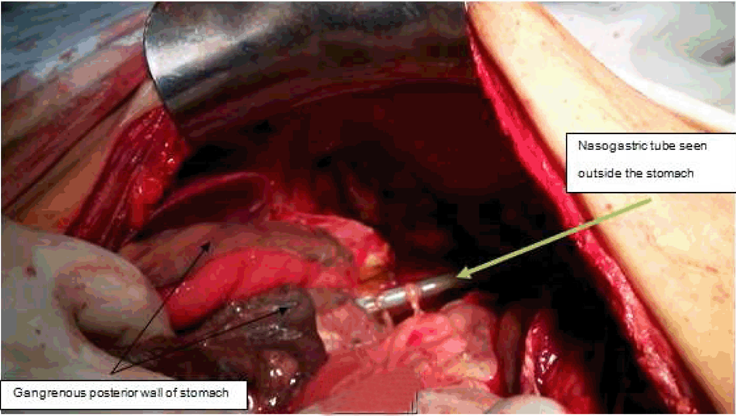
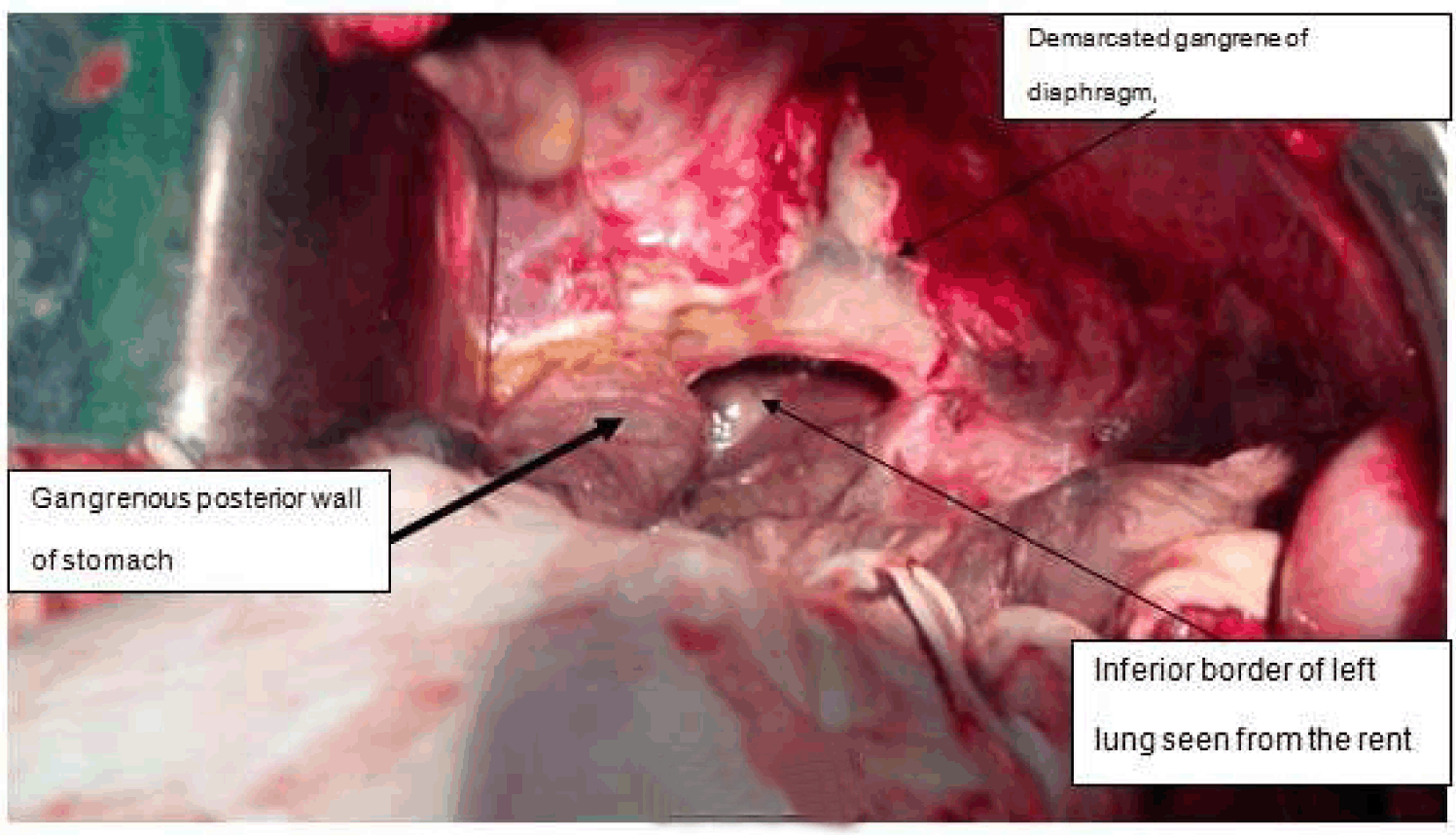
A 50-year-old male, known diabetic, presented to the emergency medical department with complaints of breathlessness and left sided chest pain since one week. Pain was continuous, dull aching and present at rest. He had no history of similar chest complaints in the past. There were no abdominal complaints. There was no significant surgical history or history of any trauma to the chest. On examination, patient was conscious and oriented, afebrile, pulse 100/minute, blood pressure 90/70 mmHg, respiratory rate was 22/minute. Air entry was decreased in left lower zones of the lung with dullness on percussion. Abdominal examination revealed mild tenderness in left hypochondriac region, otherwise unremarkable. Chest X-ray revealed left hydro-pneumothorax, abdominal ultrasound revealed free fluid in left sub-phrenic space. Computed tomography (CT) scan of chest and abdomen revealed perforation of fundus of stomach with extravasation into left pleural space, rent in left hemi-diaphragm and absent spleen (Figure 1). After initial resuscitation, patient underwent an emergency exploratory laparotomy which showed gangrene of posterior wall of stomach with demarcation, with a 1x1 cm perforation. A left diaphragmatic rent about 6x6 cm with gangrenous edges, absent spleen and moderate perigastric collection were other findings (Figure 2) and (Figure 3). Rest of the bowel was normal. Posterior gangrenous segment of stomach was excised and wall was closed using 55 mm linear staplers. Gangrenous diaphragmatic edges were excised, left intercostal drain placed and defect plugged with 15x15 cm composite mesh, as edges could not be approximated. Washes were given and distal feeding jejunostomy was done. Histopathology of resected stomach revealed necrosis with mucormycosis (Figure 4) and (Figure 5). Postoperatively, patient complicated with staple line leak and sepsis, and expired on postoperative day-3. | ||||||
| ||||||
| ||||||
|
| ||||||
| ||||||
| ||||||
|
Discussion
| ||||||
|
The gastrointestinal tract is a rare site of mucormycosis. Gastrointestinal mucormycosis was seen mainly in neonatal age group, often in association with disseminated disease [3] [4]. Other rare cases of gastrointestinal mucormycosis were previously described in association with other immunocompromised states, like HIV, systemic lupus erythematosus, and organ transplantation [5]. Cases of hepatic mucormycosis have also been associated with ingestion of herbal medications [6]. As this infection is acute and rapidly fatal; it is often diagnosed with post-mortem. Mucormycosis of gastrointestinal tract, depending on the site of pathology, may present as pain, distention, nausea and vomiting commonly [7]. Fever, hematemesis, melena or hematochezia may also occur. The patient is often misdiagnosed to have an intra-abdominal abscess. The diagnosis may be made by biopsy of the suspected area during surgery or endoscopy [7]. Our patient had an unusual presentation with mainly chest symptoms due to hydropneumothorax. In a case reported by Morton et al., a patient had Crohn's disease, who presented with perforative peritonitis and there was no specific reason to suspect mucormycosis [8]. Patient was on steroid therapy for Crohn's disease. Only the appearance of the fungi on histopathology caused the diagnosis to be made and appropriate therapy to be initiated. This case highlights the need to maintain a high index of suspicion for invasive fungal infections like mucormycosis, in immunosuppressed patients. Mucormycosis is difficult to treat with antifungals alone, like Amphotericin B and triazoles like Posaconazole. In addition, surgery is required to remove all infected tissue. Mucormycosis shows angioinvasion and tissue necrosis which results in poor local penetration of anti-fungal agents. With an ongoing epidemic of obesity and diabetes, and the increasing population of patients receiving immunosuppressive therapy for inflammatory diseases, solid organ or stem cell transplantation, it is not surprising that recent studies have reported alarming increases in the incidence of mucormycosis [9]. It is likely that the clinicians will encounter this disease more frequently in the coming years, especially in the nosocomial setting. | ||||||
|
Conclusion
| ||||||
|
Invasive mucormycosis is a life-threatening infection, with varied clinical presentations including gastrointestinal involvement. Clinical suspicion in immunosuppressed to aid diagnosis and adequate treatment in the form of antifungal agents and surgery may help to improve outcome. | ||||||
|
Acknowledgements
| ||||||
|
We are thankful to Dr. Nagsen and Dr. Samruddhi from department of Surgical Pathology for providing the histopathology images. | ||||||
|
References
| ||||||
| ||||||
|
[HTML Abstract]
[PDF Full Text]
|
|
Author Contributions
Amrit Manik Nasta – Conception and design, Acquisition of data, Analysis and interpretation of data, Drafting the article, Critical revision of the article, Final approval of the version to be published Kushal Bairoliya – Conception and design, Acquisition of data, Analysis and interpretation of data, Drafting the article, Critical revision of the article, Final approval of the version to be published Shashi Ranjan – Acquisition of data, Analysis and interpretation of data, Final approval of the version to be published |
|
Guarantor of submission
The corresponding author is the guarantor of submission. |
|
Source of support
None |
|
Conflict of interest
Authors declare no conflict of interest. |
|
Copyright
© 2015 Amrit Manik Nasta et al. This article is distributed under the terms of Creative Commons Attribution License which permits unrestricted use, distribution and reproduction in any medium provided the original author(s) and original publisher are properly credited. Please see the copyright policy on the journal website for more information. |
|
|
|
About The Authors
| |||
| |||
| |||
| |||