  |

|
 |
|
Case Report
| ||||||
| Successful tenecteplase use in sudden shock from CT-proven massive pulmonary embolism | ||||||
| Jason P. Stopyra1, William P. Bozeman1 | ||||||
|
1MD, FACEP, Wake Forest University School of Medicine, Department of Emergency Medicine Winston-Salem, NC.
| ||||||
| ||||||
|
[HTML Abstract]
[PDF Full Text]
[Print This Article]
[Similar article in Pumed] [Similar article in Google Scholar] 
|


| How to cite this article |
| Stopyra JP, Bozeman WP. Successful tenecteplase use in sudden shock from CT-proven massive pulmonary embolism. Int J Case Rep Images 2015;6(2):86–89. |
|
Abstract
|
|
Introduction:
Emergency, inpatient, and critical care physicians frequently evaluate for and treat patients with deep venous thrombosis (DVT) and pulmonary embolism. While most patients with diagnosed pulmonary embolism remain stable and do not require aggressive therapy, hemodynamic compromise due to pulmonary embolism can occur suddenly and without warning.
Case Report: In the community emergency department fibrinolysis is the only available lifesaving option for massive pulmonary embolism. In this report, we describe a patient who suddenly became profoundly unstable moments after establishing the diagnosis by computed tomography scan. Systemic fibrinolysis was administered using the single bolus agent tenecteplase; the patient stabilized and recovered without bleeding complications, neurologic or cardiovascular deficits. Conclusion: All physicians, especially those in a community setting who treat patients with venous thromboembolic disease should be familiar with and prepared to give these agents. | |
|
Keywords:
CT-proven, Fibrinolysis, Pulmonary embolism, Thrombolysis
| |
|
Introduction
| ||||||
|
Venous thromboembolic disease includes deep venous thrombosis (DVT) and pulmonary embolism (PE), and can present initially with non-specific signs and symptoms. Many patients have DVT/PE in their working differential diagnosis and clinicians in many settings must maintain a high level of suspicion to diagnose this life-threatening condition. After diagnosis, stable patients with PE are typically treated with parenteral anticoagulation and simple monitoring during conversion to oral anticoagulation therapy [1]. In the setting of hemodynamic instability, due to large PE an immediate reduction in clot burden is needed. In most settings systemic fibrinolysis is the most readily available of the several treatment options and should be undertaken emergently [1] [2]. Fibrinolytic agents are commonly and successfully used in patients with suspected PE who initially present with hemodynamic compromise, but these are most often treated empirically based on clinical suspicion since confirmatory testing is precluded by clinical instability. In some cases, however, an initially stable patient with suspected or confirmed PE may suddenly become unstable and require fibrinolysis. Emergency, critical care, and inpatient physicians in particular must be prepared for this situation and comfortable with the use of systemic fibrinolysis during this pivotal time. Though academic and international studies have been published on this topic, the use of fibrinolysis is even more important because it is the clinician's only option to prevent mortality [3] [4] [5]. | ||||||
|
Case Report
| ||||||
|
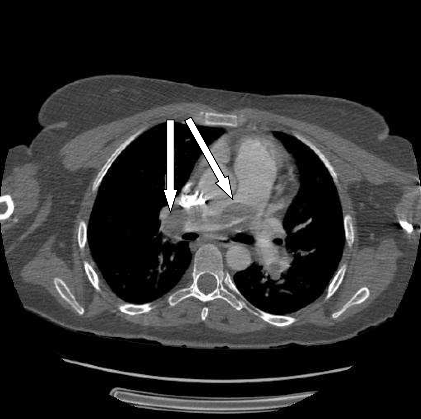
A 33-year-old female presented via ambulance to small community hospital emergency department complaining of feeling weak and dizzy. Two hours prior to calling EMS she began to feel "a funny fluttering in chest" associated with weakness, sweating and near syncope upon standing. She recalled two similar, but less severe episodes over the past week. On questioning, she also reported several episodes of left sided chest pain and dyspnea on exertion over the previous week. Two weeks prior, she had an open reduction and internal fixation of a fractured left metatarsal, with cast immobilization since that time. Other medical history included hypertension, gastroesophageal reflux, and hypercholesterolemia. Medications included lovastatin, lisinopril, esomeprazole, and oxycodone/acetaminophen. She was a non-smoker and family history was unremarkable. Initial examination showed a conscious, alert female in no distress. Her vital signs on arrival included a heart rate of 145 bpm, respiratory rate of 20 bpm, blood pressure 106/60 mmHg, and oxygen saturation of 97% on oxygen via nasal cannula. Oxygen saturations on room air were not recorded prior to EMS providers initiating oxygen therapy. The physical examination revealed a pleasant young woman with mild tachypnea, tachycardia, normal heart and lung examinations, and her left leg in a cast. An electrocardiogram (EKG) showed sinus tachycardia with right axis deviation, but no dysrhythmias or acute ischemic changes. A chest X-ray, complete blood count, electrolytes, and cardiac enzymes (troponin I and creatine phosphokinase) were all within normal limits. The risk of PE was considered to be high so a subcutaneous dose of enoxaparin (1 mg/kg) was administered empirically. She was given a fluid bolus and morphine for chest discomfort. She was transported to the radiology area of the emergency department and spiral computed tomography (CT) angiography was performed of her pulmonary vessels. After completion of the scan, just as the patient was moved back to her stretcher, she lost consciousness and displayed a brief episode of generalized tonic-clonic motor activity. Carotid pulses were not palpable during the episode indicating either cardiac arrest or profound hypotension. During the following 30–60 seconds she was rapidly moved to the emergency department treatment area for resuscitation. However, within those seconds her pulses became palpable, and her mental status and blood pressure rapidly improved and returned to baseline over the next three to five minutes. Review of the CT images obtained moments before showed a large central pulmonary embolism. (Figure 1). A heparin drip was initiated and consent was obtained for systemic fibrinolysis. Tenecteplase 45mg (0.5 mg/kg) was administered intravenously. Approximately fifteen minutes later, the patient had another sudden episode of increased dyspnea, anxiety and chest pressure, followed by a 45-second loss of consciousness with myoclonus and cyanosis of the face and neck. Blood pressure could not be measured but central pulses remained palpable. The cardiac monitor showed sinus tachycardia throughout the episode. Ventilations were assisted with bag valve mask (BVM). The patient again regained consciousness and subsequently stabilized. She was promptly transferred to the intensive care unit of a nearby tertiary care facility by helicopter. Upon arrival at the tertiary facility's ICU the patient displayed normal vital signs and had no further episodes of syncope or hypotension. A transthoracic ultrasound was performed on the next day, showing a dilated right ventricle with paradoxical septal motion. Left ventricular size and function were normal. Lower extremity Doppler studies revealed widespread deep venous thrombosis of the distal left leg and an inferior vena caval filter was placed. Serial cardiac enzymes showed a peak troponin I of 0.39 ng/mL on the second hospital day. Warfarin therapy was initiated and she was discharged home after nine days without neurologic deficits or bleeding complications. On follow-up one year later she continued to do well. | ||||||
| ||||||
|
Discussion
| ||||||
|
This case demonstrates an initially stable patient who presented with findings worrisome for pulmonary embolism (PE) that rapidly became unstable. Traditional risk stratification indicators of massive pulmonary embolism that may benefit from fibrinolysis, including hypotension, hypoxia, and elevated cardiac enzymes, were absent prior to her sudden clinical decompensation. After prompt treatment with systemic fibrinolysis in the emergency department, she stabilized and was later discharged without neurologic deficit or other adverse outcome. Without aggressive treatment the patient may well have had additional hemodynamic decompensation and a poor or fatal outcome. Despite the patient's non-specific initial chief complaint of feeling weak and dizzy, careful questioning and physical examination made the physician strongly suspect pulmonary embolism. Her rapidly evolving process gave no warning of its change from stable to unstable with the need for aggressive measures. It was fortunate and unusual that the first episode of hemodynamic instability occurred just after the CT scan was completed, allowing confirmation of the suspected diagnosis. Fibrinolytic agents are widely accepted as first line therapy in the setting of hemodynamic compromise due to massive PE. There is ongoing discussion whether this therapy should be expanded to other high risk patients without hemodynamic compromise, such as those with elevated cardiac biomarker or right ventricular dysfunction on EKG, echocardiogram, or CT angiography [1] [2] [6] [7]. Other treatments such as catheter-guided thrombectomy or surgical embolectomy, if available emergently, should also be considered to treat massive PE [1]. In this case, tenecteplase (TNKase®) was used for systemic fibrinolysis. This agent is administered as a single weight-based bolus injection; it is immediately available in our community emergency department for ST elevation myocardial infarction (STEMI) and has also been used for PE [4]. Other commonly available fibrinolytic agents include reteplase (Reteplase®), which is administered in two 10 mg doses 30 minutes apart, and alteplase (recombinant tissue plasminogen activator, r-tPA or Activase®) which is typically given as a 100 mg infusion over 2 hours. All three agents are approved by the United States Food and Drug Administration for STEMI. Alteplase and the older agents streptokinase and urokinase are formally approved for PE [2] [7]. The choice of fibrinolytic agent for PE remains one of physician judgment, preference, and availability. The tenecteplase used in this case has several potential advantages over other agents. These include single bolus dosing, which makes it attractive for resuscitation situations and may alleviate confusion about infusion protocols and co-administration of heparin, and may result in more rapid plasmin formation and clinical effects [7]. In addition, the possibility of bleeding complications is always of concern when considering systemic fibrinolysis. In previous STEMI trials, tenecteplase has had fewer non-cerebral bleeding complications than alteplase [8]. Most emergency physicians and cardiologists practicing in community hospitals are comfortable giving fibrinolytic agents to STEMI patients, in part because EKG criteria clearly show confirmatory evidence to indicate their use. The diagnosis of massive PE is frequently less clear, since unstable patients needing the therapy are frequently too unstable to undergo CT scanning. Other imaging modalities such as transesophageal ultrasound and formal angiography which can confirm the diagnosis may not be available in a timely manner. Consequently, in the setting of massive PE, systemic fibrinolytics frequently must be administered based on clinical suspicion alone. As demonstrated in this case, even an initially stable patient can decompensate suddenly and without warning. Therefore, all physicians who may treat patients with known or suspected DVT or PE should be familiar with fibrinolytic therapy and prepared to administer these agents rapidly when indicated. A fibrinolytic eligibility checklist to exclude contraindications should be completed in some form even in stable patients at the time the diagnosis is entertained since the necessary information may be unavailable or too time-consuming to collect if a patient becomes unstable and requires rapid treatment. | ||||||
|
Conclusion
| ||||||
|
All emergency and inpatient physicians, especially those in a community setting should maintain a high level of suspicion for pulmonary embolism and should be prepared to administer systemic fibrinolytic agents when needed in the setting of massive pulmonary embolism, even if patients are initially stable upon presentation. | ||||||
|
Key Points
| ||||||
| ||||||
|
References
| ||||||
| ||||||

|
[HTML Abstract]
[PDF Full Text]
|
|
Author Contributions
Jason P. Stopyra – Substantial contributions to conception and design, Drafting of article, Revising it critically for important intellectual content, Final approval of the version to be published William P. Bozeman – Substantial contributions to conception and design, Drafting of article, Revising it critically for important intellectual content, Final approval of the version to be published |
|
Guarantor of submission
The corresponding author is the guarantor of submission. |
|
Source of support
None |
|
Conflict of interest
Authors declare no conflict of interest. |
|
Copyright
© 2015 Jason P. Stopyra et al. This article is distributed under the terms of Creative Commons Attribution License which permits unrestricted use, distribution and reproduction in any medium provided the original author(s) and original publisher are properly credited. Please see the copyright policy on the journal website for more information. |
|
|