|

|
 |
|
Case Report
| ||||||
| A rare case of congenital esophagobronchial fistula in an adult | ||||||
| V. YaminiChitra1, K. N. Paramesh1, Alamelu Haran2, Nitin D. Tengli4 | ||||||
|
1MS, (General Surgery), DNB, MCH (Surgical Gastroenterology), Associate Professor, Dept of Surgical Gastroenterology and Bariatric centre, Vydehi Institute of Medical Sciences and Research Centre, Bangalore, Karnataka, India.
2MS, (General Surgery), (DNB Surgical Gastroenterology), Senior Resident, Dept of Surgical Gastroenterology and Bariatric Centre, Vydehi Institute of Medical Sciences and Research Centre, Bangalore, Karnataka, India. 3MD (Tuberculosis and Chest Diseases), Professor and HOD, Department of Pulmonary Medicine Vydehi Institute of Medical Sciences and Research Centre, Bangalore, Karnataka, India. 4MS, (General Surgery) Senior Resident, Department of Surgical Gastroenterology and Bariatric Centre, Vydehi Institute of Medical Sciences and Research Centre, Bangalore, Karnataka, India. | ||||||
| ||||||
|
[HTML Abstract]
[PDF Full Text]
[Print This Article]
[Similar article in Pumed] [Similar article in Google Scholar] 
|


| How to cite this article |
| YaminiChitra V, Paramesh KN, Haran A, Tengli ND. A rare case of congenital esophagobronchial fistula in an adult. Int J Case Rep Images 2015;6(2):70–75. |
|
Abstract
|
|
Introduction:
Congenital esophagobronchial fistulas in adults are extremely rare, acquired fistulas being more common. The aim of this paper was to present a rare case of congenital esophagobronchial fistula in a 37-year-old male of type II Braimbridge's classification and to emphasize on the diagnostic modality of choice and the appropriate mode of treatment.
Case Report: A 37-year-old male presented with chronic cough with ingestion of food, especially liquids of 13 years duration and recent onset hemoptysis. He was evaluated with upper gastrointestinal endoscopy, bronchoscopy, computed tomography scan of chest and the definitive test was barium swallow which confirmed it. He underwent transthoracic excision of the fistula with repair of both esophageal and bronchial ends. A peroperative endoscopy helped localization of the tract. Postoperative outcome was excellent with no leak and patient is totally asymptomatic after 12 weeks of surgery. Conclusion: Congenital esophagobronchial fistulas in adults, due to insidious nature need high index of suspicion as early diagnosis by barium swallow and surgical treatment gives excellent results. Peroperative endoscopy is mandatory to localize the tracts, helps do an intraoperative leak test and avoid esophageal stenosis during repair. | |
|
Keywords:
Barium swallow, Congenital esophagobronchial fistula, Peroperative endoscopy, Transthoracic excision
| |
|
Introduction
| ||||||
|
Fistula between esophagus and bronchus may be congenital or acquired. Acquired fistulas can be inflammatory, traumatic or neoplastic. Congenital esophago bronchial fistula (EBF) with atresia present immediately in infancy, are sudden and are diagnosed early and treated [1]. Congenital EBF without atresia are insidious, can present in adulthood also or missed often especially if they communicate with a lobar bronchus [1]. To differentiate between congenital and acquired EBF in an adult is usually difficult [2]. Adult EBF may present acutely with sudden respiratory distress, chronically with repeated respiratory infections or can be totally asymptomatic. Less than 200 cases of adult EBF are reported so far in literature [2]. Herein, we report a rare case of 37-year-old male with congenital EBF without atresia (type 2 according to Braimbridge's classification) [1] which was diagnosed by barium esophagogram. Patient underwent transthoracic fistulectomy with the repair of both esophageal and bronchial ends. Postoperative outcome was successful with complete resolution of symptoms and closure of fistula. | ||||||
|
Case Report
| ||||||
|
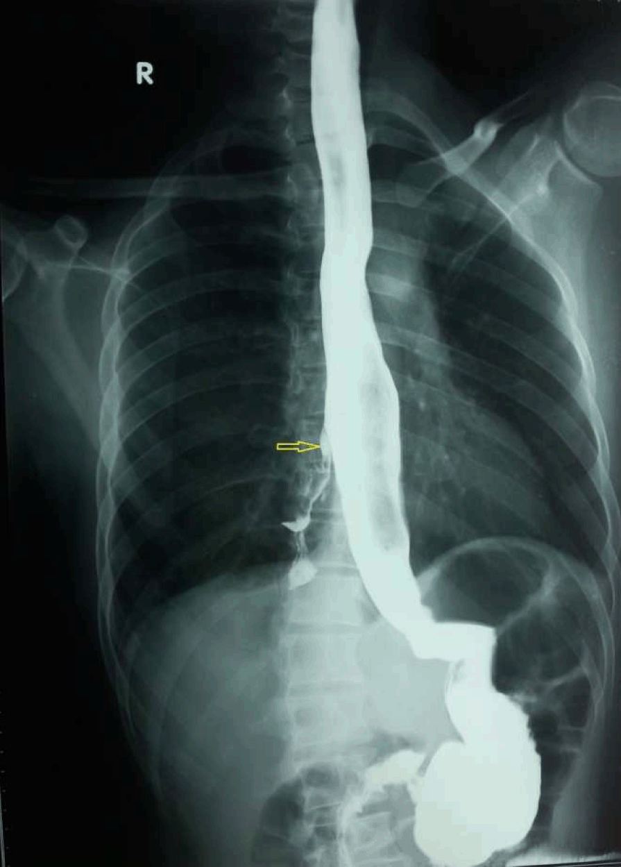
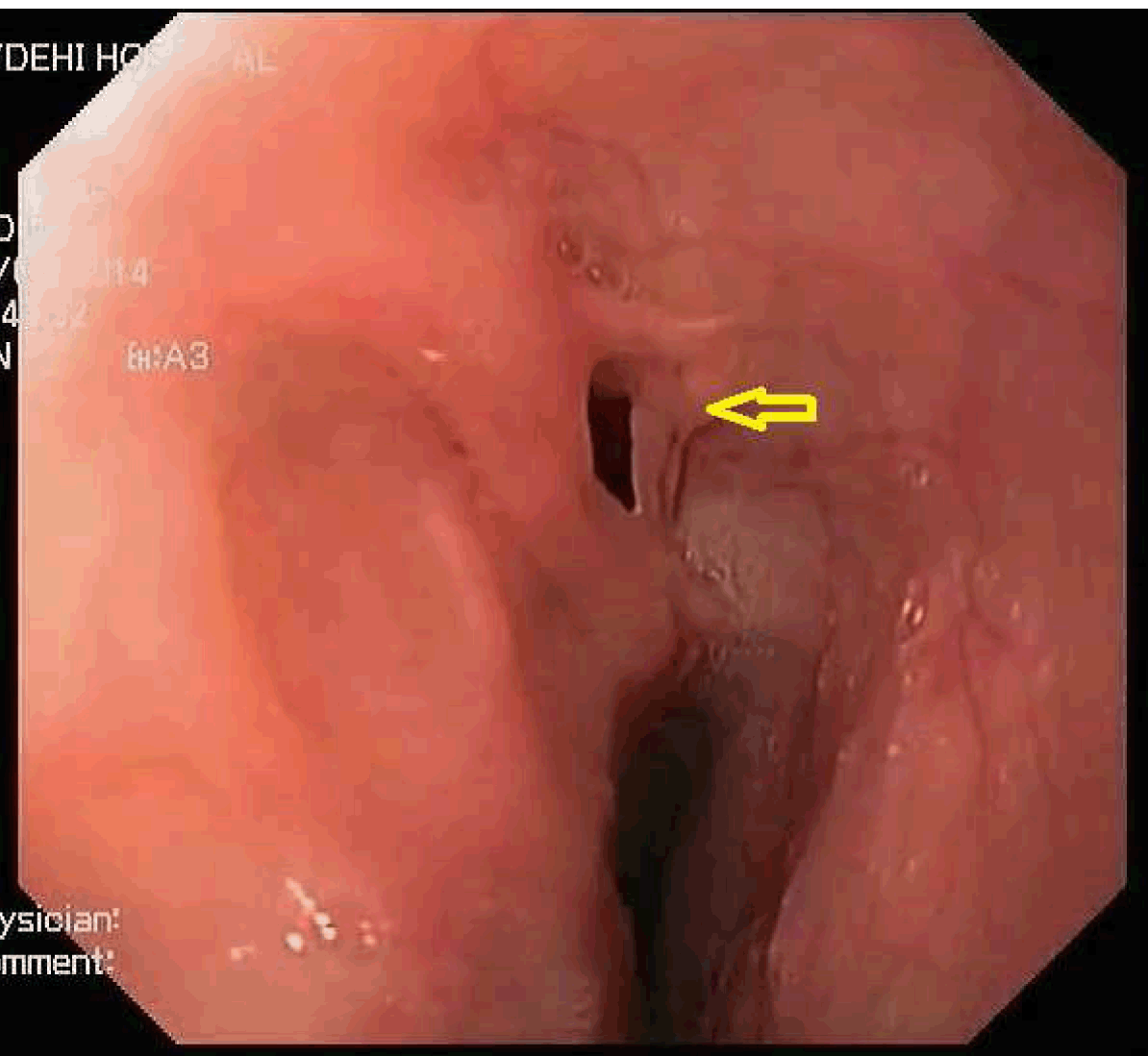
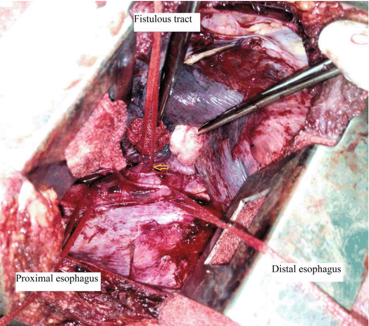
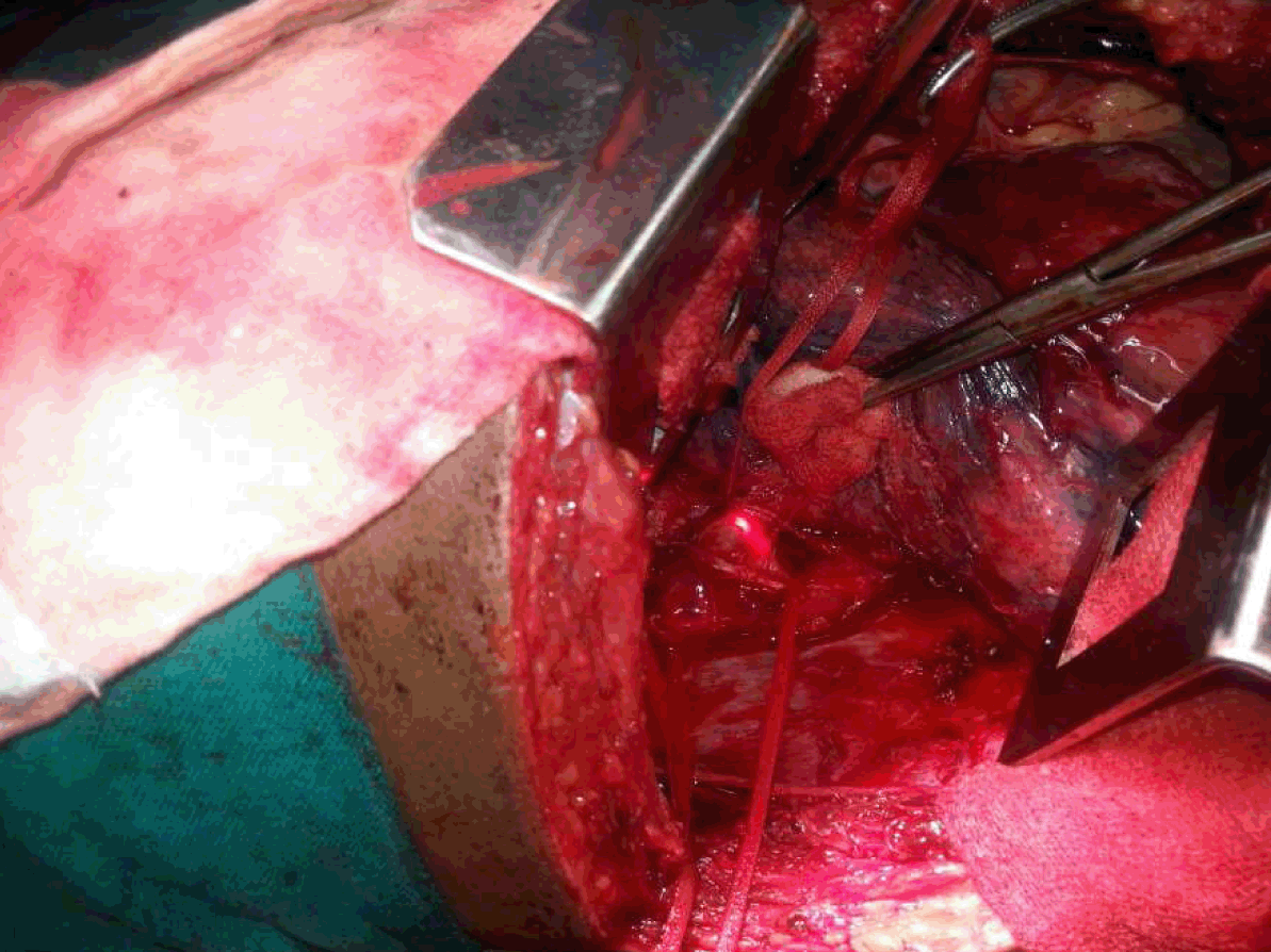
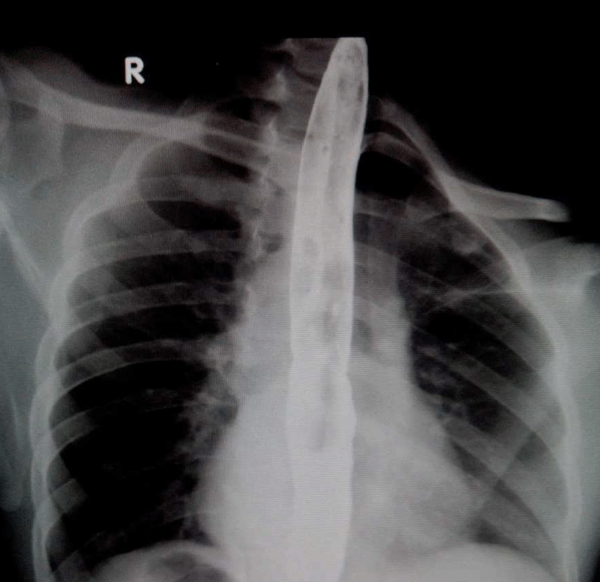
A 37-year-old male presented to our department with history of cough with expectoration immediately after taking food, especially liquids for last 13 years. He had two bouts of hemoptysis in the last 15 days which had made him seek medical attention. Clinically, chest on auscultation had crackles in right infra-scapular region. Chest X-ray revealed patchy opacities in the right lower zone paracardiac region. Computed tomography (CT) scan of thorax revealed consolidation in posterior segment of the right lower lobe with no evidence of lung sequestration or cyst. Upper gastrointestinal endoscopy showed a fistulous opening in the mid esophagus 32 cm from the incisor teeth. It did not show any evidence of malignancy, granulomatous disease or any other acquired basis for the fistula. Simultaneously, methylene blue was injected into the fistulous tract and bronchoscopy was done which was normal. Barium swallow showed fistulous communication between mid esophagus and right lower lobe bronchus at lower border of T7 with barium passing downward into the right lung (Figure 1). Preoperative evaluation done included pulmonary function tests, echocardiogram and electrocardiogram. Incentive spirometry was started for better postoperative outcome. Preoperatively, 1 fr size guide wire was introduced endoscopically into the fistulous tract to aid identification of fistula. Patient underwent right posterolateral thoracotomy. There were minimal adhesions around the fistulous site which was identified about 5 cm below the level of entry of azygos vein into superior vena cava. Azygos vein was isolated, ligated and cut to aid esophagus to be encircled, to localize the fistulous tract. As the guide wire was not palpable through the tract, intraoperative endoscopy was done and fistula tract location was confirmed. The fistulous tract was dissected, it was 10 mm long. The tract was excised, both the esophageal and bronchial ends were healthy and were closed with 4–0 Vicryl. An intraoperative leak test was done using endoscopy which confirmed the integrity of the repair and an intercostal drain placed (Figure 2) (Figure 3) (Figure 4). Postoperative period was uneventful. Intercostal drain was removed on postoperative day-4 after a barium swallow to confirm that there was no leak and patient was discharged on postoperative day-6, after starting oral soft diet (Figure 5). Histopathology showed the mucosa to be lined by stratified squamous epithelium. There was no evidence of inflammation, granuloma or carcinoma confirming the congenital nature of the fistula. Patient is totally asymptomatic after 12 weeks of surgery. | ||||||
| ||||||
| ||||||
| ||||||
| ||||||
| ||||||
|
Discussion
| ||||||
|
The majority of EBF in adults are acquired. The usual causes are inflammatory like tuberculosis, trauma and neoplasms. The EBF was first described as early as in 1916 by Heiderich [3]. Congenital EBF are rare, three times more common on the right than the left [4]. According to Braimbridge's classification, type I is a fistula associated with a wide-necked congenital diverticulum of the esophagus with inflammation at the tip. Type II, which is the simplest and most common, consists of a short tract running directly from the esophagus to the lobar or segmental bronchus. In type III, the fistulous tract connects the esophagus to a cystic pulmonary change, and in type IV a fistula runs into a sequestered pulmonary segment. The patient described here is of type 2 according to this classification [1]. Criteria for congenital EBF are suggested pathologically by the absence of surrounding inflammation and adherent lymph nodes along with the presence of a mucosa and a definitive muscularis mucosa within the fistulous tract. Surgically, uncomplicated and easy dissection of the fistula and absence of inflammation suggests a congenital fistula [5]. Reasons for delay in the onset of symptoms and presentation in the adult may be due to
Both sexes are affected almost equally (male 53% and females 47%) [6]. Symptoms are often intermittent, due to chronic bronchopulmonary infection. They are chronic cough (96%), pneumonia (56%), hemoptysis (17%). Ohno's sign characterized by symptoms of strangulation and paroxysmal coughing with swallowing liquids occurs in the presence of a very large communication [7]. The diagnosis is usually made by barium esophagogram [1]. A thin barium is given, in a position where patient gets most of the symptoms. Endoscopy and bronchoscopy should be done but will not always demonstrate the opening in esophagus and bronchus [4]. Ramo et al. have shown that bronchoscopy is negative in 67% of cases and esophagoscopy in 40% [4]. Bronchoscopy can be normal, if the communication is in the distal segmental portion of the tract as in our case [7]. Dynamic bronchoscopy with pediatric bronchoscope may help [7]. Right bronchus is most commonly affected [7]. Computed tomography scan may help to rule out type 4 fistulas and aortography can document the sequestrated lung [8] and aid in pulmonary resection if needed. Peroperative endoscopy helps accurately in identifying the tracts [2], to confirm patency of the esophagus if the repair is very proximal or on the esophageal wall itself, as in wide short tracts and also aids in doing an intraoperative leak test as done in our case. Thoracotomy and resection of the fistulous tract with primary repair of both the bronchial and esophageal defects with 4–0 Vicryl the ideal treatment of choice. Interposition of pleural/diaphragmatic flap helps reduce the recurrence. In our case, it was not done as the tract was long and the bronchial and esophageal tissue post repair were healthy. The diseased lung tissue and sequestrated lobes if any should be resected at the same time. Postoperative barium swallow helps document the healed defect in the esophagus. Endoscopic management by submucosal dissection and isolation of the fistulous tract with clipping has been tried in tracheoesophageal fistula but has been unsuccessful [9]. Other endoscopic techniques like histoacryl glue injection have been tried in recurrent congenital trachea esophageal fistulas, or when patient has refused surgery with varied results. Thoracoscopic repair assisted by peroperative endoscopy and stapling of the fistulous tract by surgeons experienced in minimally invasive surgery can reduce the morbidity associated with open thoracotomy [10]. Either thoracoscopic or open thoracotomy repair with excision of the fistulous tract and good repair of the esophageal and bronchial defects with tissue interposition when needed, gives permanent cure and relief of symptoms in the patients with EBF. | ||||||
|
Conclusion
| ||||||
|
Congenital esophagobronchial fistulas in adults, due to rare occurrence and insidious nature need high index of suspicion as early diagnosis by barium swallow and surgical treatment by open or minimally invasive technique gives excellent results. | ||||||
|
References
| ||||||
| ||||||

|
[HTML Abstract]
[PDF Full Text]
|
|
Author Contributions
V. Yamini Chitra – Substantial contributions to conception and design, Acquisition of data, Analysis and interpretation of data, Drafting the article, Revising it critically for important intellectual content, Final approval of the version to be published K. N. Paramesh – Substantial contributions to conception and design, Acquisition of data, Analysis and interpretation of data, Drafting the article, Final approval of the version to be published Alamelu Haran – Substantial contributions to conception and design, Acquisition of data, Analysis and interpretation of data, Drafting the article, Final approval of the version to be published Nitin Tengli – Substantial contributions to conception and design, Acquisition of data, Analysis and interpretation of data, Drafting the article, Final approval of the version to be published |
|
Guarantor of submission
The corresponding author is the guarantor of submission. |
|
Source of support
None |
|
Conflict of interest
Authors declare no conflict of interest. |
|
Copyright
© 2015 V. Yamini Chitra et al. This article is distributed under the terms of Creative Commons Attribution License which permits unrestricted use, distribution and reproduction in any medium provided the original author(s) and original publisher are properly credited. Please see the copyright policy on the journal website for more information. |
|
|
|
About The Authors
| |||
| |||
| |||
| |||
| |||