 |

|
 |
|
Clinical Image
| ||||||
| Acute organoaxial gastric volvulus | ||||||
| James Heaton1, Andrew Gilliam2 | ||||||
|
1MBBS, Surgical House Officer, Department of Surgery, County Durham & Darlington NHS Foundation Trust, Darlington, County Durham, UK.
2DM, FRCS, Consultant Surgeon, Department of Surgery, County Durham & Darlington NHS Foundation Trust, Darlington, County Durham, UK. | ||||||
| ||||||
|
[HTML Abstract]
[PDF Full Text]
[Print This Article]
[Similar article in Pumed] [Similar article in Google Scholar] 
|


| How to cite this article |
| Heaton J, Gilliam A. Acute organoaxial gastric volvulus. Int J Case Rep Images 2015;6(2):124–126. |
|
Case Report
| ||||||
|
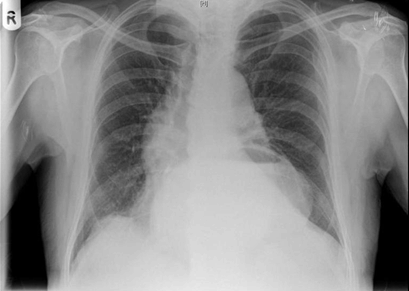
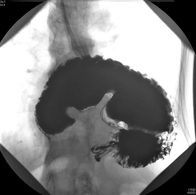
A 73-year-old male presented to the emergency department with a one-day history of severe epigastric pain with vomiting progressing to dry retching. He admitted to a long history of esophageal reflux symptoms treated with oral omeprazole. His initial examination revealed a tachycardia and a swollen, tender epigastrium but no other signs of note. A nasogastric tube was passed with difficulty, he was made nil by mouth and treated with intravenous crystalloids. Laboratory blood results were unremarkable including normal amylase and liver function tests. A chest X-ray showed a large retrocardiac viscus reported as a large hiatus hernia while his abdominal film demonstrated a paucity of bowel gas (Figure 1). An abdominal and thoracic computed tomography (CT) scan revealed a moderate hiatus hernia without obvious perforation and the possible appearance of a rotational component of the stomach with dilatation to the pylorus and no fluid beyond this point. A subsequent upper gastrointestinal contrast study clearly demonstrated an 'upside-down stomach' sign and established the diagnosis of an obstructing organoaxial volvulus secondary to a paraesophageal hiatus hernia (Figure 2) [1]. On transfer to our facility the patient went on to have definitive laparoscopic gastropexy surgery involving reduction of the volvulus, excision of the hernia sac, re-approximation of the diaphragmatic crura then placement of four sutures anchoring the greater curvature of the stomach to the abdominal wall. He has had no recurrence of the volvulus and was symptom free when followed-up in clinic for six months. | ||||||
| ||||||
|
| ||||||
| ||||||
|
Discussion
| ||||||
|
Gastric volvulus is an abnormal rotation of the stomach through more than 180 degrees, first described by Berti in 1866 [2]. This can lead to ulceration, perforation, hemorrhage, ischemia or necrosis [1]. The non-operative mortality rate is as high as 80% [3]. Adults with acute gastric volvulus typically present with epigastric pain and distension, unproductive vomiting and difficulty with nasogastric tube insertion. A constellation known as Borchardt's triad [4]. About 10–20% of cases occur in children, in adults it can occur at any age but is more common after the fourth decade of life [1] [5] . Gastric volvulus can be classified according to the axis around which the stomach rotates. In organoaxial volvulus, the stomach rotates around an axis connecting the gastroesophageal junction with the pylorus. This is the most common type of gastric volvulus occurring in approximately 60% of cases and commonly leads to strangulation and necrosis [6]. In mesenteroaxial volvulus, there is a transverse axis and the antrum rotates antero-superiorly so that the posterior surface of the stomach lies anteriorly. It is also possible to have a combined type volvulus. The most common causes of gastric volvulus in adults are diaphragmatic defects. In the case of paraesophageal hernia related volvulus, as we report, the gastroesophageal junction remains in the abdomen, whereas the stomach ascends adjacent to the esophagus, resulting in a horizontally lying, upside down stomach [2]. X-ray appearances include a retrocardiac gas/fluid filled viscus on chest film if the stomach is in the thorax and a paucity of distal gas on plain abdominal film [7]. Several authors recommend computed tomography imaging as the diagnostic method of choice, this may show a torted bi-lobular stomach with a transition line [2] [8] [9]. However, the diagnosis of gastric volvulus is classically based on upper gastrointestinal contrast studies using barium or Gastrografin. These studies are both sensitive and specific if performed in the twisted state and classically show an upside-down stomach' sign as well as illustrating the degree of obstruction [8]. Endoscopic reduction of gastric volvuli is possible but recurrence rates are high if this is performed as an isolated procedure [2]. Surgical repair was traditionally based on an open approach but this has been superseded by modern minimally invasive techniques. Laparoscopic suture gastropexy, as described in our case, is safe and effective for both acute and chronic gastric volvulus [1] [2] [3] . | ||||||
|
Conclusion
| ||||||
|
Acute gastric volvulus is a rare surgical emergency with high rates of non-operative mortality. Prompt diagnosis and urgent surgery is crucial to avoid life-threatening complications associated with this condition. A purely clinical diagnosis is challenging but the condition should be suspected in a patient who presents with abdominal pain and distension, unproductive vomiting and a difficult to place nasogastric tube. Although a computed tomography scan may prove useful our case report clearly demonstrates the power of upper gastrointestinal contrast studies in establishing a definitive diagnosis. | ||||||
|
References
| ||||||
| ||||||

|
[HTML Abstract]
[PDF Full Text]
|
|
Author Contributions
James Heaton – Substantial contributions to conception and design, Acquisition of data, Analysis and interpretation of data, Drafting the article, Final approval of the version to be published Andrew Gilliam – Acquisition of data, Analysis and interpretation of data, Drafting of the article, Final approval of the version to be published |
|
Guarantor of submission
The corresponding author is the guarantor of submission. |
|
Source of support
None |
|
Conflict of interest
Authors declare no conflict of interest. |
|
Copyright
© 2015 James Heaton et al. This article is distributed under the terms of Creative Commons Attribution License which permits unrestricted use, distribution and reproduction in any medium provided the original author(s) and original publisher are properly credited. Please see the copyright policy on the journal website for more information. |
|
|