|

|
 |
|
Clinical Image
| ||||||
| Renal carcinoma complicating autosomal dominant polycystic kidney disease | ||||||
| Francisco Rivera1, Celia López Redondo2 | ||||||
|
11Department of Nephrology, Hospital General Universitario de Ciudad Real, Spain.
2Department of Radiology, Hospital Santa Bárbara, Puertollano, Ciudad Real, Spain. | ||||||
| ||||||
|
[HTML Abstract]
[PDF Full Text]
[Print This Article]
[Similar article in Pumed] [Similar article in Google Scholar] 
|


| How to cite this article |
| Rivera F, Redondo CL. Renal carcinoma complicating autosomal dominant polycystic kidney disease. Int J Case Rep Images 2015;6(2):115–117. |
|
Case Report
| ||||||
|
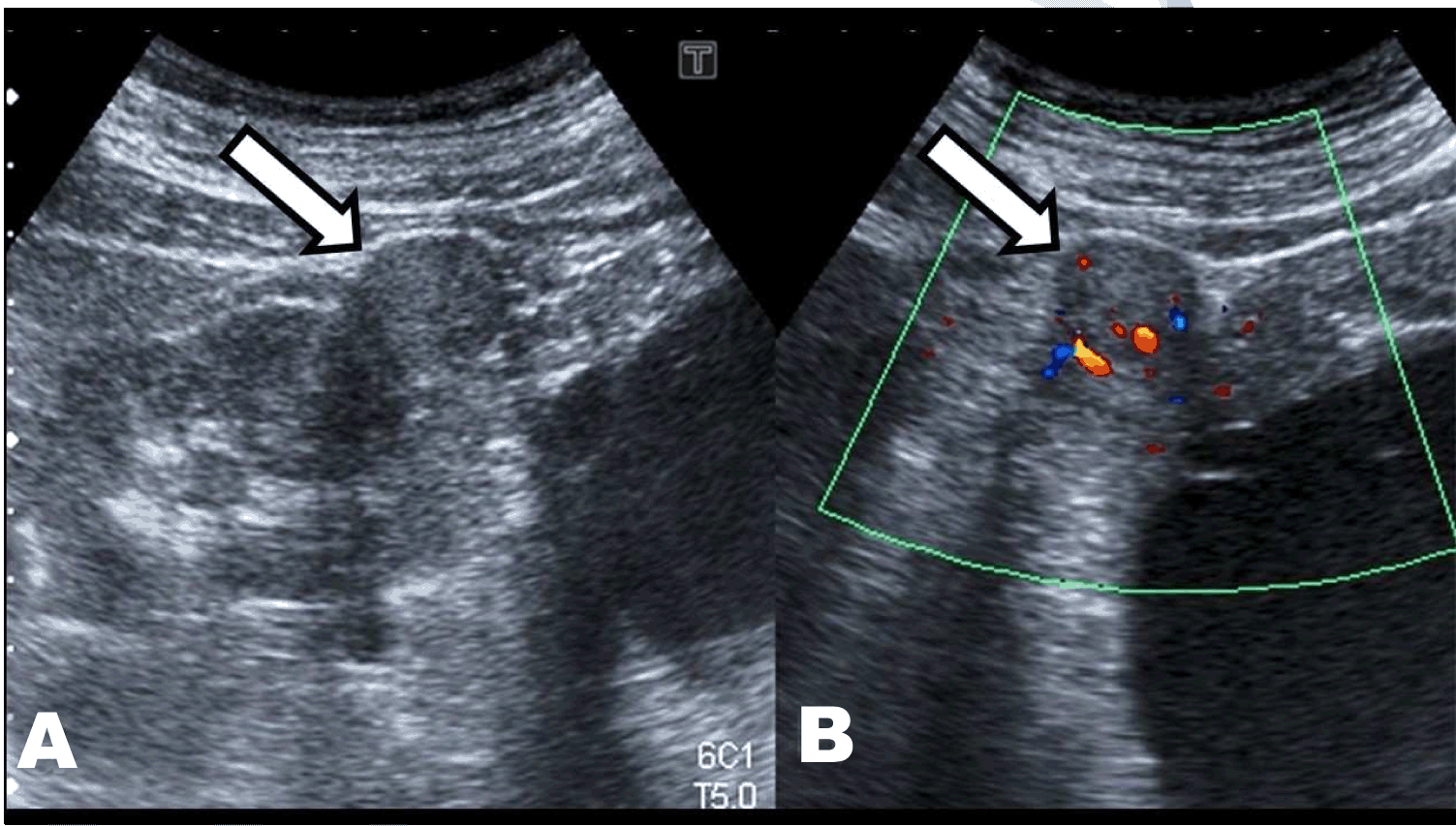
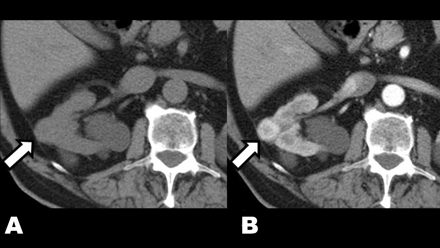
On February 2013, a 76-year-old male was sent to our nephrology unit to evaluate renal disease. He was asymptomatic and had been diagnosed with hypertension, overweight, benign prostatic hypertrophy and chronic obstructive pulmonary disease several years ago. His one daughter was also diagnosed with a renal cyst. His serum creatinine was 1.6 mg/dL, eGFR 42 mL/min, albumin/creatinine ratio in spot urine sample 2.8 mg/g and normal urinary sediment. Ultrasonography showed a slight enlargement of both kidneys and the presence of multiple bilateral cysts, predominantly with cortical distribution, classified as Bosniak I. No complex cysts that required monitoring or solid lesions were found (Figure 1). Therefore, he was diagnosed with Autosomal Dominant Polycystic Kidney Disease (ADPKD) and follow-up was drawn. He was successfully treated with losartan 50 mg/day. On July 2014, a new ultrasound control revealed the appearance of an echogenic nodule on the upper pole of the right kidney with vascularization in Doppler mode (Figure 2). The study was completed by computed tomography scan that confirmed the presence of a solid nodule of 23 mm on the right kidney with early contrast enhancement after i.v. iodine contrast administration. These findings strongly suggested the existence of superimposed renal cell carcinoma (RCC) (Figure 3). Considering his age and co-morbidities conservative treatment was planned. | ||||||
| ||||||
|
| ||||||
|
| ||||||
|
| ||||||
|
Discussion
| ||||||
|
The association between RCC and ADPKD has been described although this association has raised some controversy. While several case reports of RCC complicating ADPKD have been described and it has been found premalignant epithelial cells in the cyst epithelia, epidemiologic and autopsy studies have not shown a significant higher incidence of RCC in ADPKD [1] [2]. On the other hand, more recent studies have described that kidney-related prevalence of RCC in patients with ADPKD and advanced chronic renal failure treated by hemodialysis or renal transplantation was high [3][4]. These apparent discrepancies could be explained by the difficulties in the diagnosis of RCC in this setting and emphasizes the importance of close clinical surveillance and the interpretation of several radiological studies such ultrasonography, CT scan and MRI scan [5][6]. Herein, we report a case of RCC complicating ADPKD who has several characteristics. Firstly, he did have neither hematuria nor symptoms of occult neoplasia. Secondly, renal function was decreased although dialysis was not needed. Finally, the combination of ultrasonography and unenhanced and contrast-enhanced CT scan studies were able to achieve a diagnosis without using RMI, as it has been recently recommended. | ||||||
|
Conclusion
| ||||||
|
Renal cell carcinoma (RCC) can appear as a complication of autosomal dominant polycystic kidney disease (ADPKD). The diagnosis of RCC in this setting needs a thoroughly radiological evaluation, which should include ultrasonography and computed tomography scan studies. | ||||||
|
References
| ||||||
| ||||||


|
[HTML Abstract]
[PDF Full Text]
|
|
Author Contributions
Francisco Rivera – Substantial contributions to conception and design, Acquisition of data, Analysis and interpretation of data, Drafting the article, Revising it critically for important intellectual content, Final approval of the version to be published Celia López Redondo – Substantial contributions to conception and design, Acquisition of data, Analysis and interpretation of data, Drafting the article, Revising it critically for important intellectual content, Final approval of the version to be published |
|
Guarantor of submission
The corresponding author is the guarantor of submission. |
|
Source of support
None |
|
Conflict of interest
Authors declare no conflict of interest. |
|
Copyright
© 2015 Francisco Rivera et al. This article is distributed under the terms of Creative Commons Attribution License which permits unrestricted use, distribution and reproduction in any medium provided the original author(s) and original publisher are properly credited. Please see the copyright policy on the journal website for more information. |
|
|
|
About The Authors
| |||
| |||
| |||