Introduction
|
|
Marginal tissue recession as a clinical entity has been documented quite elaborately in literature [1] [2]. The indications for surgical intervention are quite well defined and it is essential to carry out root coverage surgery whenever concerns such as aesthetics, sensitivity, susceptibility to root caries pulpal symptoms due to root exposure, food lodgment and plaque accumulation exists [3]. Surgical techniques for root coverage have been invented and modified over the last few decades with encouraging results and most of the procedures followed today result in 80–90% root coverage. Currently, accepted procedures for root coverage include coronally advanced flap, sub-epithelial connective tissue graft, guided tissue regeneration and acellular dermal matrix. The tunnel technique followed in this case report is the one described by Langer et al. where multiple adjacent recessions can be covered in a single surgery using connective tissue graft from the palate.
|
Case Series
|
|
The three cases selected for root coverage using the pouch and tunnel technique had multiple adjacent recessions. The most important criterion for selecting this procedure was sufficient thickness of the marginal and papillary gingival at the recipient site so as to facilitate complete undermining for creating the pouch and tunnel without detaching the papillary tip.
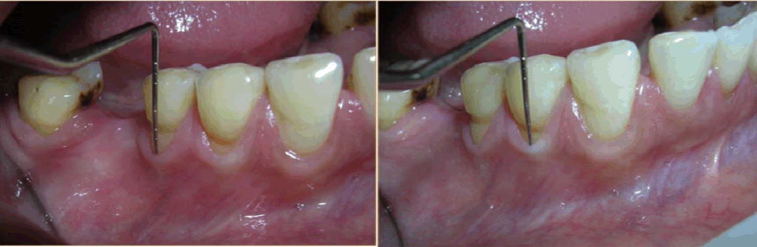
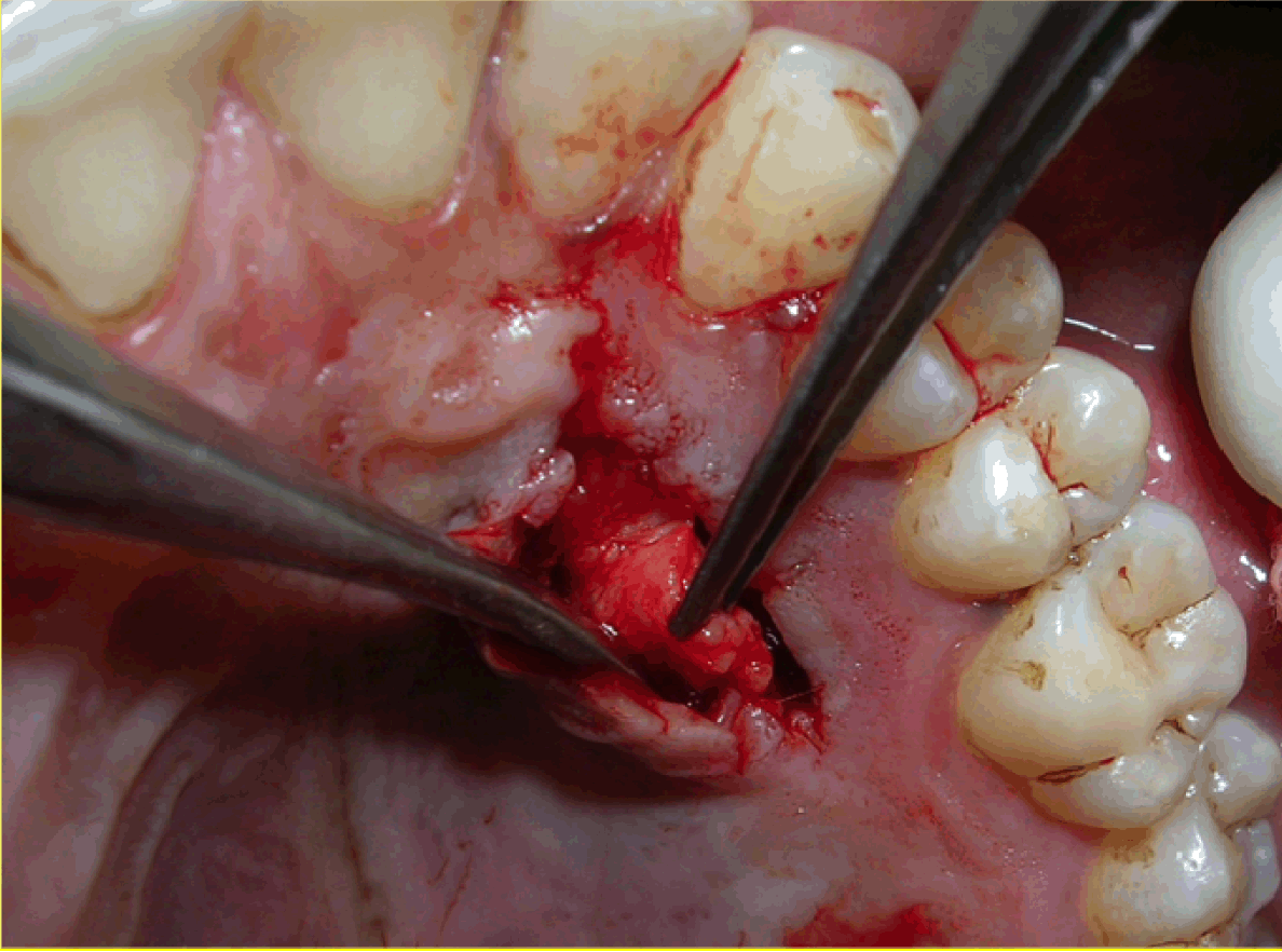
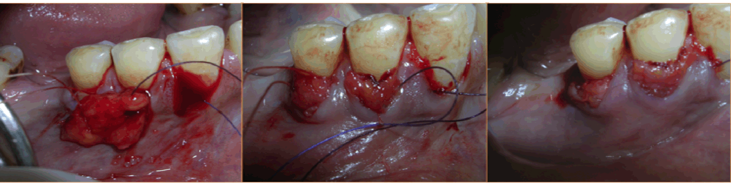
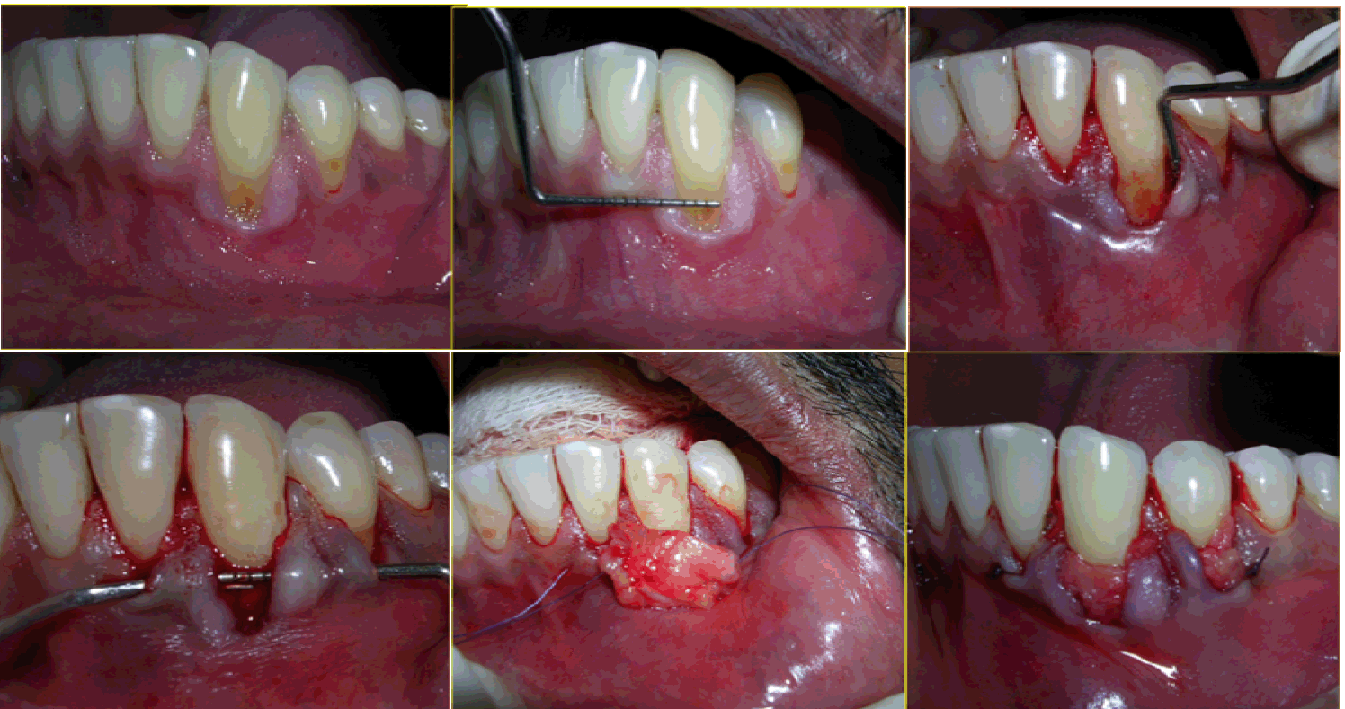
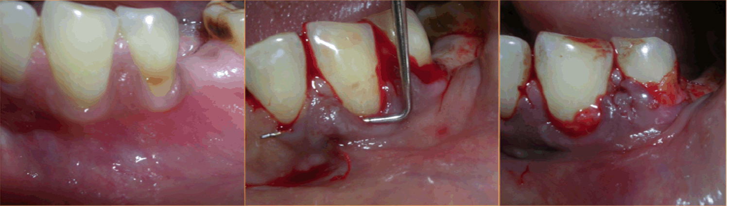
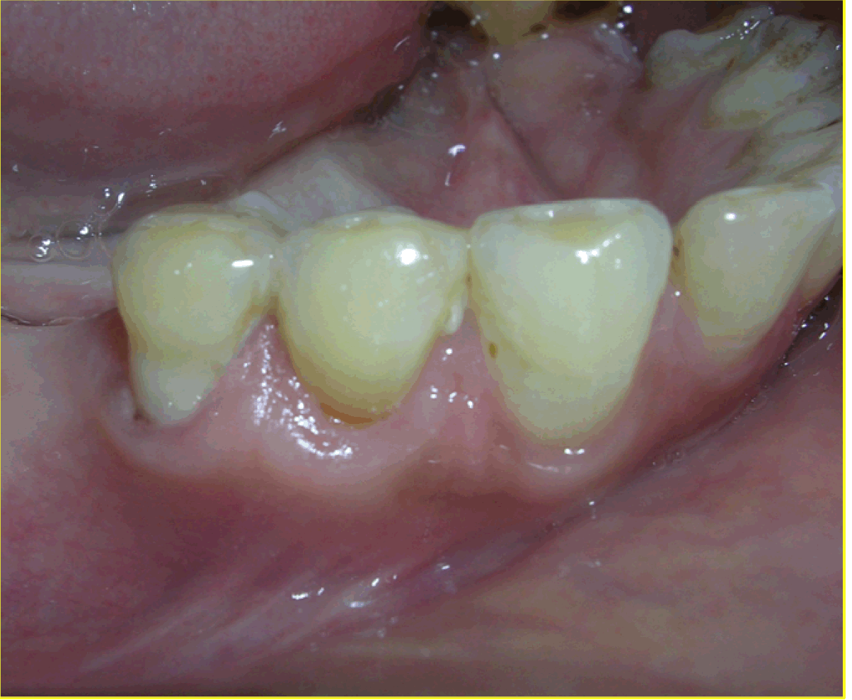
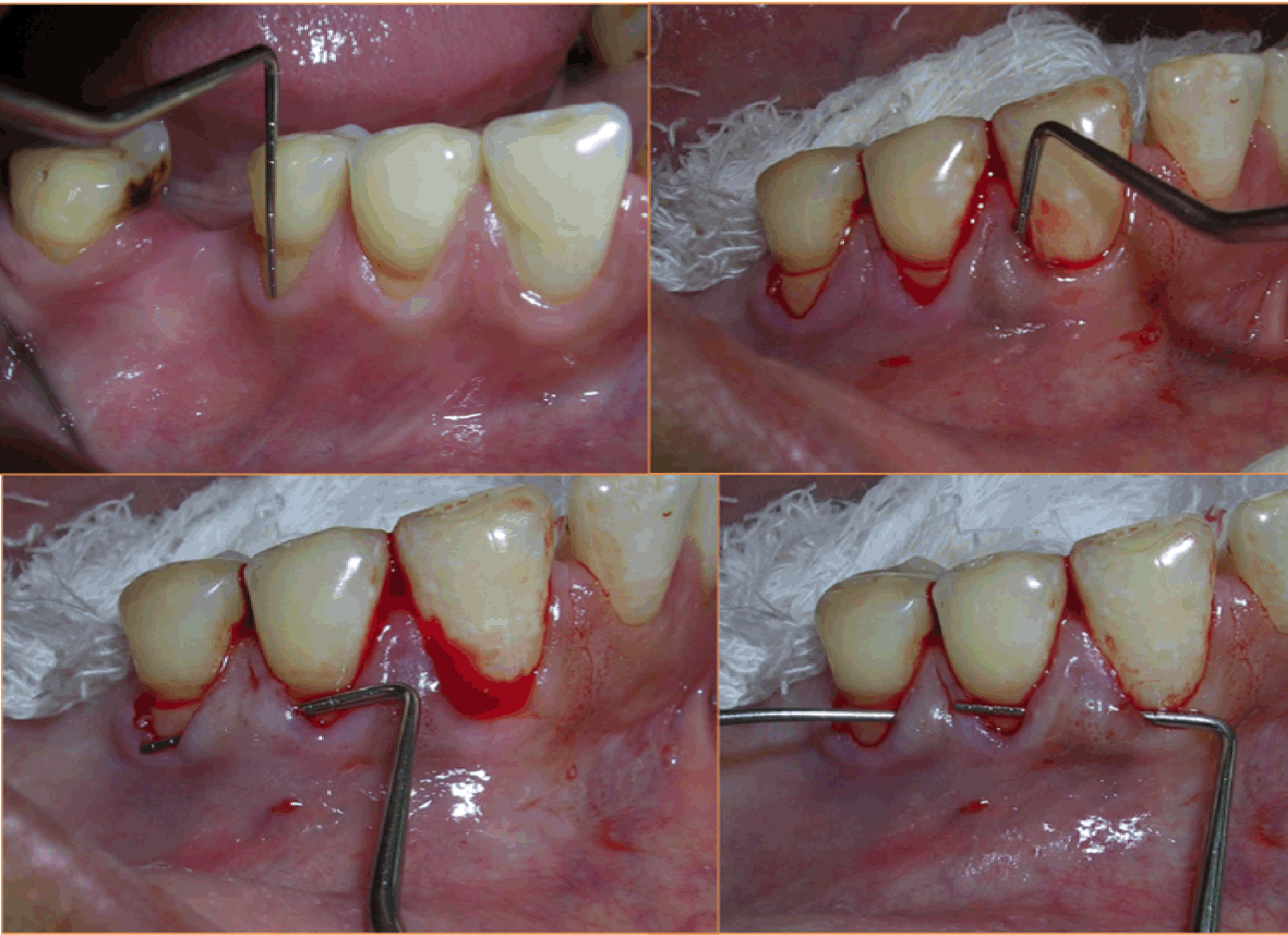
Case 1: A 36-year-old female reported with the chief complaint of sensitivity in lower right posterior teeth. On examination there was Millers Class III recession in relation to 44 and 45 (Figure 1). The width of attached gingiva was found to be inadequate since the tension test was positive. After phase I therapy, a pouch and tunnel technique utilizing a connective tissue graft was planned for root coverage. A sulcular incision was made through each recession area and the tissues gradually undermined including the base of the interdental papilla without the tip and the undermining extended up to the mucogingival junction so as to relax the flap sufficiently to allow placement of the connective tissue graft. Thus gradually a pouch and tunnel was prepared connecting the recipient sites for placement of the graft (Figure 2). The connective tissue graft was harvested from the palate using Liu's Class 1a incision (Figure 3). This graft was then placed using a technique described by Zabaluigi et al. where two resorbable sutures of different colors were placed, one on either side of the graft. Using these sutures, the graft was gradually manipulated into the pouch and through the tunnel to cover the adjacent recipient sites. Once the graft was completely inside the tunnel, it was positioned coronal to the cemento-enamel junction. The ends were sutured with a simple square knot (Figure 4). A periodontal pack was placed both at the recipient site as well as the donor site using an acrylic stent for the palatal placement.
|
|
|
|
| Cursor on image to zoom/Click text to open image |
| |
Figure 2: Pouch and tunnel preparation (Case 1).
|
|
|
|
|
|
|
|
|
Case 2: A 31-year-old male presented with the chief complaint of sensitivity in the lower teeth and was clinically diagnosed as a case of Millers Class II recession in relation to 33 and 34 (Figure 5). The same surgical technique, as in Case 1, was used to place the connective tissue graft in the pouch created in relation to 33 and 34.
|
|
|
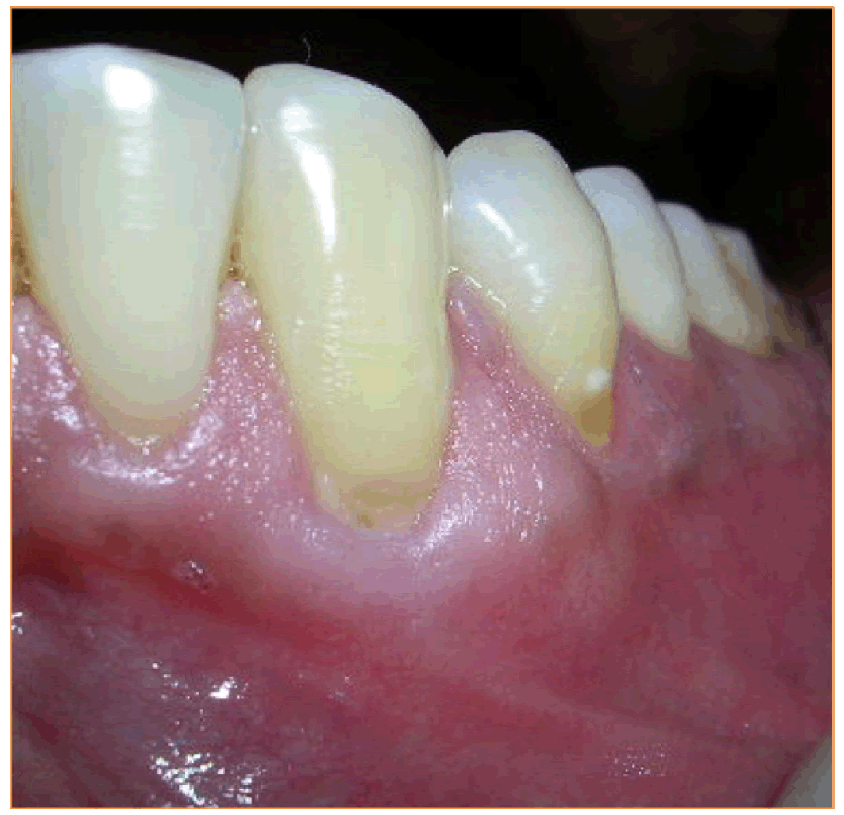
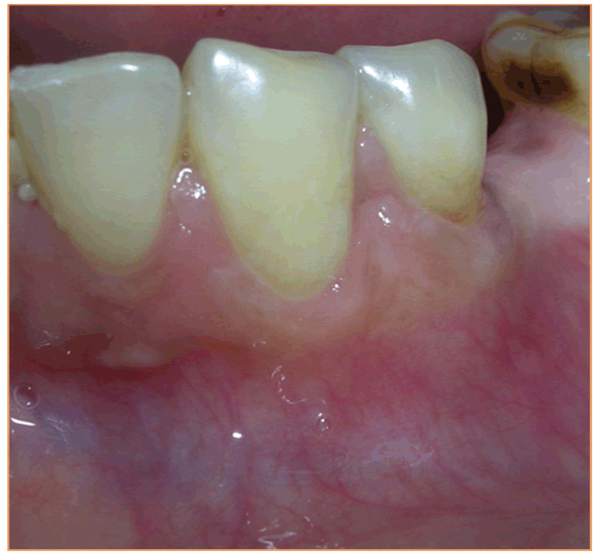
Case 3: A 35-year-old female patient presented and her main concern was the gums 'going down' in relation to the lower teeth on the left side. She was diagnosed as a case of Millers Class II recession in relation to 34 and 35 (Figure 6). This case too was treated using the pouch and tunnel technique with placement of CTG procured from the palate. The donor site appeared normal in color and healthy after four weeks and the recipient site was healthy with excellent color match with adjacent tissues in all four cases. These results were stable and maintained at the time of review, 12 months in Case 1 (Figure 7), 18 months in Case 2 (Figure 8), six months in Case 3 (Figure 9). The mean root coverage achieved in these cases was an average 90% which is close to the mean root coverage of 91.6% in the Zabaluigi study.
|
|
|
|
|
|
|
|
|
|
Discussion
|
|
The concerns of hypersensitivity, root caries, fear of tooth loss and an increasing interest in aesthetics have led to development and modifications of surgical procedures that not only enhance the width of attached gingival but also achieve maximum root coverage addressing these concerns. The subepithelial connective tissue graft technique gives the dual advantage of excellent healing of the donor site as well as excellent color match of tissues [4]. The tunnel technique was developed as a modification of the envelope technique to manage multiple adjacent recessions using a single surgical procedure and this technique has resulted in consistently favorable results [5].
The pouch and tunnel technique of connective tissue grafting, if performed correctly, is the most predictable periodontal plastic surgery procedure. It is a very sensitive procedure and requires use of operating microscope, microsurgical instruments, delicate handling of the tissues and ample patience while undermining the interdental papilla. The common complications that can occur are detachment of the interdental papilla, thinning of the flap, too thick connective tissue graft harvested which can lead to ischemic necrosis of the inter dental papilla or too thin connective tissue graft harvested which can lead to insufficient coverage of the recession defect [6].
The use of tunnel procedure preserves the interdental papilla and this facilitates an early and accelerated initial wound healing. The tunneling also applies less traction and preserves the gingival height [7]. The elimination of vertical incision which is used in subepithelial connective tissue grafting, ensures complete coverage of the connective tissue graft by the flap thus aids in faster healing as well as excellent color matching. The pouch and tunnel procedure may be of advantage in multiple adjacent recessions as compared to coronally repositioned flap as there is minimum trauma to the recipient site and there is predictable root coverage [8].
|
Conclusion
|
|
The pouch and tunnel technique combines the advantages of subepithelial connective tissue grafting as well as the envelope technique thus making it an ideal choice for treatment of multiple adjacent recessions in a single surgical procedure demonstrating early healing and highly predictable root coverage results.
|
References
|
-
Miller PD Jr. A classification of marginal tissue recession. Int J Periodontics Restorative Dent 1985;5(2):8–13.
[Pubmed]

-
Wennström JL. Lack of association between width of attached gingiva and development of soft tissue recession: A 5-year longitudinal study. J Clin Periodontol 1987 Mar;14(3):181–4.
[CrossRef]
[Pubmed]
-
Oates TW, Robinson M, Gunsolley JC. Surgical Therapies for the Treatment of Gingival Recession. A Systematic Review. Ann Periodontol 2003 Dec;8(1):303–20.
[CrossRef]
[Pubmed]
-
Liu CL, Weisgold AS. Connective tissue graft: A classification of incision design from the palatal site and clinical case reports. Int J Periodontics Restorative Dent 2002 Aug;22(4):373–9.
[Pubmed]
-
Langer B, Langer L. Subepithelial connective tissue graft technique for root coverage. J Periodontol 1985 Dec;56(12):715–20.
[CrossRef]
[Pubmed]
-
Saadoun AP. Current trends in gingival recession coverage--Part 1: Tunnel connective tissue graft. Pract Proced Aesthet Dent 2006 Aug;18(7):433–8.
[Pubmed]
-
Zabaluigi I, Sicilia J, Cambra J, Gil J, Sanz M. Treatment of multiple adjacent gingival recessions with the tunnel subepithelial connective tissue graft: A clinical report. Int J Periodontics Restorative Dent 1999 Apr;19(2):199–206.
[Pubmed]
-
Singh S, Roy S, Mandlik VB, Thapliyal GK, Prasanth L. Pouch & Tunnel technique for root coverage using palatal connective tissue graft. MJAFI 2008;64:191–2.
[CrossRef]
|
[HTML Abstract]
[PDF Full Text]
|
|
Author Contributions:
Sangeeta Singh – Substantial contributions to conception and design, Acquisition of data, Analysis and interpretation of data, Drafting the article, Revising it critically for important intellectual content, Final approval of the version to be published
|
Guarantor of submission
The corresponding author is the guarantor of submission.
|
Source of support
None
|
Conflict of interest
Authors declare no conflict of interest.
|
Copyright
©
2015 Sangeeta Singh. This article is distributed under the terms of Creative Commons Attribution License which permits unrestricted use, distribution and reproduction in any medium provided the original author(s) and original publisher are properly credited. Please see the copyright policy on the journal website for more information.
|
|