  |

|
 |
|
Case Report
| ||||||
| Ameloblastic carcinoma of the mandible: A case report | ||||||
| Soumithran C.S.1, Sudha S.2, Ikram Bin Ismail P.T.3, Ambadas4, Jibin Jose Tom3, Seeja P.5 | ||||||
|
1MDS, Professor and Head, Department of Oral and Maxillofacial Surgery, Govt. Dental College, Kozhikode, Kerala, India.
2MDS, Professor and Head, Department of Oral and Maxillofacial Pathology, Govt. Dental College, Kozhikode, Kerala, India. 3Junior Resident (Post Graduate Student), Department of Oral and Maxillofacial Surgery, Govt. Dental College, Kozhikode, Kerala, India. 4MDS, Senior Resident/Lecturer, Department of Oral and Maxillofacial Surgery, Govt. Dental College, Kozhikode, Kerala, India. 5Lecturer Trainee, Department of Oral and Maxillofacial Surgery, Govt. Dental College, Kozhikode, Kerala, India. | ||||||
| ||||||
|
[HTML Abstract]
[PDF Full Text]
[Print This Article]
[Similar article in Pumed] [Similar article in Google Scholar] 
|


| How to cite this article |
| Soumithran CS, Sudha S, Ikram Bin Ismail PT, Ambadas, Tom JJ, Seeja P. Ameloblastic carcinoma of the mandible: A case report. Int J Case Rep Images 2015;6(1):11–15. |
|
Abstract
|
|
Introduction:
Ameloblastic carcinoma is an extremely rare, malignant epithelial tumor of the jaws with a poor prognosis. The most common site of occurrence is the posterior mandible. Clinically, it is very aggressive and has potential for extensive local destruction. Majority of the cases arise de novo (primary type), but a few cases arise from a pre-existing ameloblastoma (secondary type).
Case Report: A 30-year-old female presented with a chief complaint of swelling on the left side of the face for the past one year. An incisional biopsy was performed and the histopathology was consistent of ameloblastic carcinoma. On the basis of the histopathology report, left hemimandibulectomy was done taking a safe margin of 2 cm and the defect was reconstructed using titanium reconstruction plate. Chemotherapy and radiotherapy were not advised. The patient is under regular follow-up. No recurrence nor metastases reported during the follow-up period. Conclusion: Early diagnosis and wide local excision on the primary site is the treatment of choice. The risk of malignant transformation should always be considered when a classic ameloblastoma is diagnosed and the prompt definitive management of an ameloblastoma is essential to eliminate this risk. Finally, meticulous, long-term follow-up is mandatory because recurrence and metastasis in the lung and regional lymph nodes have been reported. | |
|
Keywords:
Ameloblastoma, Ameloblastic carcinoma, Carcinoma, Odontogenic tumor
| |
|
Introduction
| ||||||
|
Ameloblastic carcinoma is an extremely rare, aggressive, malignant neoplasm of the jaws with a poor prognosis. It belongs to the family of malignant epithelial odontogenic tumors [1]. It combines the histological features of ameloblastoma with features of cytological atypia regardless of whether it has metastasized [2] [3]. In contrast to ameloblastoma, ameloblastic carcinoma exhibits more aggressive clinical behavior, such as rapid growth, perforation of the cortex and painful swelling [3] [4]. It may metastasize to the regional lymph nodes or lung [3]. In rare cases, brain or multiple bone metastases have been reported [3]. Ameloblastic carcinoma occurs in a wide age range of 15–84 years, but the mean age is approximately 30 years [5] [6]. There is no apparent sex predilection [5]. The most commonly involved area is the posterior portion of the mandible [5] [6]. Majority of the cases of ameloblastic carcinoma appear to arise de novo, and are termed primary ameloblastic carcinoma. Secondary ameloblastic carcinoma is defined as a tumor with malignant transformation within a pre-existing benign ameloblastoma, regardless of the presence of metastasis [4]. Secondary ameloblastic carcinoma is extremely rare. We report a case of secondary ameloblastic carcinoma of mandible in a 30-year-old Indian female. | ||||||
|
Case Report
| ||||||
|
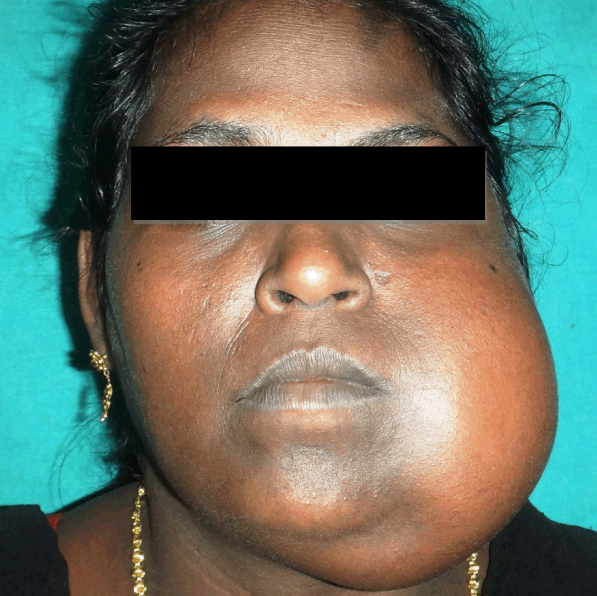
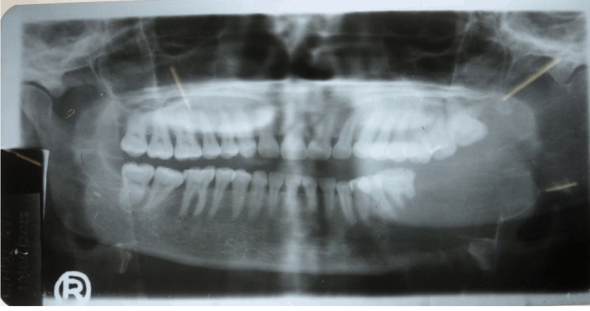
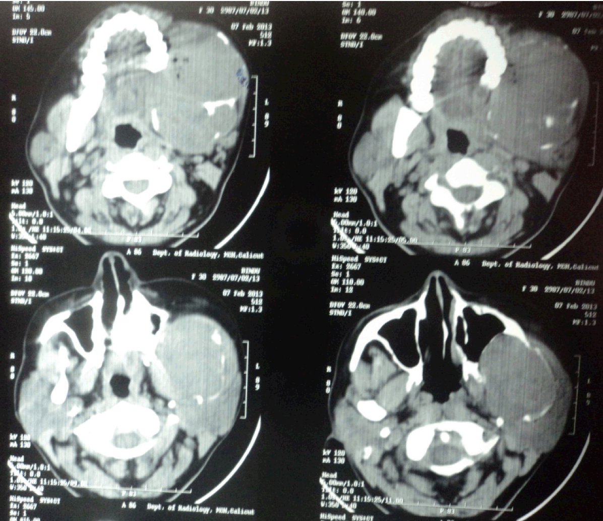
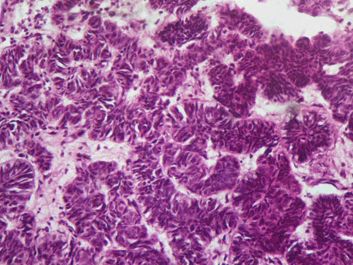
A 30-year-old female presented to our department with a chief complaint of swelling on the left side of the face for the last one year (Figure 1). The swelling was associated with mild pain and the patient had difficulty in mastication and mouth opening. The patient gave a history of mild swelling, pain and mobility of teeth in left lower back region four years ago and she reported to the same centre. Her mandibular left 2nd and 3rd molars were extracted and an incisional biopsy was carried out. The biopsy report was suggestive of ameloblastoma and surgical excision of the lesion was advised. But the patient was not willing for the same and she did not report for the review. She noticed an increase in size of swelling and reported to our department after three years. Clinical examination revealed a diffuse swelling over the left body-ramus region of the mandible, which extended antero-posteriorly from the left corner of mouth to the pre-auricular region. Superiorly, the extent was up to the infra-orbital region and inferiorly to the submandibular region. The overlying skin was normal in color and texture. On palpation, the swelling was mildly tender, smooth, and uniformly bony hard in consistency, with no local rise in temperature. The margins were ill defined. No paresthesia was associated with the swelling and no palpable regional lymph nodes noticed. Mouth opening was 32 mm and no deviation of mandible noted. Intra-oral examination revealed a diffuse swelling of the left lower buccal vestibule extending from the canine region to the retromolar area. Posterior extension was not visualized clinically. A discontinuity of the overlying mucosa was noted with irregular rolled margins posteriorly (Figure 2). On intra-oral palpation, the swelling was firm to hard in consistency and mildly tender. The orthopantomograph (OPG) showed a well-defined radiolucency involving the left body, angle and ramus of the mandible (Figure 3). The OPG revealed root resorption of left lower first molar tooth. Plain and contrast axial and coronal computed tomography (CT) scans showed a lobulated expansile lytic lesion involving body, ramus, and coronoid process of mandible measuring 6.3x8.3x10 cm in size, with thinning of the cortices (Figure 4). A chest radiograph ruled out the presence of any metastatic deposits. An incisional biopsy was done and the tissue was sent for histopathologic examination. The histopathology was consistent of ameloblastic carcinoma (Figure 5). On the basis of the histopathology report, left hemimandibulectomy was done taking a safe margin of 2 cm and the defect was reconstructed using Titanium reconstruction plate. Chemotherapy and radiotherapy was not advised. The patient is under regular follow-up. No recurrence or metastases reported during the follow-up period. | ||||||
| ||||||
| ||||||
| ||||||
| ||||||
| ||||||
|
Discussion
| ||||||
|
Ameloblastic carcinoma is a rare odontogenic tumor that poses a real challenge to the clinician in diagnosis, treatment planning and prognosis [7]. It is a neoplasm demonstrating histological evidence of malignant transformation of the ameloblastoma-like epithelial component in the primary tumor whether or not it has metastasized [8]. WHO defined ameloblastic carcinoma as a rare odontogenic malignancy that combines the histological features of ameloblastoma with cytological atypia even in the absence of metastases [7]. In the updated histologic classification of the WHO in 2005 [7], ameloblastic carcinoma is classified as primary type and secondary type. Primary ameloblastic carcinomas are those arising de novo whereas the secondary type arise from a pre-existing ameloblastoma. Ameloblastic carcinoma, secondary type is extremely rare. The exact mechanism for the malignant transformation of ameloblastoma is currently unknown because of the limited number of cases [4]. Clinically, ameloblastic carcinoma is well known for its aggressiveness and extensive destruction of local structures [8]. The most common sign described has been swelling, although others include associated pain, rapid growth, trismus and dysphonia [5]. In most cases, radiographic findings show ill-defined radiolucency, however, focal radiopacity may be detected in radiolucent lesions [3]. Microscopically, it resembles the features of conventional ameloblastoma, except for the epithelium which shows various cytological features of malignancy [8]. Histologically, ameloblastic carcinomas have characters of both a benign ameloblastoma and carcinoma [9]. A palisade arrangement of epithelial cells with nuclei away from the basement membrane (reverse polarity) is a common feature of benign ameloblastomas [9]. The epithelial cells of ameloblastic carcinomas have features of hyperchromatism, a high mitotic rate, and a high nuclear-to-cytoplasmic ratio [9]. Recommended surgical treatment is jaw resection with 2–3 cm bony margin [5]. Ameloblastic carcinoma can recur locally 0.5–11 years after definitive therapy. Distant metastasis may occur as early as 4 months or as late as 12 years postoperatively and it is usually fatal [10] . Metastatic deposits of ameloblastic carcinoma are usually found in the lung, followed by bone, liver and brain [10]. Metastasis can occur even without any evidence of local recurrence [10] . The prognosis is much worse when distant metastasis occurs [9]. Cervical lymph node dissection should be considered when there is obvious lymphadenopathy [7]. The efficacy of adjuvant radiation or chemotherapy as a post-surgical treatment is not clear, because there is insufficient evidence that either provides a significant advantage [3]. However, radiotherapy and chemotherapy need to be considered in locally advanced cases and metastatic lesions not amenable to surgical resection [3]. The prognosis of ameloblastic carcinomas is poor [9]. The fatal factor includes the rapid growth of the primary tumor and distant metastasis [9]. Close periodic reassessment with a long-period of follow-up (at least 10 years) is mandatory [7]. Radical surgical resection, strict screening for and early detection of metastatic lesions, and periodic follow-up after surgery are needed to improve patient prognosis [3]. | ||||||
|
Conclusion
| ||||||
|
Ameloblastic carcinoma is a rare entity of jaw neoplasm that combines the histological features of an ameloblastoma with features of cytological atypia, with a poor prognosis. This case report described a secondary type, ameloblastic carcinoma of mandible that originated from a pre-existing ameloblastoma which was left untreated. | ||||||
|
Acknowledgements
| ||||||
|
Dr. Martin C.P., Dr. Pallav Kumar Kinra | ||||||
|
References
| ||||||
| ||||||


|
[HTML Abstract]
[PDF Full Text]
|
|
Author Contributions
Soumithran C.S. – Substantial contributions to conception and design, Acquisition of data, Analysis and interpretation of data, Drafting the article, Revising it critically for important intellectual content, Final approval of the version to be published Sudha S. – Substantial contributions to conception and design, Acquisition of data, Analysis and interpretation of data, Drafting the article, Revising it critically for important intellectual content, Final approval of the version to be published Ikram Bin Ismail P.T. – Substantial contributions to conception and design, Acquisition of data, Analysis and interpretation of data, Drafting the article, Revising it critically for important intellectual content, Final approval of the version to be published Ambadas – Substantial contributions to conception and design, Acquisition of data, Analysis and interpretation of data, Drafting the article, Revising it critically for important intellectual content, Final approval of the version to be published Jibin Jose Tom – Substantial contributions to conception and design, Acquisition of data, Analysis and interpretation of data, Drafting the article, Revising it critically for important intellectual content, Final approval of the version to be published Seeja P. – Substantial contributions to conception and design, Acquisition of data, Analysis and interpretation of data, Drafting the article, Revising it critically for important intellectual content, Final approval of the version to be published |
|
Guarantor of submission
The corresponding author is the guarantor of submission. |
|
Source of support
None |
|
Conflict of interest
Authors declare no conflict of interest. |
|
Copyright
© 2015 Soumithran C.S. et al. This article is distributed under the terms of Creative Commons Attribution License which permits unrestricted use, distribution and reproduction in any medium provided the original author(s) and original publisher are properly credited. Please see the copyright policy on the journal website for more information. |
|
|