| |
|
|
|
Case Report
| ||||||
| Clamping of external carotid artery rather than embolization during surgical removal of a huge carotid body tumor | ||||||
| Abubakr Hashim Elrofaie Sayed Ali1, Saif Eldin Mohammed Ali Ibrahim2, Ashraf Mohamed Mokhtar Ali3 | ||||||
|
1MBBS, Ibn Sina Specialized Hospital - Senior House-officer, Unit of Vascular and Endovascular Surgery, Ibn Sina Specialized Hospital, Khartoum, Sudan.
2MBBS, MD, MRCS (ENG); D.MAS; F.MAS; F. Vasc/Endovasc (MAL), Ibn Sina Specialized Hospital - Head, Unit of Vascular and Endovascular Surgery, Ibn Sina Specialized Hospital, Khartoum, Sudan. 3MBBS, MD General Surgeon, Ibn Sina Specialized Hospital - Specialist, Unit of Vascular and Endovascular Surgery, Ibn Sina Specialized Hospital, Khartoum, Sudan. | ||||||
| ||||||
|
[HTML Abstract]
[PDF Full Text]
[Print This Article]
[Similar article in Pumed] [Similar article in Google Scholar] 
|
| How to cite this article |
| Ali AHES, Ibrahim SEM, Ali AMM. Clamping of external carotid artery rather than embolization during surgical removal of a huge carotid body tumor. Int J Case Rep Images 2015;6(1):6–10. |
|
Abstract
|
|
Introduction:
Carotid body tumors (CBT) are neoplasms that develop in the carotid body and are usually benign tumors. Malignant forms are less frequently. Carotid body tumors are widely known as paragangliomas. The carotid body is a gland located behind the carotid artery at the site of its bifurcation on either sides of the neck and originates from the neural crest and acts as a peripheral chemoreceptor.
Case Report: A 42-year-old female presented with progressively enlarging neck swelling over five years which was associated with a recent difficulty in swallowing and hoarseness of voice. On examination there was anterolateral pulsatile neck swelling, ovoid in shape, about 6×6×7 cm in size, mobile from side to side only and firm in consistency. Computed tomography angiography (CTA) and magnetic resonance angiography (MRA) showed a mass arising at the bifurcation of common carotid artery (CCA), which has rich blood supply, splaying the external carotid artery (ECA). Blood test for vanillylmandelic acid (VMA) was negative. The patient underwent surgical excision of the tumor through an endarterectomy approach after clamping of the ECA. We report this case where clamping of ECA is being applied rather than endovascular embolization of the feeding vessels to minimize bleeding during removal of the tumor, to avoid embolization related complications and to lessen the cost of the procedure. Conclusion: Carotid body tumors are rare, however, early diagnosis and prompt treatment is essential. In a large tumor, preoperative endovascular embolization is widely used for devascularization. Clamping of the external carotid artery is a good alternative way to avoid embolization related complication with better control of bleeding, less operative time and improved cost-effectiveness. | |
|
Keywords:
Carotid body tumor, Endarterectomy approach, Glomus gland, External carotid artery clamping, Paraganglioma
| |
|
Introduction
| ||||||
|
The carotid body (also known as glomus gland) is a gland located behind the carotid artery at the site of its bifurcation on either side of the neck, normally about 5 mm in size and covered with a fibrous capsule. The gland acts as peripheral chemoreceptor which releases neurotransmitter primarily in response to decrease in the arterial partial pressure of oxygen and by a lesser extent to increase in the partial pressure of CO2 and decrease in arterial pH. The gland receives afferent nerve fibers from the glossopharyngeal nerve which transmits impulses from the gland to the respiratory center in the medulla. Carotid body tumors (CBT) are neoplasms that develop in the carotid body and are usually benign tumors. Malignant forms are less frequent. CBT's are widely known as paragangliomas. Carotid body tumors are common in 45-year females and in small portion are related to multiple primary tumor syndrome [1]. | ||||||
|
Case Report
| ||||||
|
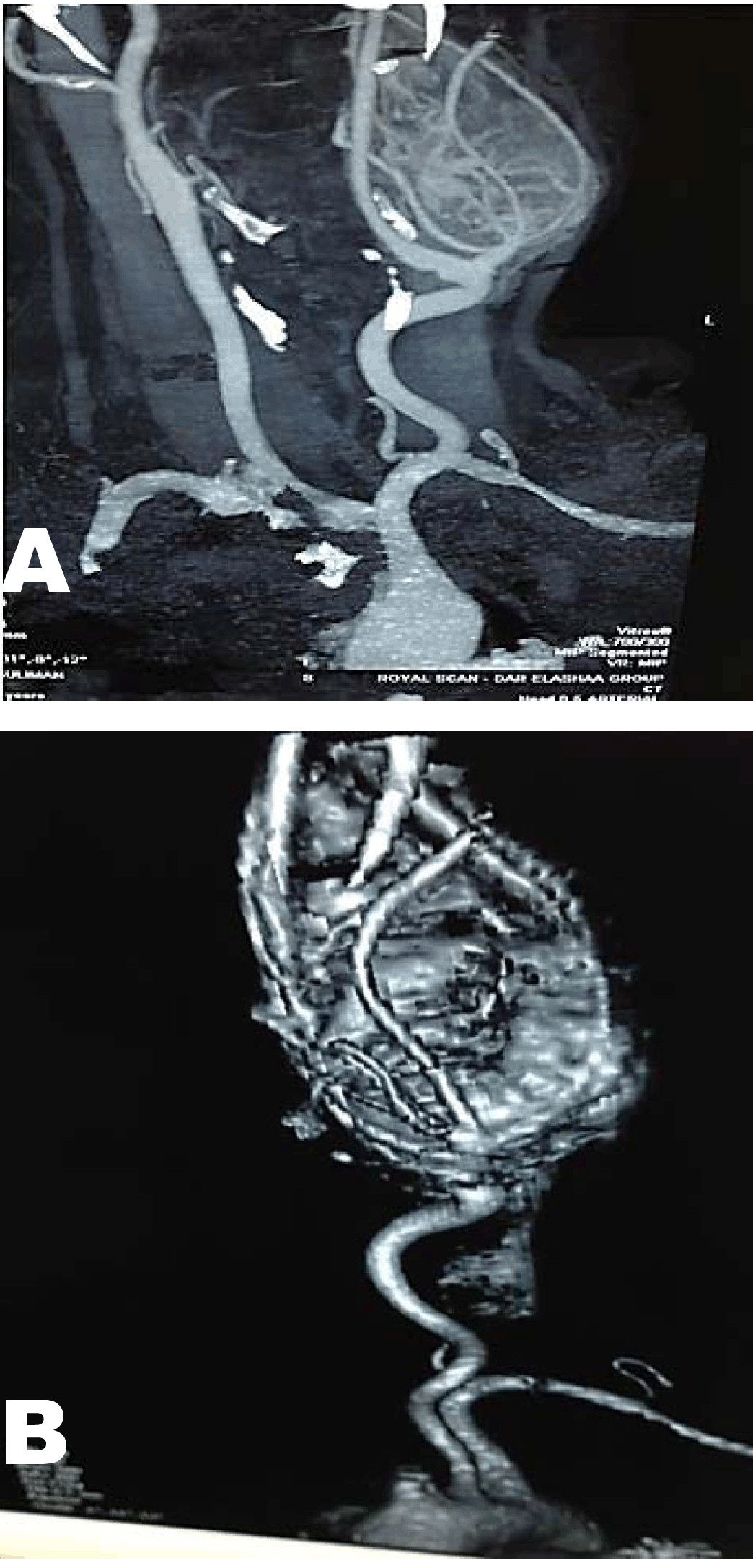
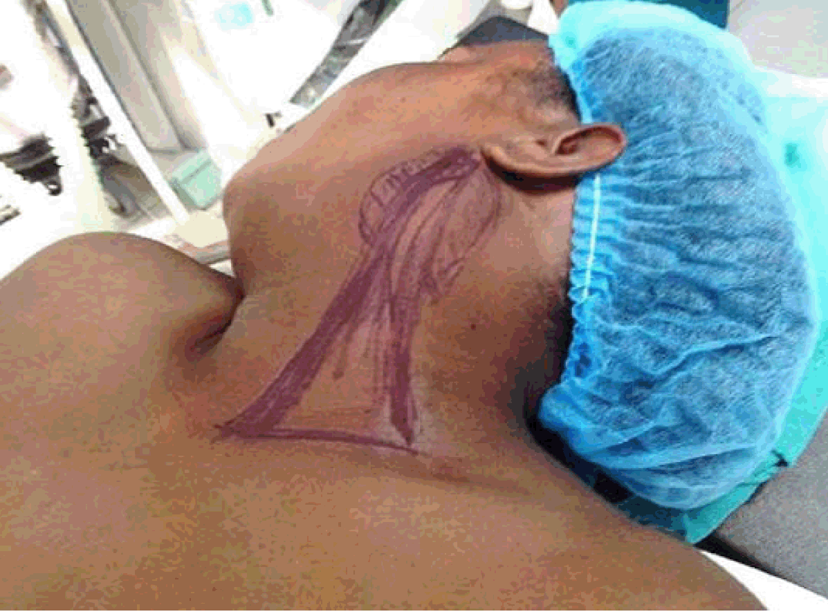
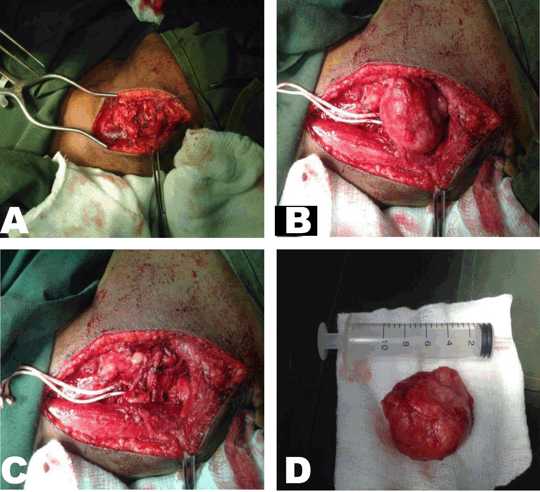
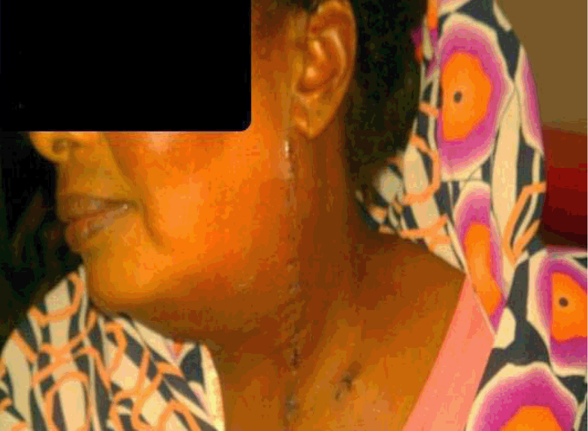
A 42-year-old female presented with a left neck swelling for five years. The swelling was small in size for the last four years. She was seen early by an ENT surgeon and advised just observation, during the last year the swelling started to increase in size with difficulty in swallowing and hoarseness of voice. There was not any other cardiovascular or neurological symptoms. On examination she was unwell, not pale, jaundice or cyanosed. Vital signs were normal. There was anterolateral neck swelling, ovoid in shape, measuring about 6×6×7 cm in size, mobile in horizontal plane but fixed in the vertical one. It was firm but not tender with transmitted pulsation, no lymph nodes were palpable. Cranial nerves examinations were intact, and both carotid arteries were palpable, with no bruit. Systemic examination revealed no signs of neurological or vascular deficit computed tomography angiography (CTA) and magnetic resonance angiography (MRA) of the carotid vessels showed a mass arising at the bifurcation of common carotid artery (CCA), which has rich blood supply, splaying the external carotid artery (ECA) (Figure 1). Blood test for vanillylmandelic acid (VMA) was negative. The patient was planned for surgical removal of the tumor under general anesthesia (Figure 2). An endarterectomy approach was used, skin, subcutaneous tissues, superficial cervical fascia and platysma muscle were dissected respectively. The sternocleidomastoid muscle and jugular vein were identified and retracted laterally. The facial vein was identified, ligated and divided to reveal the bifurcation of the CCA. The carotid sheath was dissected until the tumor was identified splaying CCA, ECA, internal carotid artery (ICA), hypoglossal and glossopharyngeal nerves. Meticulous dissection and retraction of the nerves was done to maintain their integrity. Vessel loops were applied for the carotid vessels plus clamping of the origin of the ECA was carried out to facilitate dissection and minimize bleeding. Complete excision of the tumor was done in the sub adventitial plane. Following de-clamping, blood flow was restored with minimal bleeding. A drain was put in place and the wound was closed in layer (Figure 3). The tumor specimen was sent for histopathology and results were consistent with paraganglioma. The postoperative period passed uneventfully without neurological or vascular complication (Figure 4). | ||||||
|
| ||||||
| ||||||
| ||||||
| ||||||
|
Discussion
| ||||||
|
Paragangliomas are chromaffin negative neuroendocrine tumors. There are three different types of CBTs: and sporadic hyperplastic familial. The sporadic type is the most common type and represents 85% of CBTs. Hyperplastic form is very common in patients with chronic hypoxia or chronic obstructive pulmonary disease; the familial type may present in a younger age group and bilaterally. Shamblin staging is commonly used to classify CBT into three types. Type I describes tumors which are small and easily resectable. Shamblin type II tumors are larger, slightly more adherent tumors and type III tumors are those which completely surround the carotid bifurcation. The patient we are reporting was staged as a Shamblin type III CBT using MRA which had more than 270 degrees circumferential contact with ICA as described by Arya et al. and for smaller tumor Shamblin type I and type II were described as being less than 180 degrees, and between 180 and 270 degrees of circumferential contact with ICA, respectively [2]. The standard management is surgery and careful preoperative assessment of vascular status and the functional status of the CBT is required. Functionally, active paraganglioma necessitate the use of alpha adrenoreceptor blockade [3] but in the case of the patient we are reporting, VMA and cardiovascular examination were normal. Much controversy lies with regards to the management of large (Shamblin grade II and III) CBT's. Some authors suggest that elderly patients with Shamblin III (large tumor more than 5 cm), which are associated with significant morbidities, should avoid surgery and receive radiotherapy instead [4]. Although surgery for CBT removal carries a 35% morbidity and a 1% mortality rate and a 19% risk of cranial nerve injury [5], surgical excision is the standard treatment as suggested by most of authors. The problem is surgical resection of large CBT is challenging, technically difficult and had an increasing risk of neurovascular injuries [6]. In order to avoid common postoperative neurovascular complication many suggested preoperative embolization of the feeding vessels could reduce blood loss and improve tumor excision [7] [8]. The ascending pharyngeal artery is the most common major feeding artery for CBTs. Tumor embolization with polyvinyl alcohol particles after super selection of feeding arteries is an option but numerous feeding arteries that could reduce the effect of embolization and the potential high risk of cerebral infarction by embolic particles is major limitation of embolization [9]. Embolization helped to some extent but it is expensive and as mentioned not suitable for all patients and is not quite safe and efficient as shown by study done by Ozay et al, of 14 patients who underwent CBT surgery and concluded that embolization before CBT surgery does not decrease blood loss or facilitate tumor removal [10]. Others suggest early ligation or deliberate resection of the ECA in Shamblin types II and III CBT resection which simple and significantly reduces the risk of stroke as opposed to percutaneous embolization which carries a significant risk of stroke [11] [12]. Spinelli et al. suggested a simpler technique for large CBT which is clamping the origin and the distal part of the ECA with heparinization, which is much easier, and effective[13]. We used similar sufficient technique in this reported patient to create a relatively blood less field and to allow for visualization and carful resection of CBT with preservation of adjacent and closely related neurovascular structures by clamping of the origin of the ECA only and without heparinization. External carotid artery clamping also used for other tumors resection as shown by a study conducted by Yadav et al., with regards to clamping of the ECA in excision of large meningiomas, they concluded that temporary clamping of ECA is a safe, simple and cost-effective alternative method to embolization [14]. Regarding the administration of heparin intraoperatively, any injury to the carotid vessels requiring clamping of the CCA, ICA or ECA needs heparinization to lower the risk of complications [15], however, we did not use heparin during clamping of the ECA because excision of the tumor was performed without arterial injury. | ||||||
|
Conclusion
| ||||||
|
Carotid body tumors are rare, however, early diagnosis and prompt treatment with complete surgical excision is essential. In a large tumor preoperative endovascular embolization is widely been used for de-vascularization. Clamping of the origin of external carotid artery is a good alternative way to avoid embolization related complication with better control of bleeding, less operative time and improved cost-effectiveness. | ||||||
|
References
| ||||||
| ||||||
|
[HTML Abstract]
[PDF Full Text]
|
|
Author Contributions
Abubakr Hashim Elrofaie Sayed Ali – Acquisition of data, Drafting the article, Final approval of the version to be published Saif-Eldin Mohamed Ali Ibrahim – Conception and design, Critical revision of the article, Final approval of the version to be published Ashraf Mohamed Mokhtar Ali – Conception and design, Critical revision of the article, Final approval of the version to be published |
|
Guarantor of submission
The corresponding author is the guarantor of submission. |
|
Source of support
None |
|
Conflict of interest
Authors declare no conflict of interest. |
|
Copyright
© 2015 Abubakr Hashim Elrofaie Sayed Ali et al. This article is distributed under the terms of Creative Commons Attribution License which permits unrestricted use, distribution and reproduction in any medium provided the original author(s) and original publisher are properly credited. Please see the copyright policy on the journal website for more information. |
|
|
|
About The Authors
| |||
| |||
| |||
| |||