| |
|
|
|
Clinical Image
| ||||||
| The potential hazards of ear self-cleaning | ||||||
| Iain R. M. Bohler1, Wickham M.1, McCaffer C1, Schiszler T.1, Chin A.1 | ||||||
|
1Department of ENT, Monklands Hospital, Monkscourt Avenue, Lanarkshire, Scotland, ML6 OJS.
| ||||||
| ||||||
|
[HTML Abstract]
[PDF Full Text]
[Print This Article]
[Similar article in Pumed] [Similar article in Google Scholar] 
|
| How to cite this article |
| Bohler IRM, Wickham M, McCaffer C, Schiszler T, Chin A. The potential hazards of ear self-cleaning. Int J Case Rep Images 2015;6(1):56–58. |
|
Case Report
|
|
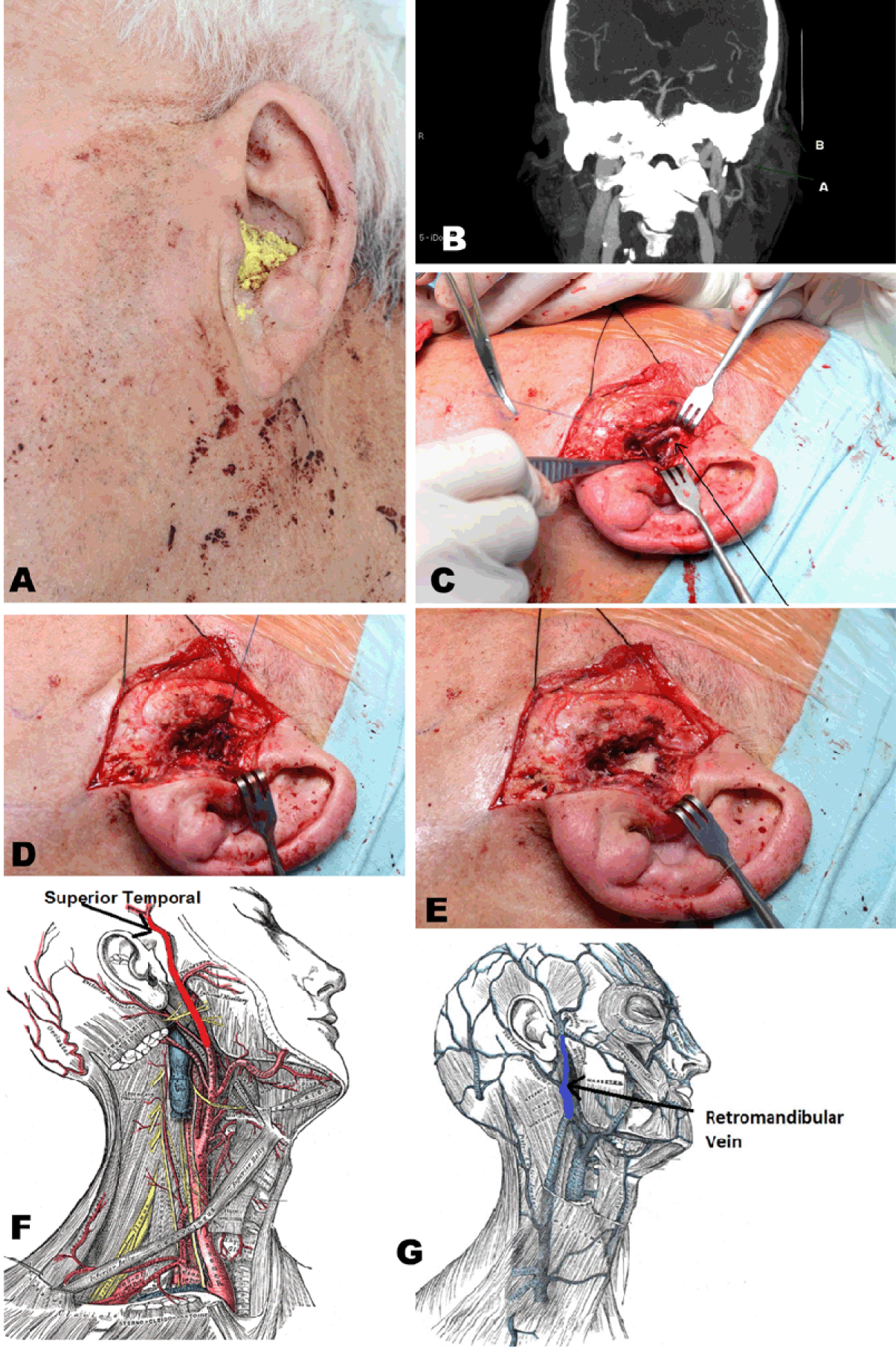
A 77-year-old male was brought in by ambulance to the resus bay of the emergency department with major bleeding from his left ear. Attempting to remove remnant cotton wool from a failed 'cleaning' attempt with tweezers, the patient had slipped and fallen on a wet bathroom floor. Tweezers in situ in the external auditory canal, the patient fell to his left side sustaining direct trauma to the auditory meatus resulting in uncontrollable hemorrhage. There was no immediate loss of consciousness. The patient was not known to suffer any coagulopathic disorder, nor prescribed anticoagulant medication. The patient suffered a syncopal episode in the ambulance, responsive to fast IV fluids, resulting on return of consciousness (GCS 15) on arrival. With continued bleeding, he subsequently deteriorated into a peri-arrest state with further syncopal episodes and an accompanying hemoglobin drop of 4 g/L. Further crystalloid resuscitation and blood (PRC) transfusion of two units stabilized the patient whilst hemostasis was achieved with a Bismuth Iodoform Paraffin Paste (BIPP) dressing (Figure 1) by the ENT team. The injury was localised to the anterior wall of the external auditory canal raising suspicion of superficial temporal artery trauma. Secondary survey revealed no injury to the facial nerve, and the patient proceeded to radiological imaging. Computed tomography (CT) scan demonstrated an intact tympanic membrane with absence of blood within the middle ear and mastoid cavity, excluding internal carotid artery and internal jugular vein injury. As suspected, the direction of trauma had sustained injury to the superior temporal artery with additional injury to the retromandibular vein to a lesser extent. The patient was observed on an ENT ward for five days with packing removed as an outpatient on day-7. Broad-spectrum antibiotic prophylaxis provided the only other active medical intervention throughout his initial admission, post resuscitation. Four weeks post trauma, the patient was seen to have a pulsatile mass on examination of the ear. Computed tomography (CT) scan showed a 1.7-cm pseudoaneurysm arising from the left superior temporary artery. The patient underwent emergency surgery to tie off the pseudoaneurysm and following a brief stay for treatment of a postoperative wound infection, was discharged. The patient has since gone on to make a full recovery. |
|
|
|
Discussion
|
|
The elegantly contoured pinna of the outer ear acts to funnel air through the external auditory meatus towards the tympanic membrane. The auditory canal is approximately 3 cm long in adults with a slight s-shaping as it meanders towards the middle ear. Cartilage supports the ear at its opening with bone predominating medially. The keratinizing stratified squamous epithelium of the external canal is continuous with the eardrum and peppered with varying numbers of sebaceous and ceruminous glands [1]. The cerumen (wax) produced by the glands of the external ear facilitates the removal of dead epithelium. Aided by the fine hairs of the auditory canal, cerumen prevents airborne pathogens reaching the middle ear, where their accumulation or growth can lead to painful and profoundly debilitating disease [2]. Although this action represents normal bodily function, cerumen is commonly misconceived to be solely a waste product and is often actively removed from the ear. This active removal and the complications associated as such are the basis for a significant number of emergency ENT referrals (foreign body in ear, impaction of wax and associated disease). This case is an infrequent consequence of a foreign body in the external auditory canal, and despite the severity of bleeding and post injury complications; the patient has gone on to make a full recovery to previous function. Computed tomography (CT) scan demonstrated an injury to the superior temporal artery. Arising from the external carotid artery, it passes anterior to the ear and is palpable superior to the zygomatic arch [3]. The retromandibular vein, also injured, is formed by the union of superficial temporal and maxillary veins. It descends through the parotid gland, deep to the facial nerve and superficial to the external carotid artery. Both vessels were damaged in a plane transverse to the external auditory meatus, as they peregrinate anterior to the ear [4]. Although this case represents a highly atypical presentation, its extremity highlights an avoidable workload on national health services (NHS). Enhanced education of patients through patient-doctor communication and health awareness campaigns, of normal bodily functions, could relieve the burden of patient numbers on emergency units. |
|
Conclusion
|
|
Although this case report demonstrates an exceptionally unusual case whereby inner ear trauma can lead to massive hemorrhage and pseudoaneurysm formation, it serves well to provide a much broader general message. Ears are self-cleaning; there is no need to insert anything into the ear. |
|
References
|
|
|
[HTML Abstract]
[PDF Full Text]
|
|
Author Contributions
Iain R. M. Bohler – Substantial contributions to conception and design, Acquisition of data, Analysis and interpretation of data, Drafting the article, Revising it critically for important intellectual content, Final approval of the version to be published Wickham M. – Substantial contributions to conception and design, Acquisition of data, Analysis and interpretation of data, Drafting the article, Revising it critically for important intellectual content, Final approval of the version to be published McCaffer C. – Substantial contributions to conception and design, Acquisition of data, Analysis and interpretation of data, Drafting the article, Revising it critically for important intellectual content, Final approval of the version to be published Schiszler T. – Acquisition of data, Analysis and interpretation of data, Revising it critically for important intellectual content, Final approval of the version to be published Chin A. – Substantial contributions to conception and design, Analysis and interpretation of data, Revising it critically for important intellectual content, Final approval of the version to be published |
|
Guarantor of submission
The corresponding author is the guarantor of submission. |
|
Source of support
None |
|
Conflict of interest
Authors declare no conflict of interest. |
|
Copyright
© 2015 Iain R. M. Bohler et al. This article is distributed under the terms of Creative Commons Attribution License which permits unrestricted use, distribution and reproduction in any medium provided the original author(s) and original publisher are properly credited. Please see the copyright policy on the journal website for more information. |
|
|