|

|
 |
|
Case Report
| ||||||
| Successful cesarean delivery following prenatal diagnosis of giant sacrococcygeal teratoma in the fetus | ||||||
| Murat Bakacak1, Salih Serin2, Aykut Urfalioğlu2, Mehmet Akif Sarica3 | ||||||
|
1Assistant Professor, Kahramanmaras Sutcu Imam University School of Medicine Department of Obstetrics and Gynecology, Kahramanmaras, Turkey.
2Assistant Professor, Kahramanmaras Sutcu Imam University School of Medicine Department of Anesthesia and Reanimation, Kahramanmaras, Turkey. 3Assistant Professor, Kahramanmaras Sutcu Imam University School of Medicine Department of Radiology, Kahramanmaras, Turkey. | ||||||
| ||||||
|
[HTML Abstract]
[PDF Full Text]
[Print This Article]
[Similar article in Pumed] [Similar article in Google Scholar] 
|


| How to cite this article |
| Bakacak M, Serin S, Urfalioglu A, Sarica MA. Successful cesarean delivery following prenatal diagnosis of giant sacrococcygeal teratoma in the fetus. Int J Case Rep Images 2014;5(11):790–794. |
|
Abstract
|
|
Introduction:
Sacrococcygeal teratomas are the most common germ cell neoplasia in the newborn, which are seen on the frontal surface of coccyx and sacrum. As a result of ultrasonographic developments in recent years, sacrococcygeal teratoma can be easily diagnosed in the prenatal period.
Case Report: We report a case of a 29 -year-old patient with a singleton pregnancy of 38 weeks, with a giant sacrococcygeal teratoma of 20x18 cm in the sacrococcygeal zone. The infant was successfully delivered by classical cesarean section. Conclusion: It is quite important to diagnose sacrococcygeal teratomas in the early weeks of pregnancy and to monitor prenatal management and prenatal complications to be able to determine the mode and place of delivery. When planning delivery, the least traumatic method should be preferred. | |
|
Keywords:
Sacrococcygeal teratoma, C-section, Delivery, Fetus, Prenatal diagnosis
| |
|
Introduction
| ||||||
|
Teratomas, which are mostly observed in the sacrococcygeal zone in the fetus, are the most common neoplasia in the newborn [1]. The prevalence rate of this disorder is 1: 40,000 live birth. The clinical significance of (SCT) results from its concurrence with other pathologies, thereby increasing prenatal and perinatal mortality and morbidity [2]. Mortality and morbidity rates are relatively high in patients suffering from SCT as a result of high output heart failure, polyhydramnios, hydrops, preterm delivery, anemia, and tumor rupture [3]. SCT can be easily diagnosed in the prenatal period due to ultrasonographic development in recent years [4]. STC is diagnosed in the early period in the form of a cystic, solid or mixed mass extending from the sacral zone to the perineum or hip [5]. Ultrasonography is valuable in monitoring the size of the tumor, early diagnosis of complications, and in the determination of the most favorable time and method of delivery as well as prenatal diagnosis [4]. In this study, a 29-year-old patient presented with a singleton pregnancy of 38 weeks, with a giant sacrococcygeal teratoma of 20x18 cm in the sacrococcygeal zone and subsequent delivery of the infant by classical cesarean section. | ||||||
|
Case Report
| ||||||
|
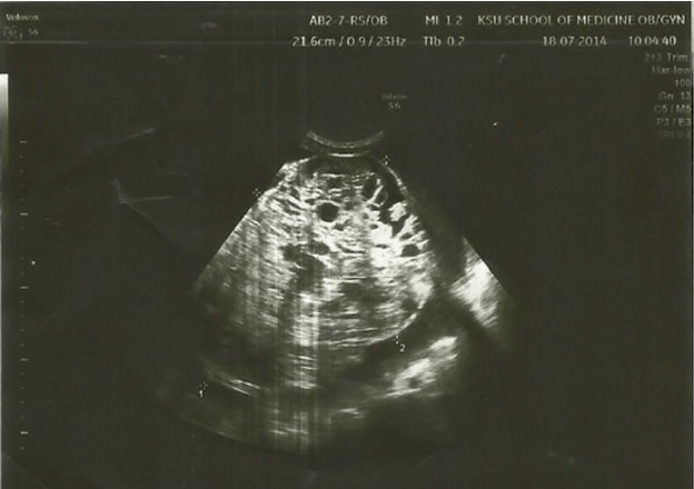
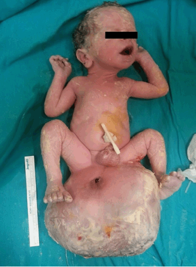
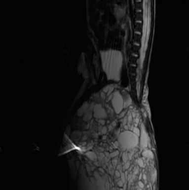
A 29-year-old Gravida 2, Parite 1 was referred to our clinic with the pre-diagnosis of intra-uterine fetal tumor. The pregnancy was calculated as 38 weeks according to the date of her last menstrual period. Antenatal follow-up had not been regular. No abnormality was observed in the medical history of her previous child. Ultrasonographic examination of the patient showed an intrauterine, single and alive fetus in transverse presentation with biometry compatible with 38 weeks. In the fetus, a heterogeneous mass was observed with a smooth surface, extending from the sacral zone to the perineum, with solid and cystic zones of 20x18 cm, and no intra-abdominal extension (Figure 1). Cesarean delivery under regional anesthesia was planned after the consideration of the complications that might arise during the removal of the fetus. Combined spinal-epidural anesthesia (CSE) was selected as the cesarean delivery anesthesia. After block application was confirmed, surgery was started. Taking into account the dimension of the fetal mass, a mid-line incision under the belly was applied to the abdomen, and a longitudinal incision was applied to the uterus in order to decrease the risk of rupturing the tumor. The head of the fetus was turned towards the opening and the fetus was delivered without complications with a mean APGAR score of 9–10. In the examination of the newborn, a mass of approximately 20x22x22 cm with a smooth surface and prominent venous congestion was detected in the fetal perineum (Figure 2). Sacrococcygeal teratoma was considered as a result of the localization and appearance of the mass. In the magnetic resonance imaging (MRI), which was applied in the postnatal period, the mass was observed arising from the sacrococcygeal region in the sagittal imaging and the mass had hypointense solid areas and hyperintense cystic septations on the T2-sequence (Figure 3). The mother was discharged from the hospital on postoperative day-2. The newborn was admitted to the pediatric surgery clinic for an operation. | ||||||
| ||||||
| ||||||
| ||||||
|
Discussion
| ||||||
|
Although teratomas, which are the most common congenital tumors, may be observed in different parts of the body, they are generally located in the sacrococcygeal zone at reported rates of approximately 50% [6]. As a result of ultrasonographic developments in recent years, sacrococcygeal teratoma is easily diagnosed in the antenatal period [7] [8]. In ultrasonography examination, a mass in cystic, solid or mixed form including both components, is observed with different echogenicity according to the content and sometimes showing intra-abdominal and pelvic extension, present especially in the sacral zone. In addition, it can also define the relationship between vascular structures and fetal circulation and can differentiate SCT from meningomyelocele by demonstrating the SCT vascular structures in the mass during Doppler examination [5] [9]. Large vascular structures in SCT and the development of arteriovenous shunts increase certain prenatal complications such as high output heart failure, fetal hydrops, polyhydramnios, and cardiomegaly [8]. Diagnosis of SCT in the prenatal period is significant for prenatal management, prenatal follow-up and planning of the delivery method. In vaginal delivery, death may occur as a result of serious dystocia and bleeding originating from a large vascular tumor. Vaginal route delivery was reported in 8 of 10 patients, but it was also noted that cystic tumor aspiration developed in one case [10]. Although vaginal delivery is possible in sacrococcygeal teratomas <5 cm and which do not include additional abnormalities, cesarean delivery is preferred for lesions >5 cm because of dystocia or hemorrhage risks [11]. Chuileannain et al. published two cases in which classical cesarean section procedure was applied to the patients, four cases in which sub-uterine segment cesarean procedure was applied, and one case in which delivery was realized through the vaginal tract, and it was stated that perinatal death was observed only in the case in which vaginal delivery was preferred [12]. On the other hand, Kay et al. reported vaginal deliveries without complication after prenatal percutaneous drainage in two cases with cystic sacrococcygeal teratomas, and percutaneous needle drainage was reported to be a potential alternative to cesarean delivery [13]. As Hoehn et al. stated, cesarean delivery should be preferred in cases with large tumors in order to avoid dystocia, tumor rupture, hemorrhage and traumatic delivery. Nevertheless, it should be remembered that difficulties may be encountered even in cesarean section. It is well-known that extensive hysterotomy applied due to a large teratoma, will increase morbidity [14] . This current case, cesarean section was applied since dystocia might occur due to the sacrococcygeal tumor having a large volume, and a classical uterine incision was preferred to prevent teratoma rupture. Since SCTs are generally diagnosed via ultrasonography in the prenatal period, intrauterine cyst aspiration, open fetal surgery, radiofrequency ablation method and thermoregulation are alternative options in the treatment. However, there are varying success rates of these operations, and more comprehensive studies are needed on those subjects [11]. Specialist physicians in the fields of gynecology, pediatrics and pediatric surgery should be present in the centers where the deliveries of patients with SCT will be performed in order to create a multi-disciplinary team approach [6]. | ||||||
|
Conclusion
| ||||||
|
In conclusion, diagnosis of sacrococcygeal teratoma in the early weeks of pregnancy may contribute to prenatal management, follow-up of prenatal complications and determination of the mode and place of delivery. When the delivery is planned, the least traumatic method should be preferred for the delivery. | ||||||
|
Acknowledgements
| ||||||
|
We would like to thanks all of colleagues who helped us in this study and Caroline Walker for English editing. | ||||||
|
References
| ||||||
| ||||||

|
[HTML Abstract]
[PDF Full Text]
|
|
Author Contributions
Murat Bakacak – Substantial contributions to conception and design, Acquisition of data, Analysis and interpretation of data, Drafting the article, Revising it critically for important intellectual content, Final approval of the version to be published Salih Serin – Substantial contributions to conception and design, Acquisition of data, Drafting the article, Final approval of the version to be published Aykut Urfalioğlu – Acquisition of data, Analysis and interpretation of data, Revising it critically for important intellectual content, Final approval of the version to be published Mehmet Akif Sarica – Acquisition of data, Analysis and interpretation of data, Revising it critically for important intellectual content, Final approval of the version to be published |
|
Guarantor of submission
The corresponding author is the guarantor of submission. |
|
Source of support
None |
|
Conflict of interest
Authors declare no conflict of interest. |
|
Copyright
© 2014 Murat Bakacak et al. This article is distributed under the terms of Creative Commons Attribution License which permits unrestricted use, distribution and reproduction in any medium provided the original author(s) and original publisher are properly credited. Please see the copyright policy on the journal website for more information. |
|
|
|
About The Authors
| |||
| |||
| |||
| |||
| |||