| |
|
|
|
Case Report
| ||||||
| Systemic lupus erythematosus presenting as erythroderma | ||||||
| Kingsuk Mukherji1, Nigel Jowett2, Julie Barber3 | ||||||
|
1Specialty Doctor, Cardiology, Withybush Hospital, Hywel Dda Health Board, Fishguard Road, Haverfordwest, United Kingdom SA61 2PZ.
2Consultant Cardiologist, Withybush Hospital, Hywel Dda Health Board, Fishguard Road, Haverfordwest, United Kingdom SA61 2PZ. 3Consultant Rheumatologist, Withybush Hospital, Hywel Dda Health Board, Fishguard Road, Haverfordwest, United Kingdom SA61 2PZ. | ||||||
| ||||||
|
[HTML Abstract]
[PDF Full Text]
[Print This Article]
[Similar article in Pumed] [Similar article in Google Scholar] 
|
| How to cite this article |
| Mukherji K, Jowett N, Barber J. Systemic lupus erythematosus presenting as erythroderma. Int J Case Rep Images 2014;5(11):772–776. |
|
Abstract
|
|
Introduction:
Systemic lupus erythematosus (SLE) is an autoimmune disorder involving multiple organs, predominantly seen in women of child-bearing age. Erythroderma is described in patients with subacute cutaneous lupus erythematosus (SCLE) but is rare in SLE. Among the cardiac manifestations, pericarditis is common but myocarditis is rare. We report a case of severe SLE presenting with erythroderma, pancytopenia, arthralgia and myocarditis.
Case Report: A 67-year-old male with a background history of hypertension, transient ischemic attack and polymyalgia rheumatica presented with severe erythroderma, malaise, arthralgia, weight loss and was found to be pancytopenic. His antinuclear antibody (ANA) was positive and double stranded DNA (dsDNA) was more than 200 IU/mL with very low complement levels. He developed lupus associated myocarditis with moderately impaired global left ventricular systolic function. He was initially started on steroids and hydroxychloroquine. But as he developed steroid induced myopathy, it was gradually tapered off and mycophenolate mofetil was started. He responded well to the treatment. He satisfied 8 of the 17 systemic lupus international collaborating clinics (SLICC) criteria establishing a diagnosis of systemic lupus erythematosus (SLE). His systemic lupus erythematosus disease activity index (SLEDAI) score came down from 17 to 2 over a period of six months of follow-up. Conclusion: Systemic lupus erythematosus may present with erythroderma. Careful clinical examination is important in all potentially multi-system diseases. | |
|
Keywords:
Erythroderma, Systemic lupus erythematosus, Myocarditis, Pancytopenia, Arthralgia
| |
|
Introduction
| ||||||
|
Systemic lupus erythematosus (SLE) is an autoimmune disease involving multiple organs. Ninety percent of patients are women of child-bearing age although both sexes, all ages and all ethnic groups may be affected. It is characterized by immunological abnormalities with production of number of antinuclear antibodies. Erythroderma is rarely associated with SLE, though described with subacute cutaneous lupus erythematosus (SCLE) [1] [2] [3]. Premature atherosclerosis is a significant cause of morbidity and mortality in SLE [4]. Pericarditis is a frequent manifestation, whereas myocarditis and Libman-Sacks endocarditis are less common but serious manifestations of SLE [5]. We report a case of severe SLE presenting with erythroderma, pancytopenia, arthralgia and myocarditis. | ||||||
|
Case Report
| ||||||
|
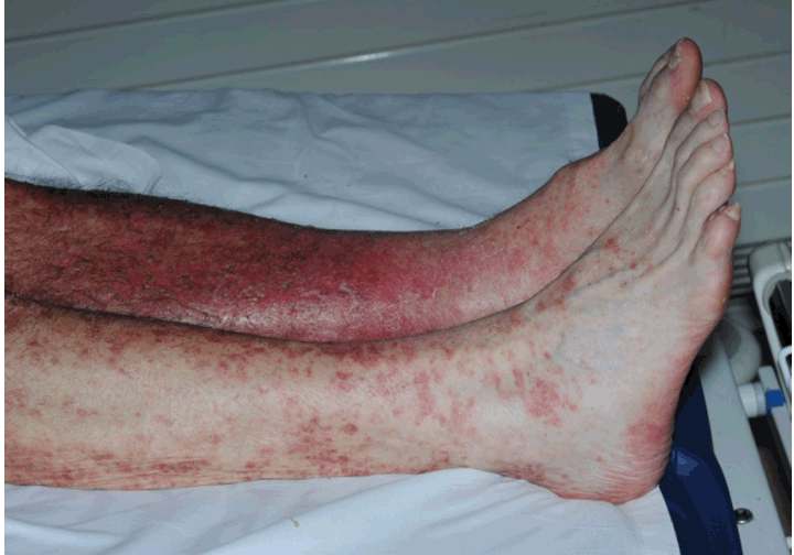
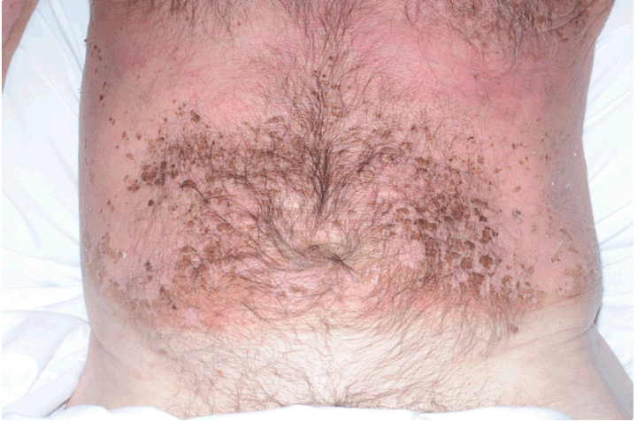
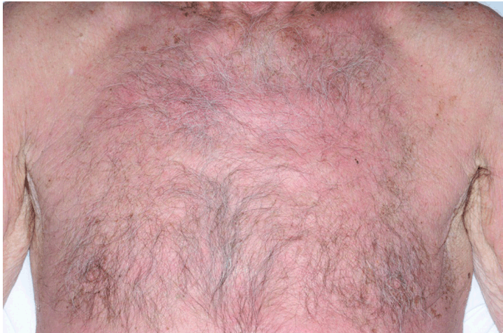
A 67-year-old male was presented with symptoms of malaise for two weeks, a generalized rash for one week and joint pain involving the ankles and knees. He also reported weight loss of 5 kg over last six months. His past medical history included hypertension, transient ischemic attack and polymyalgia rheumatica for 16 years on continuous steroids. He was a smoker but did not drink alcohol. There was no significant family history. On clinical examination, he had widespread erythematous rash which initially started on the back and then involved trunk and limbs sparing hands, feet and mucosa. The rashes were photosensitive with a clear demarcation between exposed and covered skin (Figure 1) (Figure 2) (Figure 3) (Figure 4). He complained of pain in the small joints of hands, both knees and left ankle. There was no lymphadenopathy and chest, heart and abdominal examination were unremarkable at the initial presentation. But later on he developed third heart sound on cardiac auscultation and an echocardiogram was requested. Although initially apyrexial, he developed high fever on second day of admission. | ||||||
| ||||||
| ||||||
| ||||||
| ||||||
|
| ||||||
|
Investigations and Differential Diagnosis
| ||||||
|
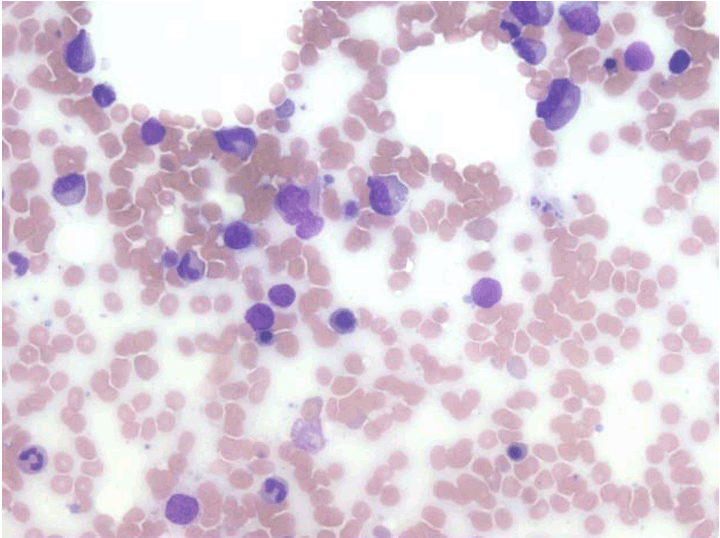
Full blood count revealed hemoglobin of 11.9 g/dL with a mean corpuscular volume (MCV) of 91 fl, white blood cell count (WBC) 3.3x10⁹/L (neutrophil 2.5x10⁹/L) and platelets 48x109/L. Blood glucose, urea and electrolytes, and liver function test were normal on admission. C-reactive protein (CRP) was 4 µg/mL on admission. He continued to spike temperature intermittently during the admission with no clear source of infection evident on clinical examination. CRP climbed to 216 µg/mL on seventh day. Four sets of blood culture were sent at different times which were all negative. Urine culture was also negative. On sixth day of admission, he became neutropenic with WBC dropping to 1.4x10⁹/L (neutrophil 0.80x10⁹/L) and he was started on piperacillin and tazobactam. Hemoglobin dropped to 9.9 g/dL on 15th day of admission. There was no history of blood loss. Direct Coombs test was mildly positive with presence of IgG and absence of C3 on the red cells. Haptoglobin and hemopexin levels were normal. LDH was normal at 548 IU/L. Bone marrow aspirate showed trilineage dysplastic changes thought to represent myelodysplastic changes (Figure 5). Bone marrow trephine biopsy revealed presence of trilineage hematopoiesis which was disorderly, but no evidence of any abnormal infiltrate. Skin biopsy showed prominent exocytosis of lymphocytes with keratinocyte necrosis present together with papillary dermal patchy lymphocytic infiltrate. Focal colloid body formation was present at the dermoepidermal interface but no lichenoid infiltrate was present. These findings were in keeping with graft versus host disease but not typical of an acute phase. The features while not classical also warrant exclusion of lupus erythematosus and dermatomyositis. Serum protein electrophoresis and urine for Bence Jones protein were negative. Urine protein/creatinine ratio was 58.5 mg/mmol (normal range <45 mg/mmol). Serum creatinine was 66 µmol/L with an eGFR of >90 mL/min. Hepatitis B&C and HIV serology were negative. ANA was positive with a titre of >1:2560 with a homogeneous pattern. Anti-dsDNA was more than 200 IU/mL (normal 10–20 IU/mL). Anticardiolipin antibodies and antibodies to extractable nuclear antigen (ENA) were negative. There was severe hypocomplementia with C3 level of 0.24g/L and C4 level of 0.02g/L. Computed tomography (CT) scan of chest, abdomen and pelvis showed two hemangiomas in liver, gallstones and indeterminate subpleural nodule in the lingular segment of the left lung. A diagnosis of SLE with erythroderma was made. His disease activity score (SLEDAI) was 17 initially [6]. Systemic steroids (prednisolone 70 mg/day) and hydroxychloroquine (400 mg OD) were started. Skin lesions started improving, white blood cell and platelets gradually normalized. Routine examination during his stay in the hospital revealed third heart sound with no signs of heart failure. Echocardiogram showed globally impaired moderate left ventricular systolic dysfunction with an ejection fraction 40% and he was started on ACE inhibitors and beta blockers. Troponin I was raised at 0.07 (normal range <0.04). These findings were suggestive of myocarditis. He developed pain and weakness of the proximal muscles of both legs within seven days of starting steroids, so his steroid dose was reduced to 40 mg/day and then gradually tapered and was started on mycophenolate mofetil (500 mg BD). His creatinine kinase (CK) was normal at 24 IU/L. Magnetic resonance imaging (MRI) scan of both thighs was done. It showed atrophy of the flexors and the adductors of both hip joints but there was no focal areas of inflammation to guide biopsy. Electromyography (EMG) was normal. Acetylcholine receptor antibodies were negative. Within a week of reducing the dose of steroids his muscle pain improved. He continued to improve while being followed-up as out-patient. The dose of mycophenolate mofetil was increased to 500 mg TDS after two weeks. His exercise tolerance significantly improved, some mild residual erythema was present on the back at second month of follow-up but cleared at sixth month. His repeat echocardiogram at sixth month showed improvement of left ventricular function with an ejection fraction of 48%. At sixth month, he was on 15 mg of prednisolone. | ||||||
| ||||||
|
Discussion
| ||||||
|
The diagnosis of SLE in this case is based on the patient fulfilling 8 out of the 17 SLICC criteria [7]. There are case reports of subacute cutaneous lupus erythematosus (SCLE) presenting with erythroderma, not SLE. Erythroderma is a severe life-threatening condition that presents with diffuse erythema and scaling involving all or most of the skin surface area. A wide range of cutaneous or systemic diseases can present with erythroderma. The most common causes of erythroderma are exacerbation of psoriasis or atopic dermatitis [8]. It can also be secondary to drugs most commonly penicillins, sulfonamides, carbamazepine, phenytoin and allopurinol. Uncommon causes of erythroderma include cutaneous T cell lymphoma and other hematologic and systemic malignancies, connective tissue diseases and infections. Among the connective tissue diseases, subacute cutaneous lupus erythematosus and dermatomyositis have been described to be associated with erythroderma. Patients with SLE frequently develop abnormalities in one or more of the three blood cell lines. In this case, the pancytopenia gradually responded with the treatment. His Direct Coombs test was mildly positive with other markers of hemolysis being normal suggestive of mild hemolytic anemia. Due to the timing of onset of muscle pain and weakness in the proximal muscles of legs after glucocorticoid exposure, he likely had steroid induced myopathy. Muscle enzymes and EMG are usually normal in glucocorticoid myopathy. There are a variety of cardiac manifestations of SLE. Pericarditis is relatively common, but myocarditis is uncommon. Verrucous (Libman-Sacks) endocarditis is usually clinically silent, but it can produce valvular insufficiency and can serve as a source of emboli. In this case, the myocarditis was severe enough to cause moderate global systolic dysfunction that produced no symptoms, but came to light by vigilant cardiovascular examination. Antimalarials such as hydroxychloroquine is helpful in patients with skin and musculoskeletal involvement. They have also shown to prevent damage to kidneys and central nervous system [9]. Systemic glucocorticoids are usually reserved for patients with organ involvement. In this case, it was considered because of the hematological and severe skin involvement. Immunosuppressive agents like methotrexate, azathioprine, cyclophosphamide and mycophenolate mofetil are considered in patients who are unresponsive or intolerant to steroids. Mycophenolate mofetil was added in this patient because of the side effects of steroids. In patients unresponsive to well-establish treatment, biologic agents like rituximab or belimumab may be considered. | ||||||
|
Conclusion
| ||||||
|
We describe a very severe case of systemic lupus erythematosus initially presenting as erythroderma and later on developed pancytopenia and myocarditis. The diagnosis was based on features fulfilling the systemic lupus international collaborating clinics (SLICC) criteria. Though rare, systemic lupus erythematosus may present with erythroderma. Careful clinical examination is important in all potentially multi-system diseases. | ||||||
|
Acknowledgements
| ||||||
|
Dr. Sumanta Kundu, Consultant Hematologist, Withybush Hospital | ||||||
|
References
| ||||||
| ||||||
|
[HTML Abstract]
[PDF Full Text]
|
|
Author Contributions
Kingsuk Mukherji – Substantial contributions to conception and design, Acquisition of data, Analysis and interpretation of data, Drafting the article, Revising it critically for important intellectual content, Final approval of the version to be published Nigel Jowett – Analysis and interpretation of data, Drafting the article, Revising it critically for important intellectual content, Final approval of the version to be published Julie Barber – Analysis and interpretation of data, Drafting the article, Revising it critically for important intellectual content, Final approval of the version to be published |
|
Guarantor of submission
The corresponding author is the guarantor of submission. |
|
Source of support
None |
|
Conflict of interest
Authors declare no conflict of interest. |
|
Copyright
© 2014 Kingsuk Mukherji et al. This article is distributed under the terms of Creative Commons Attribution License which permits unrestricted use, distribution and reproduction in any medium provided the original author(s) and original publisher are properly credited. Please see the copyright policy on the journal website for more information. |
|
|