| |
|
|
|
Case Report
| ||||||
| Epidermolysis bullosa in a small for gestational age preterm male neonate with two affected siblings: A case report | ||||||
| Swathi Chacham1, Jakkampudi Nagasravani2, Uppin Narayan Reddy3, Jillalla Narsing Rao4, S. Pratap Rao5, Kumar AS6 | ||||||
|
1MD Pediatrics, DM Neonatology (from Post Graduate Institute of Medical Education and Research, Chandigarh, India), Assistant Professor, Department of Pediatrics, Princess Esra Hospital, Deccan College of Medical Sciences, Hyderabad, Andhra Pradesh, India.
2MBBS, (MD, Pediatrics), Junior Resident Doctor, Department of Pediatrics Princess Esra Hospital, Deccan College of Medical Sciences, Hyderabad, Andhra Pradesh, India. 3MD Pediatrics, Professor and Head, Department of Pediatrics Princess Esra Hospital, Deccan College of Medical Sciences, Hyderabad, Andhra Pradesh, India. 4MD Pediatrics, Professor, Department of Pediatrics, Princess Esra Hospital, Deccan College of Medical Sciences, Hyderabad, Andhra Pradesh, India. 5MD Pediatrics, Professor, Department of Pediatrics, Princess Esra Hospital, Deccan College of Medical Sciences, Hyderabad, Andhra Pradesh, India. 6MD Dermatology (From All India Institute Of Medical Sciences, New Delhi), Professor and Head, Department of Dermatology, Princess Esra Hospital, Deccan College of Medical Sciences, Hyderabad, Andhra Pradesh, India. | ||||||
| ||||||
|
[HTML Abstract]
[PDF Full Text]
[Print This Article]
[Similar article in Pumed] [Similar article in Google Scholar] 
|
| How to cite this article |
| Chacham S, Nagasravani J, Reddy UN, Rao JN, Rao SP, Kumar AS. Epidermolysis bullosa in a small for gestational age preterm male neonate with two affected siblings: A case report. Int J Case Rep Images 2014;5(10):691–694. |
|
Abstract
|
|
Introduction:
Epidermolysis bullosa is a rare group of hereditary disorders, which are characterized by blistering of the skin and mucosa due to little or no apparent trauma. The severity can range from mild, localized skin blisters to generalized, systemic life-threatening disease and the treatment is mainly supportive. Dystrophic type of epidermolysis bullosa is one of the rarer forms. Skin fragility often leads to secondary skin infections and in turn generalized infection which can be lethal. Hence, the major challenge encountered in the care of a neonate with epidermolysis bullosa is optimum skin care and expert nursing care. A preterm, small for gestational age (SGA) neonate, has immature skin physiologically, which is a portal for systemic infection when compared to a term appropriate for gestational age neonate. The presence of blistering lesions in a preterm, SGA neonate further challenges the management.
Case Report: We report a preterm, low birth weight (small for gestational age) male neonate, who had extensive bullous lesions and dystrophic nails in the first week of life. These lesions healed by scarring leading to stiffness of underlying joints and were complicated by secondary infection. There was history of consanguinity and previous two siblings also had similar lesions with contractures. Conclusion: A preterm male small for gestational age neonate born of consanguineous marriage, with the history of cutaneous bullous lesions in two siblings, presented with extensive bullous lesions and dystrophic nails, suggestive of dystrophic epidermolysis bullosa. | |
|
Keywords:
Blistering, Dystrophic, Neonate, Epidermolysis bullosa, Low birth weight, Preterm, Small for gestational age (SGA), Infant
| |
|
Introduction
| ||||||
|
Epidermolysis bullosa (EB) is an intractable, rare and often lethal skin condition. It is characterized by the development of vesicles and bullae either spontaneously or as a result of minimal insignificant trauma. Epidermolysis bullosa is due to a congenital defect in the skin structure and is often hereditary. There are three major types of EB described, namely: simplex, junctional and dystrophic [1]. Reported prevalence of EB varies from 10–20 cases per 1 million live births as per various EB registries [2] [3] [4] [5]. A preterm, small for gestational age (SGA) neonate, has immature skin physiologically, which is a portal for systemic infection when compared to a term appropriate for gestational age neonate [6]. | ||||||
|
Case Report
| ||||||
|
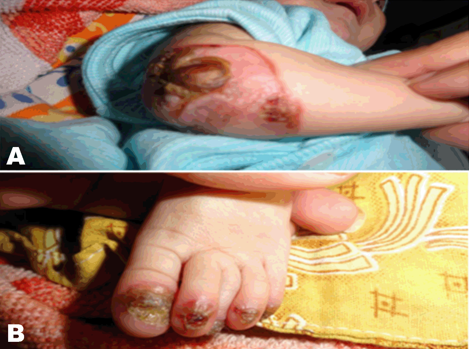
A 36-week male neonate, weighing 1800 grams was born to a gravida four mother by emergency cesarean section for absent end diastolic flow in antenatal Doppler ultrasound. The neonate had normal extra uterine transition with normal APGAR scores. The birth weight was less than third centile, hence it was a SGA neonate. The neonate had mild respiratory distress requiring supplemental oxygen and antibiotics. Sepsis screen was negative and initial blood glucose levels were low. Blood culture was sterile and the chest X-ray showed features of transient tachypnea of newborn. Respiratory distress subsided by 48 hours and baby developed mild jaundice requiring phototherapy. Feeds were started on second day of life and full feeds were established by fourth day of life. There was history of second degree consanguinity and history of bullous lesions in two previous siblings in the neonatal period. They succumbed to death at second and seven months of life, respectively due to repeated skin and respiratory infections. The neonate developed fluid filled bullae over both elbows and gluteal region on the fourth day of life. Basic interventions such as intravenous cannulation provoked blistering. The bullous lesions subsequently spread all over the body and healed by scarring (Figure 1A). However, there was no mucosal involvement. Hence, EB was diagnosed and the dystrophic variety was considered in view of extensive scarring and dystrophic nails (Figure 1B). The neonate was discharged with topical emollients, skin protective measures and nutritional supplements for catch up growth. The infant had good weight gain, and was thriving well till four months, despite frequent blistering episodes and scarring. Frequent scarring had lead to stiffening of fingers and toes. Though the skin biopsy could not be performed due to the financial constraints, clinically dystrophic EB was considered in view of stiffening of fingers and toes in the index case and contractures in the previous siblings. Subsequently, the infant had secondary skin infection which was associated with generalized sepsis and pneumonia. Sepsis screen was positive and antibiotics were started. However, worsening pneumonia had lead to respiratory failure and death. | ||||||
| ||||||
|
Discussion
| ||||||
|
Epidermolysis bullosa is a mucocutaneous blistering disorder of hereditary nature. Blistering lesions are provoked by little or no apparent trauma. It has a wide spectrum of manifestations as well as complications. Severity ranges from localized skin blisters to neonatal mortality [2]. Three major phenotypes have been described depending on the level of cleavage of the basement membrane at dermoepidermal junction, namely simplex, junctional and dystrophic [1] [7]. The level of separation in EB simplex is intraepidermal and is at lamina densa in junctional EB. However, in dystrophic EB the separation is below the basement membrane [2] [8]. Each of these three main phenotypes have both autosomal dominant and autosomal recessive form of inheritance. Epidermolysis bullosa simplex has predominantly autosomal dominant inheritance, while the mode of inheritance is autosomal recessive in majority of junctional EB [7] [8]. The wide range of genetic abnormalities occur in various types of EB. K5 or K14 gene mutations result in bullous formation by disrupting the basal cells in dominant EB simplex as well as some forms of recessive EB simplex. While, type XVII collagen, laminin 5 and a6, β4 integrin gene mutations are seen in junctional EB and dystrophic variant is linked with type VII collagen gene abnormalities [7]. The index neonate had developed the lesions in the first week of life, suggestive of autosomal recessive inheritance. Along with the history of consanguinity and similar lesions in siblings in the neonatal period, the recessive mode of inheritance was further confirmed. Though the skin biopsy is confirmatory to differentiate various forms of epidermolysis bullosa, deep scarring with contractures is peculiar to dystrophic EB. The index case had stiffening of fingers and toes, though the neonate did not survive longer to develop contractures. However, there was history of contractures in the previous siblings. The clinical severity of the different forms of EB varies widely and internal organ involvement can occur [4]. The incidence and prevalence rates expressed as number of cases per 1 million live births, for EB simplex: 10.75, junctional EB: 2.04, dystrophic EB dominant type: 2.86 and recessive dystrophic EB: 2.04. Epidermolysis bullosa has no gender, geographical or racial preponderance. Epidermolysis bullosa simplex is the most prevalent phenotype, where as autosomal recessive dystrophic and junctional EB are rare [2] [3] [4] [5]. In view of blisters affecting the whole body with scarring and disfigurement, recessive type of dystrophic epidermolysis bullosa (Hallopeau-Siemens type) was considered in the index case [9]. A preterm, SGA neonate has immature skin physiologically, which is a portal for systemic infection when compared to a term appropriate for gestational age neonate. The need for invasive monitoring and invasive procedures hampers the skin integrity. The presence of blistering lesions in a preterm, SGA neonate further challenges the management. Currently, there is no definitive and curative treatment for EB. Hence, the mainstay of treatment is symptomatic, supportive and preventive [10]. The major challenge encountered in the care of a neonate with EB is optimum skin care and expert nursing care more so, with a preterm, low birth weight neonate who undergoes many invasive skin procedures as in the index case [6]. The key measures that need to be taken in neonates and children with EB are preventing trauma induced blisters and avoiding unnecessary antibiotics. These can be achieved by nursing the infants on soft foam pads and restricting the use of topical antimicrobials, which were implemented in the index case [10]. The lesions were cleaned with sterile normal saline and covered with non-adhesive dressings. Topical antimicrobials were avoided, unless needed, to prevent the emergence of resistant bacteria. Soft, non-adhesive dressing materials soaked with emollients such as vaseline (which are preferred) were used as a preventive measure. Parents were counseled and psychological support was given to them, which plays a significant role in the care of these neonates [10]. Adequate nutritional support is necessary for optimum growth and development and to aid in wound healing, more so in a preterm, SGA neonate who is challenged nutritionally otherwise. Genetic counseling is required for families of affected children along with prenatal diagnosis (using molecular techniques) [10]. Most common cause of death is secondary bacterial infection resulting from frequent blisters. Similarly, index case developed skin infection which lead to pneumonia, respiratory failure and death subsequently. | ||||||
|
Conclusion
| ||||||
|
A preterm male small for gestational age neonate born of consanguineous marriage, presented with extensive bullous lesions and dystrophic nails, suggestive of epidermolysis bullosa. The lesions were complicated by scarring, which lead to stiffening of fingers. Similar lesions in the two previous siblings along with contractures suggests dystrophic variant of epidermolysis bullosa. | ||||||
|
Acknowledgements
| ||||||
|
Thank Mrs. Shashi vadana for helping with graphics and Dr. Taha, Dr. Muzammil Hassan and Muzahid for helping with drafting. | ||||||
|
Abbreviations
| ||||||
|
APGAR, BM, (Basement membrane), EB, (epidermolysis bullosa), SGA, (small for gestational age). | ||||||
|
References
| ||||||
| ||||||
|
[HTML Abstract]
[PDF Full Text]
|
|
Author Contributions
Swathi Chacham – Conception and design, Acquisition of data, Analysis and interpretation of data, Drafting the article, Critical revision of the article, Final approval of the version to be published Jakkampudi NagaSravani – Conception and design, Acquisition of data, Analysis and interpretation of data, Drafting the article, Critical revision of the article, Final approval of the version to be published Uppin Narayan Reddy – Conception and design, Acquisition of data, Analysis and interpretation of data, Critical revision of the article, Final approval of the version to be published Jillalla Narsing Rao – Conception and design, Acquisition of data, Analysis and interpretation of data, Critical revision of the article, Final approval of the version to be published S. Pratap Rao – Conception and design, Acquisition of data, Analysis and interpretation of data, Critical revision of the article, Final approval of the version to be published Kumar A. S. – Conception and design, Acquisition of data, Analysis and interpretation of data, Critical revision of the article, Final approval of the version to be published |
|
Guarantor of submission
The corresponding author is the guarantor of submission. |
|
Source of support
None |
|
Conflict of interest
Authors declare no conflict of interest. |
|
Copyright
© 2014 Swathi Chacham et al. This article is distributed under the terms of Creative Commons Attribution License which permits unrestricted use, distribution and reproduction in any medium provided the original author(s) and original publisher are properly credited. Please see the copyright policy on the journal website for more information. |
|
|