|

|
 |
|
Case Report
| ||||||
| Harlequin ichthyosis: A rare dermatological disorder | ||||||
| Alok Kumar Dubey1, Ilisapeci Vereti Tuibeqa2, Nina Baivou Pio3 | ||||||
|
1MD,Pediatrics,MPhil (HHSM), Professor, Department of Pediatrics, College of Medicine, Nursing & Health Sciences, Suva, Fiji.
2MCH, PGDCH, MBBS, Acting Consultant Department of Pediatrics, Colonial War Memorial Hospital, Suva, Fiji. 3MBBS, Registrar Obstetrics & Gynecology Colonial War Memorial Hospital, Suva, Fiji. | ||||||
| ||||||
|
[HTML Abstract]
[PDF Full Text]
[Print This Article]
[Similar article in Pumed] [Similar article in Google Scholar] 
|


| How to cite this article |
| Dubey AK, Tuibeqa IV, Pio NB. Harlequin ichthyosis: A rare dermatological disorder. Int J Case Rep Images 2014;5(8):590–594. |
|
Abstract
|
|
Introduction:
Harlequin ichthyosis is one of the most devastating of the genodermatoses. Neonates usually die within the first few days of life from infection or dehydration related complications. Prenatal diagnosis remains difficult but may be possible in high risk pregnancies by performing a fetal skin biopsy or by three-dimensional ultrasonography.
Case Report: We report the case of Harlequin ichthyosis for its rarity and briefly review literature. Conclusion: This case has been reported for the rarity of Harlequin ichthyosis and to create awareness among pediatricians to identify the condition promptly. | |
|
Keywords:
Harlequin ichthyosis, ABCA12 gene, Ectropion, Eclabium
| |
|
Introduction
| ||||||
|
Harlequin ichthyosis is extremely rare, and is the most severe form of the keratinizing disorders characterized by profound thickening of stratum corneum [1]. A dense armor like scale covers the body. The newborn appears to be encased in a tight thin membrane which allows little movement and holds the limbs in semi-flexed position (the harlequin fetus). Other features include underdeveloped external ears, nasal hypoplasia, bilateral ectropion with occlusion of the eyes and eclabium. Neonates usually die within the first few days of life from infections and dehydration related complications [2]. The first known report of this is in the diary of Reverend Lover Hart in 1750 [3]. | ||||||
|
Case Report
| ||||||
|
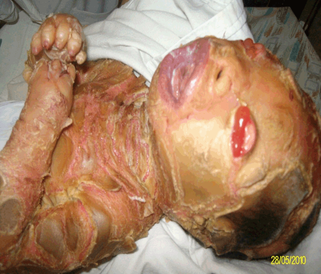
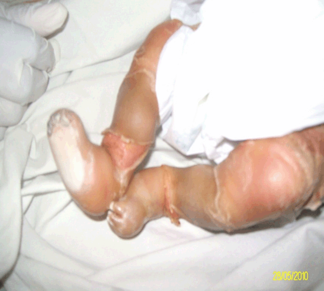
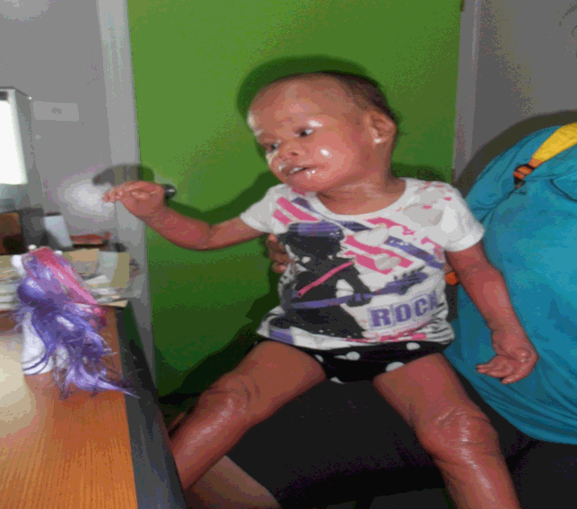
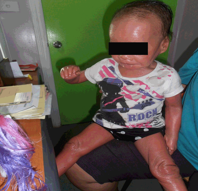
A 15-year-old primipara delivered a term, appropriate for gestational age female neonate with a birth weight 2.8 kg, 50 cm length and head circumference of 36 cm, respectively. The mother was seronegative and had attended antenatal clinics at a peripheral center. She was referred to the main hospital for delivery being a young primipara. There is a history of third generation consanguinity. Her antenatal ultrasound scans showed a single viable fetus with no anomalies. She had a normal spontaneous delivery of a live female infant with APGAR scores of 8, 9, and 9. On examination, the skin of infant looked like dried cracked earth, and was pale. She also had hyperkeratotic scales with hair loss, severe ectropion, eclabium, small pinnae and hypoplasia and contracture of all digits (Figure 1). There was depressed bridge of the nose with contractures of both upper and lower limbs. She had good sucking reflex, however, was fed by syringe because mother was reluctant to handle the baby. Apart from these striking dermatological abnormalities, the rest of the systemic examinations were normal. Clinically, she was diagnosed as harlequin ichthyosis. A skin biopsy revealed irregular thickening of epidermis with hyper and parakeratosis. Epidermal cells showed a clear space around the nuclei. Intradermal cleavage was visible with evidence of bacterial infection. Findings were consistent with the clinical diagnosis. However, due to non-availability of the high resolution microscopy, and special stains sub typing of the harlequin ichthyosis was not possible. Genetic testing could also not be done as facilities for karyotyping and cytogenetics were not available. The baby was managed with intravenous fluids through an umbilical line and with expressed breast milk by mouth. She was kept under a neonatal warmer, with paraffin applied all over the skin paraffin along with artificial tears (Lacri-Lube). Ophthalmic ointments 1% chloramphenicol was used six-hourly for seven days. The ectropion was noticed to be reducing spontaneously, that precluded any surgical intervention and within four weeks artificial tears were also ceased. Initially, injection penicillin 50 mg/kg intravenous (IV) twice daily and injection gentamycin 5 mg/kg once a day were commenced empirically, prophylactically. Despite this baby developed clinically and microbiologically confirmed sepsis with Acinetobacter baumannii which was then managed with injection ampicillin 200 mg/kg/day in four divided doses by IV route and injection gentamycin 5 mg/kg/day IV for 14 days, with due monitoring of the renal functions, as indicated by the culture and sensitivity reports. Physiotherapy and soft splinting of the contractures was done in hospital and later continued at home. Sterile gauze balls were kept in the fists to prevent hand contractures. Further management in hospital comprised continued care of the eyes, maintenance of the hydration and nutrition, and paraffin emollient application. Movement improving exercises were carried out, care givers were trained and educated about the importance of "skin to skin" contact and they all carried out the same whilst observing all infection control precautions. The radiant warmer was also used to maintain thermo neutrality. The baby was discharged after four months, after thorough education of the care givers about the ongoing care of the baby. They were, specifically, educated about the danger signs that warrant urgent medical attention. At present baby is four years old and is being regularly followed-up at the pediatric unit of the local hospital. Almost all the hyperkeratotic scales have shed off and been replaced by neo-epithelial tissue, is able to sit with support and is thriving well (Figure 2) (Figure 3) (Figure 4). Baby, however, has not been subjected to a formal hearing test and a psychological evaluation. | ||||||
| ||||||
| ||||||
| ||||||
| ||||||
|
Discussion
| ||||||
|
Harlequin ichthyosis has an incidence of about 1 in 300,000 births [4]. As per scientific literature reports in 2007, there have been reports of 101 cases in worldwide medical literature [5]. Harlequin ichthyosis is an inherited autosomal recessive trait disease [6] [7] caused by mutations of the ABCA12 gene (adenosine triphosphate-binding cassette A12), resulting in defective lipid transport significantly impacting the normal development of the skin barrier [8] [9]. Diffuse hyperkeratinization and desquamation are characteristic of harlequin ichthyosis [10]. The development of harlequin ichthyosis phenotype is initiated by the onset of hair canal keratinization at 17 weeks of gestation and is expressed in the entire hair carrying skin from 20 weeks of gestation onwards [11]. The ABCA12 gene (chromosome 2q35) product is a protein that functions in the intracellular lipid transfer system in keratinocytes in stratum corneum. In the stratum corneum, granules are there which secrete a barrier forming lipid layer between granular and cornified layers [7] [12] [13]. Most cases from families with a negative family history are diagnosed clinically after birth. The first antenatal diagnosis was reported by Blanchet-Bardon et al. in 1983 [12]. Prenatal diagnosis remains difficult but is possible by fetal skin biopsy or three-dimensional ultrasonography. Cells from the peripheral blood smear or the skin can be obtained and complete sequential analysis of the coding region of the ABCA12 gene can be carried out to identify the specific mutations, which lead to the development of harlequin ichthyosis. Once the mutation in the proband is recognized, the relatives can also be screened for carrier status. Facility for prenatal diagnosis of harlequin ichthyosis in suspected fetuses is also available [14]. A three-dimensional ultrasonogram and the electron microscopic examination of the fetal skin biopsy permit the possibility of prenatal diagnosis of this disorder [10] Babies with harlequin ichthyosis have such a striking appearance due to alligator like skin, ectropion, eclabium and dysplastic ears that the diagnosis is obvious clinically. Laboratory tests are necessary only for the associated metabolic derangements such as dyselectrolytemia and for possible secondary infection/sepsis. An abdominal ultrasonography was done to rule out renal anomalies which are described in literature. The baby in this case did not have any renal anomalies [15]. The other type of Icthyosis Colloidian baby may have similar physical features but the thin shining layer that covers these babies clearly enables the clinician to make the differentiation. In this case though the diagnosis was clinically obvious, we still got it confirmed by histopathological examination of the skin. Non-availability of the electron microscope and special stains precluded keratinizing hyaline granules, profilaggrin and filaggrin which are essential for sub typing the harlequin ichthyosis. Genetic testing could not be done as facilities for karyotyping and cytogenetics do not exist in Fiji. The management of such cases basically involves stabilization of airway, breathing and circulatory compromise due to armor like encasing of the thorax by hyperkeratotic skin. The protection of eyes from exposure by artificial tears and antibiotic ophthalmic ointments. At later stage, the ectropion can be corrected by surgery. Skin should be covered with sterile lubricants to soften it, thus facilitating desquamation. The baby should be nursed in a humidified crib to ensure thermo-neutrality. Umbilical vein access should be established for administration of the fluids, nutrients and medications as it is extremely difficult to get a peripheral vascular access in these babies. The babies with harlequin ichthyosis being prone to dehydration, and electrolyte disturbances should be vigilantly monitored for the same and should be followed-up periodically. The role of prophylactic antibiotic is questionable, however, it is a prudent practice, in particular in the developing world to cover the baby with broad spectrum antibiotics to which local microbiological flora are known to be sensitive. Ideally, a team comprising pediatrician, dermatologist, geneticist, ophthalmologist, reconstructive surgeon, should make an individualized plan for management along with full involvement of the parents or the care givers. The most effective class of the drugs in harlequin ichthyosis is retinoids and their derivatives, which prevent cracking of the skin and facilitate desquamation and this hastens up pliability. Pliability is an important variable that renders immense benefit in improving the movement range, prevention and early correction of contractures, including ectropion and eclabium. Pain control is another important aspect of the management, that occurs on account of the cracks and it is advisable to keep the babies with harlequin ichthyosis sedated. Duration has to be individualized. The other important area of management comprises genetic counseling about recurrence in other sibs. An ongoing service of a social worker is an integral part of the management. Abnormal appearance of the babies may make them subject of ridicule adversely affecting their psyche and eroding self-esteem. Social and professional psychological support therefore is also an essential part of the holistic management of such babies [16]. Treatment of babies with harlequin has been attempted with a battery of medications ranging from oral retinoids, topical paraffin, antiseptics, and emollients to soften the skin. In general, harlequin fetuses do not survive the neonatal period. Death occurs due to dehydration, systemic infection or impaired respiration [17]. This baby is still alive at the age of four years. The parents of the proband should undergo genetic testing for ABCA12 gene defect detection and be appropriately counseled. Furthermore, the fetal skin biopsy and a three-dimensional ultrasonogram may be offered in subsequent pregnancies to save the family from having yet another baby with this potentially lethal and rare disorder. | ||||||
|
Conclusion
| ||||||
|
This case has been reported for the rarity of Harlequin ichthyosis and to create awareness among pediatricians to identify the condition promptly. | ||||||
|
| ||||||
|
Acknowledgements
| ||||||
|
The authors would like to thank Dr. Abha Gupta, for histopathological confirmation of the diagnosis, Dr. Joseph Flear for critically reviewing the manuscript and Swaran Lata for helping in typing and computer work | ||||||
|
References
| ||||||
| ||||||

|
[HTML Abstract]
[PDF Full Text]
|
|
Author Contributions
Alok Kumar Dubey – Conception and design, Analysis and interpretation of data, Drafting the article, Critical revision of the article, Final approval of the version to be published Ilisapeci Vereti Tuibeqa – Acquisition of data, Analysis and interpretation of data, Critical revision of the article, Final approval of the version to be published Nina BaivouPio – Acquisition of data, Drafting the article, Final approval of the version to be published |
|
Guarantor of submission
The corresponding author is the guarantor of submission. |
|
Source of support
None |
|
Conflict of interest
Authors declare no conflict of interest. |
|
Copyright
© 2014 Alok Kumar Dubey et al. This article is distributed under the terms of Creative Commons Attribution License which permits unrestricted use, distribution and reproduction in any medium provided the original author(s) and original publisher are properly credited. Please see the copyright policy on the journal website for more information. |
|
|
|
About The Authors
| |||
| |||
| |||
| |||