  |

|
 |
|
Case Report
| ||||||
| The effect of photoactivated platelet-rich plasma injections in the novel treatment of shoulder osteoarthritis | ||||||
| Julien Freitag | ||||||
|
MBBS, BMedSci, Sports Medicine Clinician, Lifecare Prahran Sports Medicine Centre.
| ||||||
| ||||||
|
[HTML Abstract]
[PDF Full Text]
[Print This Article]
[Similar article in Pumed] [Similar article in Google Scholar] 
|


| How to cite this article |
| Freitag J. The effect of photoactivated platelet-rich plasma injections in the novel treatment of shoulder osteoarthritis. Int J Case Rep Images 2014;5(8):546–552. |
|
Abstract
|
|
Introduction:
Shoulder osteoarthritis can cause significant patient morbidity and current conservative treatment is limited. We present a case report of shoulder osteoarthritis treated with photoactivated platelet-rich plasma injections (PAPRP).
Case Report: A 62-year-old female was presented with chronic shoulder pain secondary to glenohumeral osteoarthritis. The patient underwent a course of intra-articular glenohumeral PAPRP. Patient outcome measures included the numerical pain rating scale (NPRS), Disabilities of arm, shoulder and hand score (DASH) and patient perceived improvement (PPI). Following treatment the patient reported improvements in both pain and function as measured by the NPRS, DASH and PPI. Repeat imaging showed no evidence of osteoarthritis progression. Conclusion: In this case report, PAPRP injections for the treatment of shoulder osteoarthritis resulted in improvement in all recorded measures of pain and function. Whilst only level 5 evidence, the promising outcome of this single case report highlights the need to further evaluate through more structured controlled trials the efficacy of PAPRP in the treatment of glenohumeral osteoarthritis. | |
|
Keywords:
Osteoarthritis, Shoulder, Platelet-rich Plasma
| |
|
Introduction
| ||||||
|
Shoulder osteoarthritis (OA) affects up to 20% of the elderly population [1]. It can lead to significant patient morbidity and have serious economical impact [2] [3] [4]. Most current medical treatment strategies are aimed at pain reduction and/or symptom control with available pharmacological treatments having limited and often unwanted side effects [5]. Noel et al. have shown significant reduction in pain scores following use of hyaluronic acid (HA) injections in glenohumeral OA [6]. Further, systematic review of the use of hyaluronic acid/visco-supplementation in the treatment of knee OA has also indicated comparable efficacy to regular use of oral anti-inflammatories [7]. However, whilst HA intra-articular injections have become accepted as an adjunctive treatment in the conservative management of knee OA, its use for shoulder OA has not become widespread. More recently there has been a focus within clinical medicine on biological therapies. There is growing evidence to support the use of platelet-rich plasma (PRP) injections for the treatment of symptomatic knee osteoarthritis. Growth factors expressed by platelets, including transforming growth factor beta and platelet derived growth factor, have?the ability to influence and direct tissue regeneration through cell proliferation and also via synthesis of extracellular matrix proteins [8]. Animal and in vitro studies have demonstrated the ability of PRP to improve cartilage matrix expression and also to stimulate the synthesis of hyaluronic acid from the synovium [8] [9]. More recent clinical case control series have indicated significant pain and functional improvement following PRP therapy, with follow-up to 12 months [10] [11] [12] [13]. Interestingly, despite the initial results of invitro studies and given the improvement in symptoms, no evidence of disease modification and/or cartilage repair has been documented [14]. Another biological medium, autologous conditioned serum (ACS), has also shown promise in symptom control [15]. Autologous conditioned serum is an injectable (IL-1) receptor antagonist (IL-1RA) medium that has been used in Europe for the treatment of musculoskeletal conditions [15]. Research has identified Interleukin-1 IL-1 as a critical mediator of cartilage loss. Interleukin-1 Receptor Antagonist (IL-1RA) successfully inhibits the intra-articular actions of IL-1 [16] [17] [18] [19] [20] [21]. Polychromatic light photoactivation of peripheral blood increases the expression of leukocyte derived anti-inflammatory cytokines (including IL-1RA) and reduces expression of pro-inflammatory cytokines (IL-2 & 6) [22] [23]. A recent publication has raised the concept of using photo-activation to combine the benefits of both PRP and IL1-RA [24]. As the shoulder is a non-weight bearing joint, we cannot arguably make a direct correlation with the results observed in the treatment of knee OA. Despite the growing use of PRP and other biological mediums within clinical practice, there has been no published data-till now-on the clinical outcome of PRP in the treatment of shoulder OA. | ||||||
|
Case Report
| ||||||
|
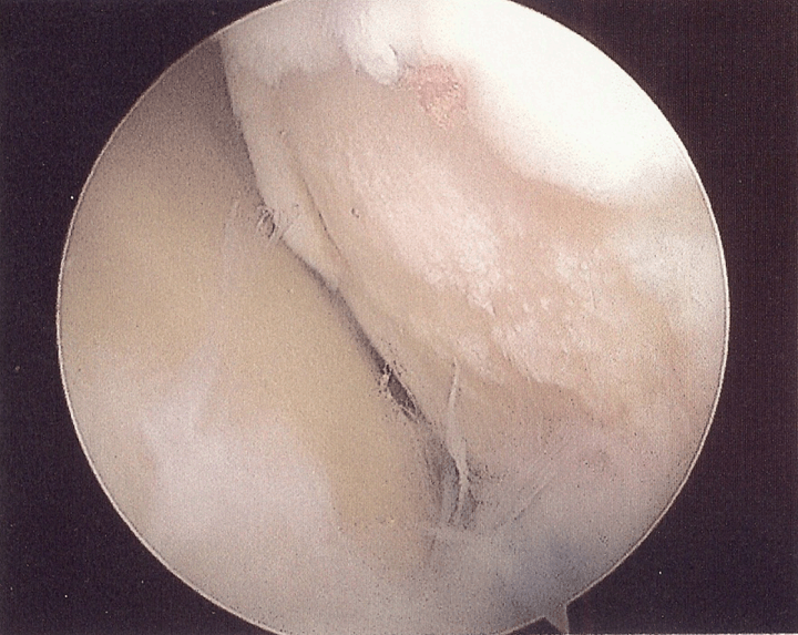
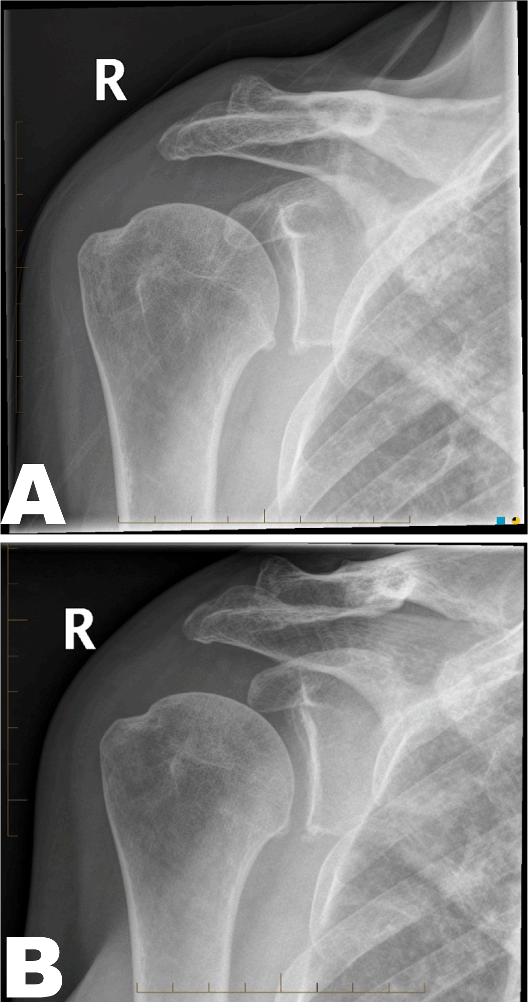
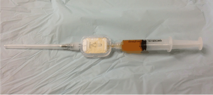
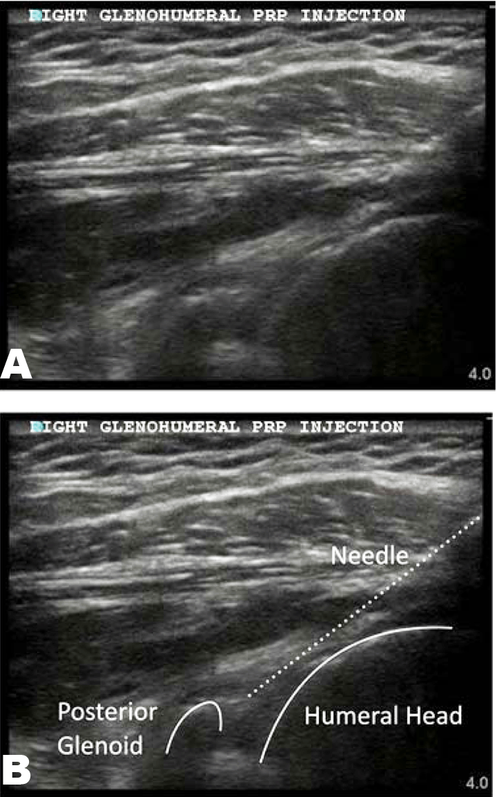
A 62-year-old female patient was presented to a Sports and Exercise Medicine Clinic with chronic and progressively worsening right shoulder pain. She had a past history of right shoulder arthroscopy four years ago and at the time a large humeral head chondral lesion was noted (Figure 1). She was taking paracetamol regularly though was unable to tolerate anti-inflammatories as she had a history of gastritis. She was otherwise fit and healthy and took no other regular medications. The patient was very active and enjoyed hiking but despite regular use of simple analgesics now found that her shoulder pain limited her ability to perform even simple activities of daily living. She also suffered from increasing night pain. Clinical examination showed tenderness over her acromio-clavicular joint and limited active range of motion (ROM) secondary to pain. Passive ROM was marginally limited. Rotator cuff testing indicated good strength and nil pain on resisted movements. Cervical spine examination was normal. Radiological examination confirmed glenohumeral osteoarthritis with inferior humeral head and glenoid osteophyte formation (Figure 2A). The patient also had degenerative changes of her acromioclavicular joint. Formal ultrasonography examination showed no evidence of rotator cuff pathology or subacromial bursitis. To determine the relevance of her glenohumeral or acromioclavicular joint degenerative changes to her pain, an ultrasound guided diagnostic local anesthetic block of her glenohumeral joint was performed. This resulted in complete resolution of her pain for a limited period of 8 hours and confirmed that her pain was primarily related to her glenohumeral joint OA. The patient sought advice on possible additional treatments for her shoulder arthritis and made specific enquiries regarding platelet-rich plasma intra-articular injections as her son had good but limited response to PRP for his bilateral knee OA. Written information and education was provided regarding PRP and its current use within osteoarthritis, including relevant alternatives and possible risks involved. Formal written consent was obtained prior to commencement of PRP therapy. Investigations Formal ultrasonography was performed to assess for rotator cuff and subacromial pathology. Differential Diagnosis Treatment Numerical pain rating scale (NPRS) was recorded prior to each injection, with follow-up intervals occurring at week 3rd, 4th, 8th, 12th, 31st and 42nd. The DASH score was recorded prior to the first injection and at weeks 8th, 12th, 31st and 42nd. Percentage perceived improvement was recorded at 42nd week. Prior to commencing PRP and despite maximal conservative management the patient had a pretreatment baseline NPRS of six out of ten. Her pretreatment DASH score of 65 out of 100 (100 being maximal disability) suggested significant disability. Photoactivated Platelet-rich Plasma Preparation Twenty five and a half milliliters of autologous blood was taken from the study participant via 3x8.5 mL BD vacutainers (BD, Franklin Lakes, NJ, USA) containing ACD (trisodium citrate 22.0g/L, citric acid, 8.0 g/L, and dextrose 24.5 g/L) to prevent clotting. The tubes were centrifuged at 1000 rpm for 10 min resulting in separation of the whole blood constituents with a platelet-poor plasma (PPP) layer, a middle buffy coat layer (high in platelets and leukocytes) and a red blood cell layer. Platelet-poor plasma (PPP) and the buffy coat was withdrawn from each tube to the level of the red blood cell layer and placed in a single sterile vacutainer (BD, Franklin Lakes, NJ, USA) which was re-centrifuged at 3500 rpm for 3 min resulting in the formation of a platelet plug and PPP. PPP was withdrawn to 40 mm and discarded. The remaining PPP and platelet plug were re-constituted using gentle manual agitation and then exposed to photo-activation for 10 minutes. Photoactivation was achieved using the commercial Adi-Light 2 device (AdiStem Ltd, Hong Kong). Four milliliters of photoactivated platelet-rich plasma was drawn up into a syringe which was then connected to a sterile 5-micron filter (GVS, Bologna, Italy) which would result in a leukocyte poor PAPRP preparation upon injection (Figure 3). Injection Method Potential Adverse Effects | ||||||
| ||||||
|
| ||||||
| ||||||
|
| ||||||
|
Results
| ||||||
|
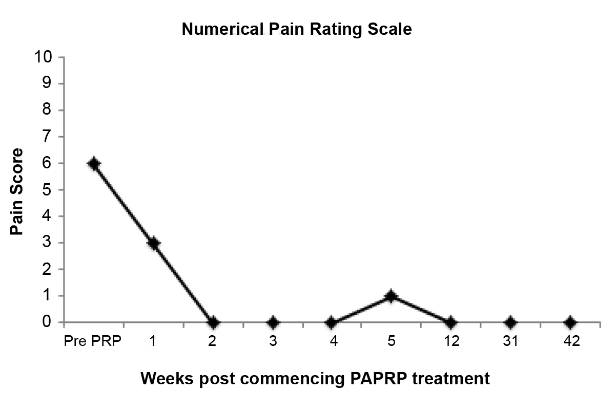
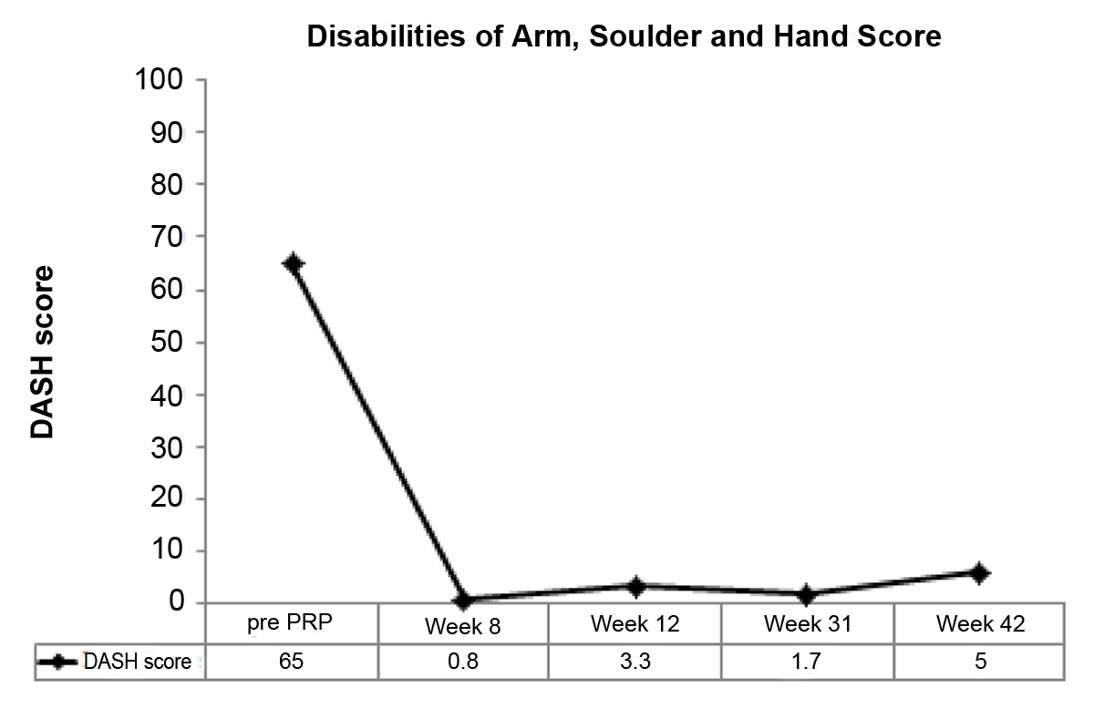
Numerical pain rating scale improved after the first injection of PAPRP and reduced to 0 by 2nd week. NPRS remained between 0 and 1 from week 2nd to final data collection in 42nd week (Figure 5). DASH score at baseline indicated significant functional impairment (DASH score 65 out of 100). At 8th week, the DASH score had improved dramatically and remained improved up to final data collection at 42nd week (Figure 6). At 42nd week following the initial PAPRP injection, a percentage perceived improvement of 90% was reported by the patient. Repeat X-ray examination at 42nd week indicated no radiological progression of glenohumeral osteoarthritis since commencing PAPRP therapy (Figure 2B). The patient noted some marginal discomfort following the second PAPRP injection, however, this was self limiting (2 days). No increase in analgesia was required. Similar short-term discomfort has been observed in other publications following PRP injection [10] [11]. No other complications were noted. | ||||||
| ||||||
| ||||||
|
Discussion
| ||||||
|
This report documents the successful treatment of symptomatic glenohumeral osteoarthritis by photoactivated PRP. Importantly, the patient demonstrated improvement in all recorded treatment outcome measures. The rapid improved in NPRS at week 1st (7 days post 1st injection) suggests an initial anti-inflammatory and analgesic benefit of PAPRP. The improvement in NPRS was observed to be sustained until completion of data collection at 42nd week. Concurrent results were observed in the DASH score. There is a growing body of published evidence on the use of autologous mediums such as PRP in the treatment of OA [10] [11] [12] [13]. These studies have, however, all been largely limited to the use of PRP in knee osteoarthritis. Most recently MieDan et al. have published the positive results of a trial using PRP in the treatment of ankle talar dome chondral lesions [27]. Our preliminary findings suggest that PAPRP may also have promise in the treatment of symptomatic shoulder OA. In a single case report, PAPRP therapy was associated with improvement in all recorded treatment outcome measures. Further, repeat radiological examination indicated no progression of osteoarthritis up to final data collection at 42nd week. Arthroscopic examination would add scientific value to determine the nature of the change to the patients symptoms though it was ethically unreasonable to perform a repeat arthroscopy on a shoulder without clinical reason to do so. Post-therapy magnetic resonance imaging (MRI) scan was not performed as there was no comparative imaging prior to commencement of PRP. Previous publications on the use of PRP in osteoarthritis have not specifically studied the progression of chondral damage/pathology and this remains an important area of investigation. Whilst radiological examination using X-ray remained unchanged over 42 weeks, it is arguable that the follow-up period is too short to make any conclusions from this. Freitag et al. showed no evidence of chondral pathology change with MRI follow-up in acute chondral injury following PAPRP therapy despite significant improvement in both pain and function [14]. It has been theorized that perhaps PRP may influence overall joint homeostasis, reducing synovial membrane hyperplasia and modulating cytokine levels, thus leading to an improvement in the clinical outcome without affecting the cartilage tissue structure and joint degenerative progression [28]. This has yet to be conclusively shown. Given that PRP may only effect pain and function and not disease progression some may argue that intra-articular corticosteroid injection is a simpler and more cost effective treatment. Evidence, however, suggests that intra-articular corticosteroids achieve good but short- term improvement only [29]. Studies that have followed-up pain and functional improvement from PRP and other biological mediums have suggested improvement between 6–24 months [10] [11] [12] [13] [15]. While recognizing the low level of evidence, the positive results of this limited case report highlight the need to further investigate, through a more structured and appropriately powered randomized controlled trial, the use of PAPRP in the treatment of shoulder osteoarthritis. Depending upon the results of further research, PAPRP may have a role in the non-surgical management of shoulder osteoarthritis. This is a promising development for the patient population who remain symptomatic despite maximal conservative management and who are unsuitable for surgical intervention. It is yet to be shown if PAPRP may have disease modifying properties and be effective in delaying need for joint replacement. | ||||||
|
Conclusion
| ||||||
|
Current conservative treatments for symptomatic shoulder osteoarthritis are limited. There is growing evidence to support the use of autologous platelet-rich plasma therapy in the treatment of knee osteoarthritis and the results of this single case study indicate that platelet-rich plasma (PRP) may similarly have promise as an adjunctive therapy in the treatment of glenohumeral osteoarthritis. Further research is required to determine if PRP has structural disease modifying properties and delays need for glenohumeral joint replacement. | ||||||
|
References
| ||||||
| ||||||

|
[HTML Abstract]
[PDF Full Text]
|
|
Author Contributions
Julien Freitag – Substantial contributions to conception and design, Acquisition of data, Analysis and interpretation of data, Drafting the article, Revising it critically for important intellectual content, Final approval of the version to be published |
|
Guarantor of submission
The corresponding author is the guarantor of submission. |
|
Source of support
None |
|
Conflict of interest
Authors declare no conflict of interest. |
|
Copyright
© 2014 Julien Freitag. This article is distributed under the terms of Creative Commons Attribution License which permits unrestricted use, distribution and reproduction in any medium provided the original author(s) and original publisher are properly credited. Please see the copyright policy on the journal website for more information. |
|
|