| |
|
|
|
Case Series
| ||||||
| Management of osseous defects in aggressive periodontitis: A report of four cases using different techniques | ||||||
| Sangeeta Singh1, Parul Sharma2 | ||||||
|
1MDS (Periodontology), Reader, Department of Dental Surgery, Armed Forces Medical College, Pune, Maharashtra, India.
2MDS (Periodontology), Officer Commanding 12 Air Force Dental Centre C/o 56 APO, India. | ||||||
| ||||||
|
[HTML Abstract]
[PDF Full Text]
[Print This Article]
[Similar article in Pumed] [Similar article in Google Scholar] 
|
| How to cite this article |
| Singh S, Sharma P. Management of osseous defects in aggressive periodontitis: A report of four cases using different techniques. Int J Case Rep Images 2014;5(7):468-473. |
|
Abstract
|
|
Introduction:
Successful treatment of aggressive periodontitis is considered to be dependent on early diagnosis, targeted antimicrobial therapy and modifying the tissue architecture that is conducive to long-term maintenance. Osseous defects present a challenge in periodontal practice and successful treatment depends primarily on selection of the correct technique and materials.
Case Series: This case series presents four cases and a total of eight defects treated with different techniques and materials. One defect was treated using a combination of platelet-rich plasma (PRP) with bone graft substitute, three defects were treated using bone graft substitute with resorbable membrane, one using autogenous bone graft combined with bone graft substitute, one using bone graft substitute alone, one using resection technique and the last one was managed as an endo-perio lesion. The results in all four cases discussed here are satisfactory and have shown long-term stability emphasizing the importance of selection of technique and material. Conclusion: There are different techniques and a variety of newer materials available for elimination of osseous defects. The key to successful management of such defects is correct diagnosis, early intervention and proper selection of technique and materials. | |
|
Keywords:
Aggressive periodontitis, Guided tissue regeneration (GTR), Platelet-rich plasma, Endo-perio lesion
| |
|
Introduction
| ||||||
|
Aggressive periodontitis by definition comprises a group of rare, often severe, rapidly progressing forms of periodontitis often characterized by an early age of clinical manifestation and a distinctive tendency to aggregate in families. The 1999 international classification workshop identified clinical and laboratory features and further sub-classified as aggressive periodontitis into localized and generalized forms [1]. Localized aggressive periodontitis cases generally present isolated osseous defects, which need early intervention to improve prognosis. The regeneration of periodontal tissues lost due to destructive inflammatory process has been the ultimate goal of any periodontal therapy. However, despite the development of a wide range of regenerative surgical techniques, this goal remains difficult if not impossible to achieve. Two techniques with the most successful documentation of periodontal regeneration are osseous grafting and guided tissue regeneration [2]. | ||||||
|
Case Series
| ||||||
|
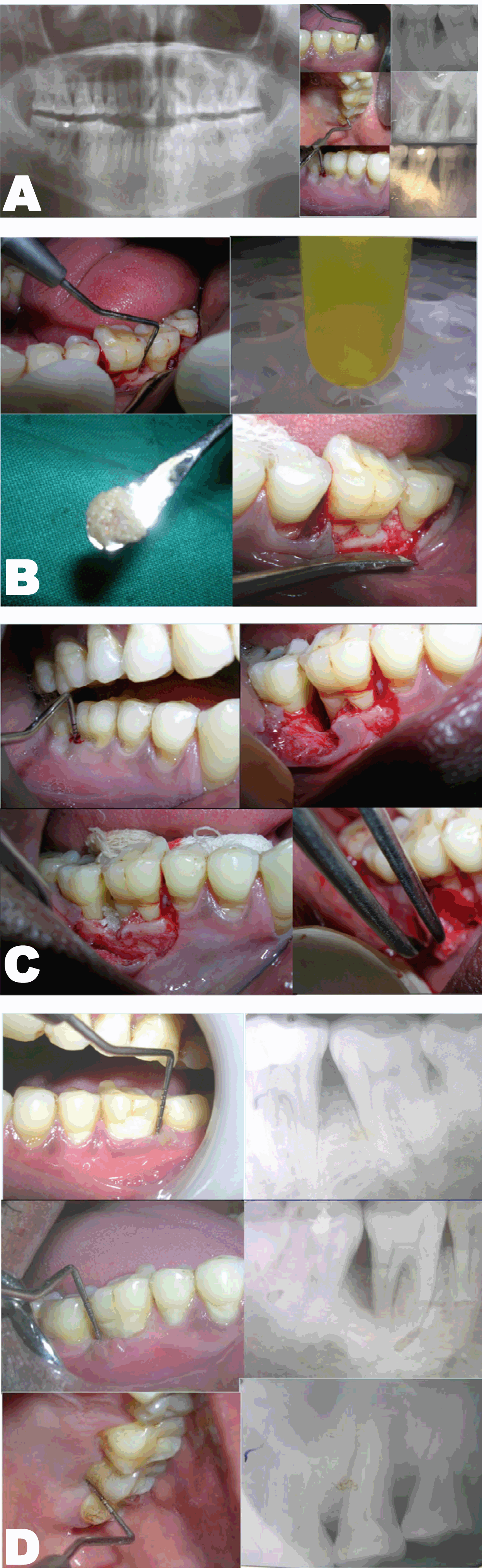
Case 1: A 36-year-old male reported with the chief complaint of pain and bleeding from gums since three months. He also complained of pus discharge from maxillary left last molar since one month. On clinical examination, there were deep pockets > 5 mm in 46, 36 and 27. There was bleeding on probing in all the three teeth involved. The tooth #27 also had Grade II mobility with purulent discharge from the pocket on probing. An orthopantomogram (OPG) revealed infrabony defects on distal aspect of 46 and 36. The tooth #27 had a large periapical radiolucency surrounding the root (Figure 1A). Based on history, clinical findings and radiographic evaluation a diagnosis of Localised Aggressive Periodontitis with an endo-perio lesion in 27 was arrived at. After phase I therapy and endodontic treatment 27, three different procedures were planned to correct the osseous defects. The first surgery was carried out in 36 region. After debridement, bone graft substitute mixed with platelet-rich plasma (PRP) was placed inside the defect (Figure 1B). In the second surgery involving 46, a bone graft substitute was placed which was covered with a resorbable membrane (Figure 1C). In 27, a complete debridement was carried out. | ||||||
|
| ||||||
|
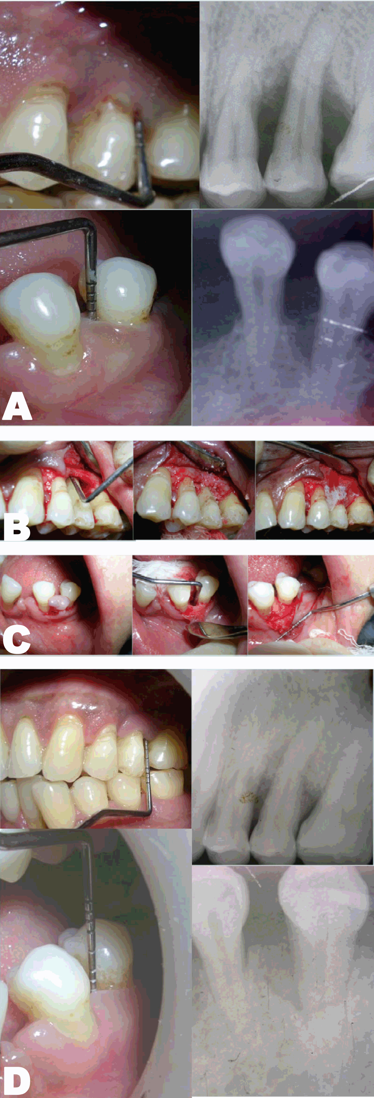
Case 2: A 29-year-old male reported with the chief complaint of pain and bleeding in relation to upper left tooth since one month. He also complained of dull pain in lower left tooth since three months. Clinically, there were deep periodontal pockets on both mesial and distal aspects of 25 and mesial aspect of 35. Radiographic picture showed osseous defects on mesial and distal aspects of 25 and on mesial aspect of 35 (Figure 2A). A diagnosis of localized aggressive periodontitis was made taking into consideration his positive family history, clinical evaluation and X-rays. After completing phase therapy, surgeries were planned for 25 and 35 using a bone graft substitute in combination with a resorbable membrane (Figure 2B-C). | ||||||
|
| ||||||
|
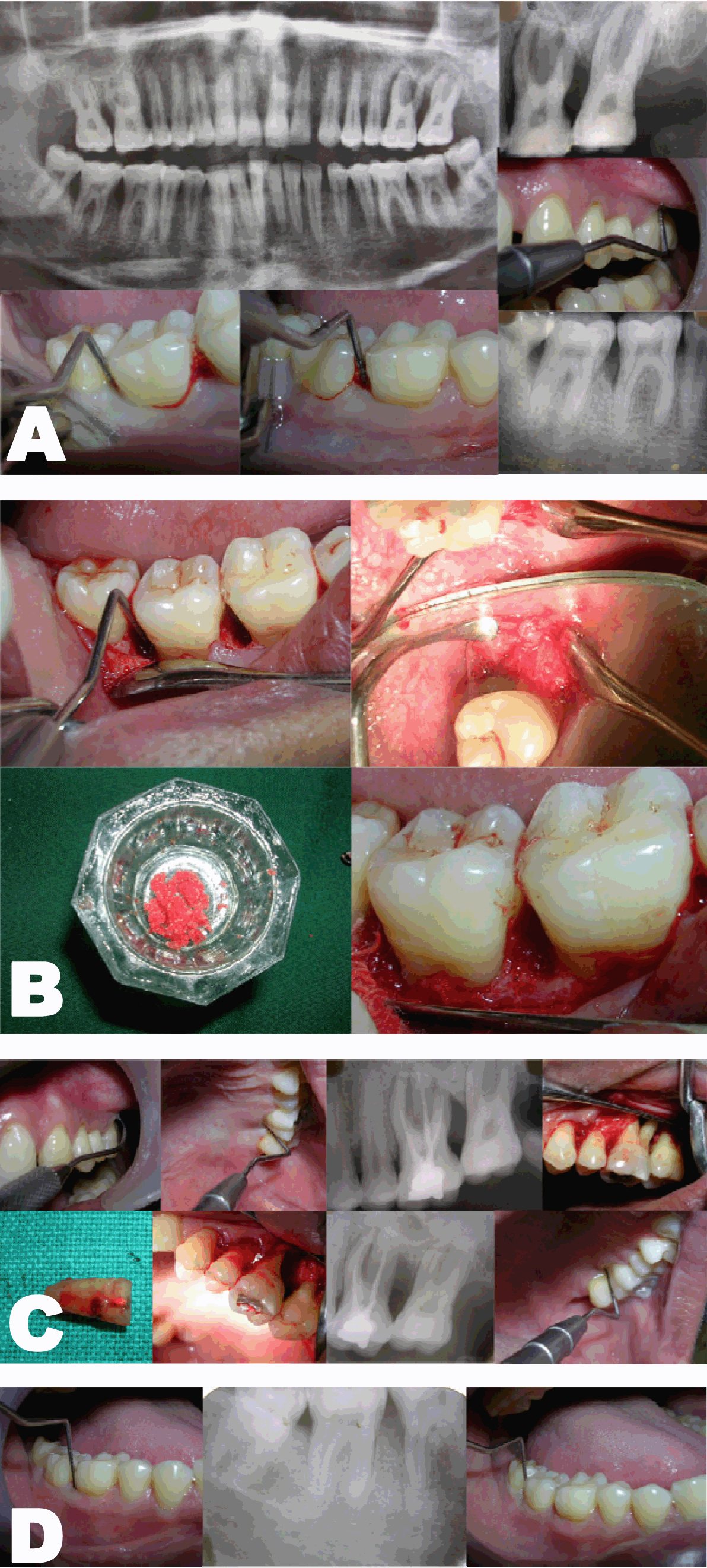
Case 3: A 34-year-old male reported with the chief complaint of pain, bleeding and pus discharge in relation to upper left tooth since two months. He also complained of bleeding from gums in lower posteriors since one year. Clinically, there was bleeding and pus discharge on probing from 26 region. There was a deep periodontal pocket and Grade III furcation involvement. There were pockets > 6 mm on distal aspect of 46, 47 and 36 regions. An OPG revealed infrabony defects on distal aspect of 46, 47 and 36. There was a large radiolucency around the roots of 26 and severe bone loss especially around the distobuccal root of 26 (Figure 3A). After phase I therapy and endodontic treatment in 26, root resective surgery was planned. The distobuccal root was resected out since there was complete absence of bone around it. After thorough debridement, a bone graft substitute was placed and flap sutured (Figure 3B). Further surgeries were planned to eliminate the osseous defects in 46, 47 and 36 regions. In 36, 46 area, the plan was to use autogenous bone chips from maxillary tuberosity mixed with bone graft substitute making a composite graft. After debridement, the defects were filled with this composite graft (Figure 3C). In 36 region, a debridement followed by placement of bone graft substitute was carried out. | ||||||
|
| ||||||
|
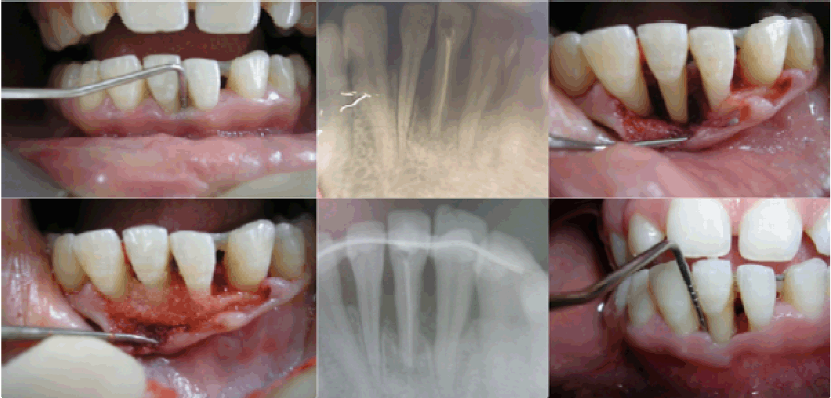
Case 4: A 17-year-old female reported with the chief complaint of pus discharge and mobility in relation to lower anterior tooth since two months. She had first noticed dull ache in the tooth six months back, which subsided after taking medication. Her main concern was loss of tooth. Clinically, there was a deep pocket on both mesial and distal aspects of the tooth # 31. There was bleeding on probing and advanced grade II mobility. Radiograph revealed a significant osseous destruction around 31, 32 and 41. After endodontice therapy I 31, splinting was done to stabilize the tooth and improve healing. After complete debridement a deep intraosseous defect was evident. A bone graft substitute was placed and flap sutured. In the Case 1, clinical as well as radiographic picture at sixth month showed reduction in pocket depth and osseous lesions in 36, 46 and 27 (Figure 1). In Case 2, at the time of evaluation at ninth month postoperative, there was significant decrease in the radiolucency in 25, 35 as well as reduction in the pocket depth (Figure 2). In Case 3, following root resection, 26 was clinically healthy and a radiograph at sixth month revealed reduction in radiolucency around the roots (Figure 3D). In Case 4 involving tooth #31, the tooth appeared healthy with no pain or pus discharge. Radiograph showed some amount of bone fill. | ||||||
| ||||||
|
Discussion
| ||||||
|
Aggressive periodontitis results in a very rapid and severe attachment loss which needs immediate corrective measures to arrest progression and prevent further loss. An infrabony defect results when the junctional epithelium is apical to the alveolar crest. These kind of defects are frequently found in areas of the mouth where the cortical bone is thick and is separated by large volume of cancellous bone. The most common location of infrabony defects is the mesial interproximal location between maxillary and mandibular second molars. Since the mandible widens anteroposteriorly, the incidence of infrabony defects increases in the posterior region [3]. Most of the defects in the case series presented here were in mandibular molars. Literature supports the concept that periodontal pockets associated with intrabony lesions are at higher risk of disease progression in patients who do not receive timely and systematic periodontal therapy [4]. A periodontally compromised patient is considered to have poor prognosis when there is moderate to advanced bone loss around one or more teeth leading to tooth mobility of grade II or I. Furcation involvement in difficult to maintain areas, doubtful patient cooperation and presence of coexisting systemic or environmental factors also lead to poor prognosis. Poor prognosis depends on a large number of factors that can interact in an unpredictable number of ways. Prognosis of individual tooth is determined after the overall prognosis and is affected by it. In patients with poor prognosis, the following points should be considered:
In the following conditions decision to save the tooth may be taken:
Numerous factors must be considered in the treatment planning process when cases present with advanced periodontal disease. The procedure is selected based the clinical findings and in cases with poor prognosis the ultimate decision must be made considering all the presenting factors in that particular case. With proper management and adequate maintenance even hopeless prognosis teeth can be maintained for a long time. Despite advent of numeraous alloplastic materials, Autogenous grafts continue to be the gold standard among graft materials with documented excellent regenerative potential proven earlier with histologic evidence [5]. In Case 3, the defects in 46, 47 were treated using autogenous graft from maxillary tuberosity and the results achieved were excellent in terms of reduction in probing depth and radiographic evidence of bony fill. Alloplastic bone graft substitutes are widely used treatment options for the correction of osseous defects [6] [7]. In this case series, these have been used in all the defects combining them with either resorbable membranes or platelet-rich plasma (PRP) or autogenous graft. The use of PRP combined with bone graft substitute has given excellent results in treatment of infrabony defects [8]. In Case 1, the tooth #36 was treated using PRP combined with a bone graft substitute. There was significant reduction in probing depth and evidence of bony fill on the radiograph. Endoperio lesions are fairly common conditions that are often difficult to diagnose and if not treated completely can recur. However with proper diagnosis and treatment planning, these lesions can be completely eliminated to give predictable results. At times, a properly done endodontic treatment is sufficient to eliminate the infection. Whenever secondary periodontal involvement exists, it requires periodontal therapy to achieve success. These lesions generally have a vertical osseous defect and periodontal regenerative therapy gives excellent results [8]. In Case 1, tooth #27 had a large endo-perio lesion with Grade II mobility. This tooth could be successfully conserved by a proper endodontic therapy and subsequent complete debridement. Postoperative follow up showed a functional tooth with elimination of periodontal pocket and bony fill evident radiographically. In Case 3, the defect in 26 was very large. There was Grade III furcation involvement and the entire distobuccal root had lost significant bone around it. The tooth was first treated endodontically and then the distobuccal root was resected out. After thorough debridement, a bone graft substitute was placed to fill the defect. In Case #4, the bone loss was quite extensive. The tooth was mobile and required splinting to stabilize it to enhance healing. After debridement, a bone graft substitute was placed to fill the defect. All four cases in this case series had isolated infrabony defects as a result of the extensive destruction associated with Aggressive periodontitis. The regenerative surgeries carried out to eliminate these defects resulted in successfully conserving the teeth with improved overall long term stability. | ||||||
|
Conclusion
| ||||||
|
Aggressive periodontitis is associated with severe and rapid destruction of the attachment apparatus leading to infrabony osseous defects. The treatment plan for these patients must address these defects with the aim of providing a stable environment to prevent recurrence. Newer treatment modalities are being introduced and researched all over the world providing the clinicians with a wide range of options to achieve a more predictable regeneration of the periodontium. | ||||||
|
References
| ||||||
| ||||||
|
[HTML Abstract]
[PDF Full Text]
|
|
Author Contributions:
Sangeeta Singh – Substantial contributions to conception and design, Acquisition of data, Analysis and interpretation of data, Drafting the article, Revising it critically for important intellectual content, Final approval of the version to be published Parul Sharma – Analysis and interpretation of data, Revising it critically for important intellectual content, Final approval of the version to be published |
|
Guarantor of submission
The corresponding author is the guarantor of submission. |
|
Source of support
None |
|
Conflict of interest
Authors declare no conflict of interest. |
|
Copyright
© 2014 Sangeeta Singh et al. This article is distributed under the terms of Creative Commons Attribution License which permits unrestricted use, distribution and reproduction in any medium provided the original author(s) and original publisher are properly credited. Please see the copyright policy on the journal website for more information. |
|
|