| |
|
|
|
Case Report
| ||||||
| Spontaneous spermatic vein thrombosis as a circumstance of discovery of the nutcracker syndrome: An exceptional entity | ||||||
| Faouzi Mallat1, Wissem Hmida1, Khaled Ben Ahmed1, Sarra Mestiri2, Faouzi Mosbah3 | ||||||
|
1MD, Urology Department, Sahloul Hospital, Sousse.
2MD, Pathology Department, Fahat Hached Hospital, Sousse. 3Professor, Urology Department, Sahloul Hospital, Sousse. | ||||||
| ||||||
|
[HTML Abstract]
[PDF Full Text]
[Print This Article]
[Similar article in Pumed] [Similar article in Google Scholar] 
|
| How to cite this article |
| Mallat F, Hmida W, Ahmed KB, Mestiri S, Mosbah F. Spontaneous spermatic vein thrombosis as a circumstance of discovery of the nutcracker syndrome: An exceptional entity. Int J Case Rep Images 2014;5(7):519–523. |
|
Abstract
|
|
Introduction:
Spontaneous thrombosis of a varicocele is an extremely rare event. Preoperatively, it may be misdiagnosed due to non-specific presentation because of its clinically indistinguishable from many other inguinal conditions.
Case Report: We report a case of left spermatic vein thrombosis extending to the nearby renal vein diagnosed by Doppler ultrasound, in a 28-year-old male. Blood congestion caused by the nutcracker syndrome, which was incidentally found by computed tomography angiography, may therefore have contributed to the pathogenesis of the thrombosis in our patient. After undergoing surgical treatment (complete resection of the spermatic vein thrombosis), our patient is asymptomatic, with complete resolution of his pathology. Watchful observation was our option for the nutcracker syndrome. Conclusion: The diagnosis of spontaneous thrombosis of varicocele is difficult and it may be misdiagnosed before ultrasound examination, and should be considered in patients without usual risk factors of thrombosis vein. Our purpose is to raise clinician's awareness for this condition so that they will be more likely to diagnose it. Our recommendation is: before classification of the left spermatic vein thrombosis as spontaneous in the absence of the usual risk factors for thrombosis, nutcracker syndrome should be sought especially among young males. | |
|
Keywords:
Varicocele, Thrombosis, Nutcracker syndrome
| |
|
Introduction
| ||||||
|
Spontaneous thrombosis of a varicocele is a rare event; approximately less than 25 cases have been reported in literature. Herein, we present a new case of spontaneous thrombosis of a varicocele in which pathogenesis is contributed to the blood congestion caused by the nutcracker syndrome. To the best of our knowledge, we present the first thromboembolic complication of this syndrome in literature, this report is the first to describe the spermatic vein thrombosis extending to the nearby renal vein as a circumstance of discovery of the nutcracker syndrome. | ||||||
|
Case Report
| ||||||
|
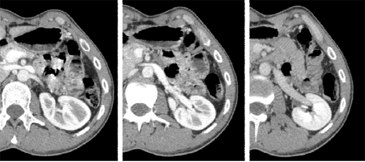
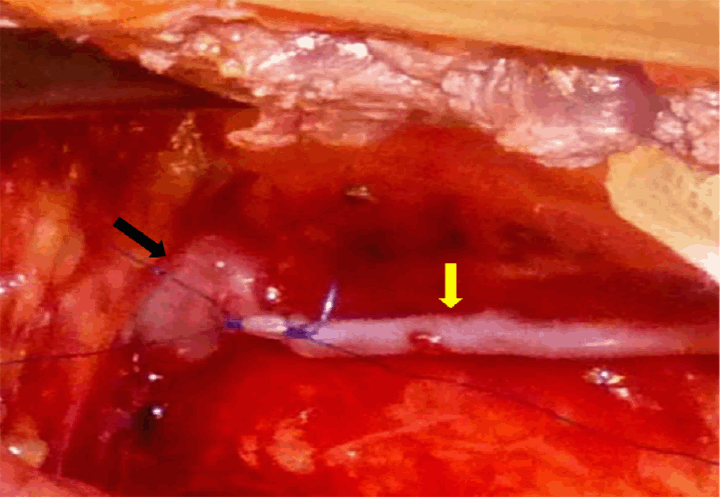
A 28-year-old male, with no previous major health problems, was presented to our department for an induration in the left inguinal region with tenderness associated with left abdominal and testicular pain. The condition had started 14 days before with moderate-in-severity dull, aching pain in the left groin and left testis. Our patient denied any history of left inguinal swellings or hernia, and has no history of surgeries, chronic illness, allergy, or trauma. He did not present any thrombogenic factors. Physical examination of the patient revealed a height of 182 cm and weight of 58 kg with a lower body mass index at 17.5, his blood pressure was 135/75 mmHg, and was significant only for tender, movable and firm to hard 7.0 cm mass extending from the left scrotum to the external inguinal region. The testes, prostate and contralateral scrotum were normal to palpation. Initial laboratory reports were within normal limits. Doppler ultrasonography (Figure 1) confirmed the diagnosis of left thrombosed varicocele with complete cessation of blood flow in the left spermatic vein. Computed tomography angiography (CTA) revealed left spermatic vein thrombosis starting at the external inguinal ring and extending retroperitoneally to the nearby renal vein (Figure 2). The CTA also demonstrated a compression of the left renal vein between the aortic-mesenteric space, the angle between aorta and superior mesenteric artery was approximately 11°. The postero-anterior diameter of the hilar portion of the LRV was 9.8 mm and that of the aortico-mesenteric stenotic portion was 2.1 mm; suggesting anterior nutcracker syndrome (Figure 3) and (Figure 4). When we asked the patient again, he reported an intermittent macroscopic hematuria, and chronic left lumbar pain aggravated by physical activity; associated with systemic signs dominated by intermittent fatigue and headache; which lasted for his childhood. Repeated laboratory tests revealed hemoglobin 12.0 mg/dL, microscopic hematuria, but 24-hour urine collection analysis did not show proteinuria. Based on clinical presentation, urinary and systemic symptoms, laboratory reports, Doppler ultrasonography and computed tomography angiography finding, a diagnosis of anterior nutcracker syndrome leading to total left spermatic vein thrombosis was confirmed. Surgical intervention was decided, and the patient consented to. Left lumbar incision was done. There was a firm-to-hard swelling inside the dilated spermatic vein. Careful dissection and complete excision of the left spermatic vein was performed (Figure 5). All the veins draining into the left renal vein were ligated and bisected. The patient had an uneventful postoperative hospital course and was discharged from the hospital four days later. With eight months follow-up, the patient is doing well. The patient remained asymptomatic and had returned to his usual life. The varicocele improved significantly after operation. | ||||||
| ||||||
|
| ||||||
|
| ||||||
| ||||||
| ||||||
|
Discussion
| ||||||
|
Spermatic vein thrombosis is a rare entity that is usually associated with a chromogenic background. In a review of literature, less than 25 cases of spontaneous thrombosis of the spermatic vein were found. The evolution of the spermatic vein thrombosis is unpredictable, it may disappear spontaneously [1], and in contrast it may be complicated by pulmonary embolism [2]. Three broad categories of factors, known as Virchow's triad, contribute to thrombosis: blood stasis, coagulation factors and mural factors. Our case had no mural factors or prothrombotic coagulation factors. The nutcracker syndrome causing blood congestion may explain the pathogenesis in our present case. The compression of the left renal vein in the aortico-mesenteric space is known as anterior nutcracker syndrome, and is responsible for the development of venous varicosities in the renal pelvis, ureter, and the gonadal vein [3]. Posterior nutcracker syndrome or entrapment of the left renal vein between the aorta and the vertebra is not rare [4] [5] . However, only a few cases showing clinical symptoms with this anomaly have been reported; and may lead to several urological problems such as varicocele, hematuria, and ureteropelvic junction obstruction [4] [5]. The extrinsic left renal vein (LRV) compression causes an impeded outflow from the LRV into the inferior vena cava, and is accompanied by LRV hypertension. Some authors contend that LRV hypertension is the usual cause of varicoceles; and it was admitted that the LRV was compressed in 50–100% of all patients with varicocele [3] [6] [7]. The pathogenesis of the varicocele thrombosis and its extension to the nearby renal vein, in our case, is explained by the chronic blood congestion in the left spermatic vein. Especially, when the diagnosis of NCS is commonly delayed. In this case, we present the first thromboembolic complication of this syndrome in literature. To our knowledge, this report is the first to describe the spermatic vein thrombosis extending to the nearby renal vein as a circumstance of discovery of the nutcracker syndrome. The higher sensitivity and specificity of Doppler examination, as well as its low cost and non-invasiveness, make this the procedure of choice in the diagnosis of the varicocele thrombosis and may have a role in differentiating this condition in men presenting with a history of acute idiopathic left inguinal pain; given that spermatic vein thrombosis is clinically indistinguishable from many other groin conditions. While CTA may help reveal whether the thrombus extends beyond the external inguinal ring, either externally or internally, and help to find aetiology such as the nutcracker syndrome especially in the young male. The management of the spermatic vein thrombosis [8] [9] [10] [11] [12] [13] [14] [15] [16] is controversial. With regard to its management, Roach et al. recommended conservative management following their experience with a postoperative complication in a patient with bilateral thrombosis which resulted in left orchiectomy despite a satisfactory outcome on the right side with anticoagulation therapy alone [17]. In addition, no case of pulmonary embolism from superficial spermatic vein thrombosis out of external inguinal ring has been reported. In contrast, Castillo et al. reported a case of pulmonary thromboembolism associated with deep-seated spermatic vein thrombosis [2], and recommended spermatic vein ligation to prevent from pulmonary embolism. From these findings, Yoko et al. [1] propose two strategies for the treatment of spontaneous thrombosis of the spermatic vein on the basis of anatomical location. Conservative management, including watchful observation, is acceptable for thrombosis of a peripheral vein localized out of external inguinal ring in the so-called pampiniform plexus. In contrast, surgical excision may prevent pulmonary embolism in deep-seated spermatic vein thrombus inside the external inguinal ring and extending to the nearby renal vein as in the present case. Treatment options of nutcracker syndrome include follow-up, conservative treatment and surgical therapy and is still controversial. The aim of treatment is to decrease LRV hypertension. The procedures include intravascular or extravascular stents and open surgical options include the transposition of left renal vein, transposition of the superior mesenteric artery and renal autotransplantation. These options should be considered only when the nutcracker syndrome was complicated by severe or persistent symptoms, such as severe pain, severe acute or chronic hematuria, not respond to conservative treatment or renal insufficiency [3]. In our case, the surgery was to prevent embolic phenomenon. Watchful waiting/observation was our strategy for the nutcracker syndrome, first because of the possible spontaneous improvement of the syndrome, and secondarily the operation is not denied of severe complications in our young and active patient with intermittent and not severe symptoms, which does not consent to. Possible intervention is considered to relieve the angulation between the aorta and superior mesenteric artery to correct LRV hypertension in case of no improvement or worsening of the symptoms or complications and when the consent of patient will be done. | ||||||
|
Conclusion
| ||||||
|
Thrombosis of varicocele is a difficult clinical diagnosis and requires a high index of suspicion. Doppler ultrasonographic examination is the procedure of choice in the diagnosis of the varicocele thrombosis with higher sensitivity and specificity and may have a role in differentiating this condition in men presenting with a history of acute idiopathic left inguinal pain; given that spermatic vein thrombosis is clinically indistinguishable from many other groin conditions. Computed tomography angiography may help reveal whether the thrombus extends beyond the external inguinal ring, either externally or internally, and help to find aetiology for the spermatic vein thrombosis classified as spontaneous or idiopathic, such as nutcracker syndrome especially in the young male. If properly diagnosed, the extensive spermatic vein thrombosis must be treated surgically. | ||||||
|
Acknowledgements
| ||||||
|
We are grateful to Professor Faouzi Mosbah for his comments on the manuscript. | ||||||
|
References
| ||||||
| ||||||
|
[HTML Abstract]
[PDF Full Text]
|
|
Author Contributions
Faouzi Mallat – Substantial contributions to conception and design, Acquisition of data, Drafting the article, Revising it critically for important intellectual content, Final approval of the version to be published Wissem Hmida – Substantial contributions to conception and design, Acquisition of data, Drafting the article, Revising it critically for important intellectual content, Final approval of the version to be published Khaled Ben Ahmed – Substantial contributions to conception and design, Acquisition of data, Drafting the article, Final approval of the version to be published Nadia Mama – Substantial contributions to conception and design, Drafting the article, revising it critically for important intellectual content, Final approval of the version to be published Faouzi Mosbah – Substantial contributions to conception and design, Acquisition of data, Drafting the article, Revising it critically for important intellectual content, Final approval of the version to be published |
|
Guarantor of submission
The corresponding author is the guarantor of submission. |
|
Source of support
None |
|
Conflict of interest
Authors declare no conflict of interest. |
|
Copyright
© 2014 Faouzi Mallat et al. This article is distributed under the terms of Creative Commons Attribution License which permits unrestricted use, distribution and reproduction in any medium provided the original author(s) and original publisher are properly credited. Please see the copyright policy on the journal website for more information. |
|
|