| |
|
|
|
Case Report
| ||||||
| Endometrial stromal sarcoma: Where only rarity prevails over obvious presentation | ||||||
| Vijayata Sangwan1, Mukesh Kumar Sangwan1, Sunita Siwach1, Amrita Duhan1, Rajeev Mahendroo1, Richa Kansal1 | ||||||
|
Assistant Professor, Department of Obst. & Gynae, B.P.S Govt. Medical College For Women, KhanpurKalan, Sonepat, Haryana 131305, India.
| ||||||
| ||||||
|
[HTML Abstract]
[PDF Full Text]
[Print This Article]
[Similar article in Pumed] [Similar article in Google Scholar] 
|
| How to cite this article |
| Sangwan V, Sangwan MK, Siwach S, Duhan A, Mahendroo R, Singh N. Endometrial stromal sarcoma: Where only rarity prevails over obvious presentation. Int J Case Rep Images 2014;5(6):435–438. |
|
Abstract
|
|
Introduction:
Endometrial stromal sarcoma (ESS) is a rare malignancy arising from the endometrial stroma. The majority (90%) of patients present with abnormal uterine bleeding (AUB) followed by mass per abdomen and pelvic pain. However, rarity of this tumor limits the clinician to consider it even as a differential diagnosis despite of having its typical symptoms and only diagnosed by biopsy as a surprise. On analysis of age of presentation, clinical findings and various diagnostic modalities, endometrial stromal sarcoma (ESS) mimics other disorders such as leiomyoma, leiomyosarcoma and endometrial polyp. The diagnostic dilemma continues even on gross and microscopic features of ESS and above mentioned disorders. The role of immunomarkers like CD10, desmin, h-caldesmon, etc. has evolved for specific diagnosis.
Case Report: A 60-year-old female was presented with a complaint of postmenopausal bleeding underwent hysterectomy with a provisional diagnosis of submucous myoma and histopathological examination revealed this as endometrial stromal sarcoma. Conclusion: Endometrial stromal sarcoma is a rare tumor and a strong clinical suspicion is the essential tool for its early diagnoses and better management. | |
|
Keywords:
Endometrium, Hysterectomy, Leiomyoma
| |
|
Introduction
|
|
In the field of medicine rare diagnosis is rarely correct. Therefore, endometrial stromal sarcoma (ESS) because of its rarity is never thought as a preoperative diagnosis and almost always diagnosed after biopsy of hysterectomy specimen. Endometrial stromal sarcoma is a rare malignant tumor that constitutes about 0.2% of all uterine malignancies and 10–15% of uterine sarcomas [1]. Exposure to tamoxifen, unopposed estrogen and polycystic ovary syndrome (PCOS) are implicated in pathogenesis of ESS [2]. Chromosomal aberration are found associated with ESS, deletion on chromosome 7p being the most common finding (55.6%) [2]. On the basis of mitotic activity, vascular invasion and observed differences in prognosis endometrial stromal tumors are divided into the following three types:
It is frequently misdiagnosed as leiomyoma or endometrial polyp and mostly revealed on histopathological examination postoperatively because of its rarity [4] [5]. |
|
Case Report
|
|
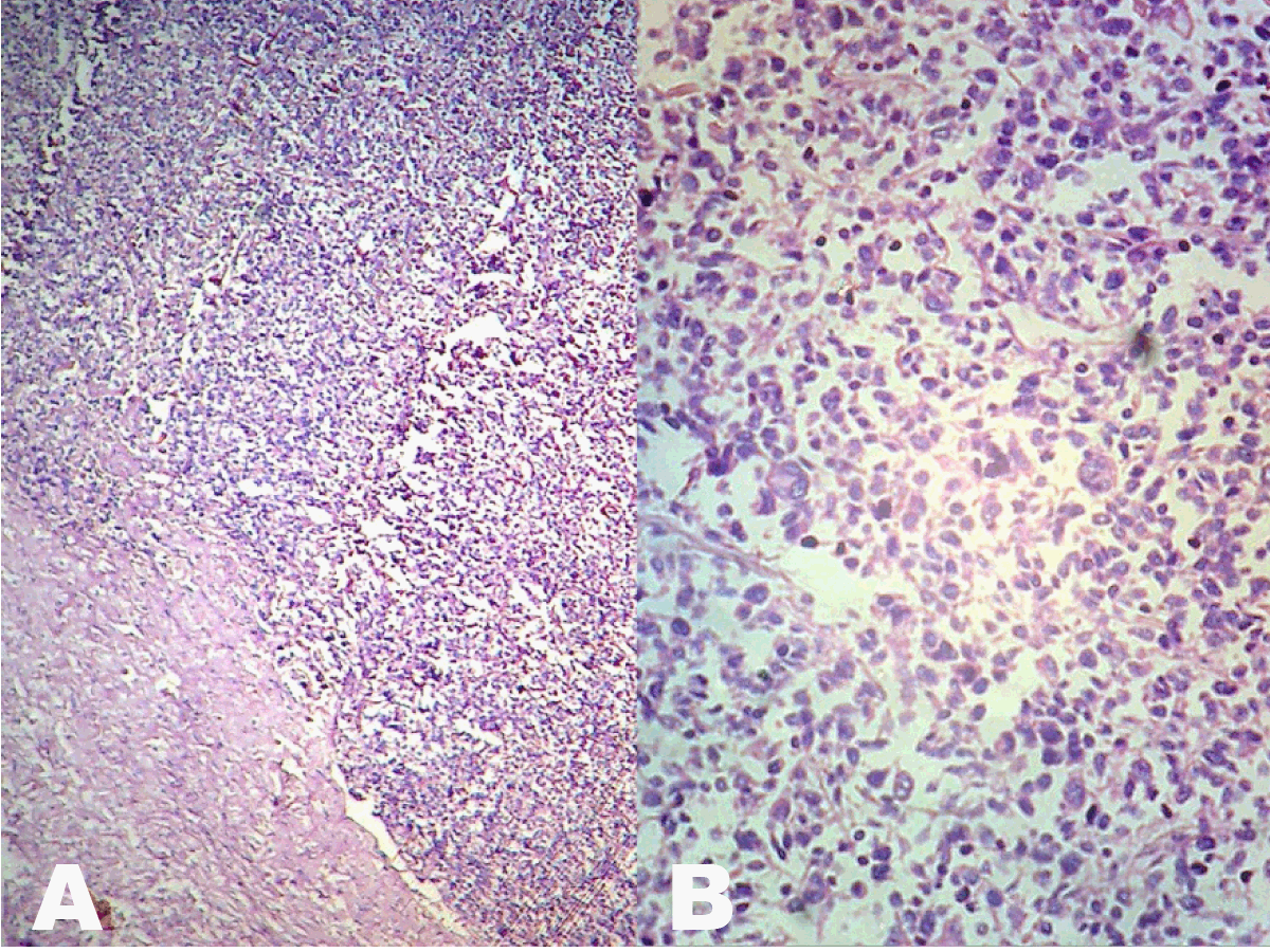
A 60-year-old multiparous (P6) lady presented with bleeding per vaginum for six months after menopause of 12 years. The bleeding was not associated with coitus, trauma or any foreign body insertion. There was no history of use of any hormonal therapy. On local examination external genital, cervix and vagina were healthy, uterus was of menopausal size. Her per rectal examination was also normal. Ultrasonography revealed a well-circumscribed lesion of 2–3 cm size in endometrial cavity suggestive of submucous fibroid. Fractional curettage revealed normal endometrium intermixed with blood clots. Patient was planned for total hysterectomy with bilateral salpingo-oophorectomy, intraoperatively uterus, bilateral tubes and ovaries were normal with no palpable lymph nodes in pelvic area. Cut section of uterus revealed a 3.4-cm size nodule in posterior wall of uterus mimicking a submucous fibroid. On gross examination there was 3.5–4.5 cm well circumscribed nodular growth, grey yellow in color with a necrotic area. Microscopic examination endometrial stromal sarcoma without involving myometrium and uterocervical junction (Figure 1). The patient was discharged on third postoperative day and is on regular follow-up for one year without any complications. |
|
|
|
Discussion
|
|
Endometrial stromal tumors are composed of cells resembling normal endometrial stroma in its proliferative phase. It affects younger age group and the mean age is 42–58 years [2]. However, slightly higher age (60 years) in present case is in accordance with recent literature which may be due to changing trends [4] [5]. Although abnormal uterine bleeding (AUB) is present in 90% cases, and 70% patient present with uterine enlargement, pelvic pain [1] [2] [6]but still is rarely suspected as first diagnosis and common causes of AUB like leiomyoma or endometrial polyp are considered as its etiology. In the present study also submucous fibroid was suspected as diagnosis in repetition with literature [7]. Although endometrial curettage is commonly used diagnostic modality for AUB but it is not sensitive for ESS specially when the lesion is completely embedded in myometrium. Moreover, because of resemblance between ESS and normal endometrial tissue curettage fragments can easily miss it [2]. Ultrasound, specially, transvaginal can be helpful in diagnosing uterine leiomyoma, adenomyosis, endometrial polyps but it is also unreliable in diagnosing ESS. Transvaginal Doppler can play a role in its diagnosed in which ESS has a low impedance flow and hence delineates it from rest of uterine tissue. Magnetic resonance imaging (MRI) scan can also diagnose it by the presence of low intensity bands within the area of myometrial invasion due to worm like permeations of tumor. This can help in preoperative diagnosis of ESS but it is not feasible to get transvaginal Doppler and MRI scans in all the patients of AUB. Abnormal uterine bleeding constitutes a major part of total patients attending Gynecology OPD, besides this lack of clinician suspicion for ESS is also a cause as happened with us in the present case. After all investigations and with a probable diagnosis of submucous fibroid patient underwent total hysterectomy and bilateral salpingo-oophorectomy. Histopathological examination revealed ESS as surprise diagnosis. Non-availability of immunomarkers limited our diagnosing ability. The review of literature also reveals similar case reports [1] [8] [9]. On gross examination ESS usually appears as an irregular mass or nodule involving endometrium or myometrium, along with focal areas of necrosis, hemorrhagic or cystic changes [1] [10]. Treatment includes debulking surgery followed by total hysterectomy with removal of adnexa. As these tumors have a tendency of late recurrence, and distant metastasis [11] long-term follow-up is essential in these tumors. It shall be once in three months for the first year and half-yearly for next four years thereafter annual follow-up is recommended [2] . Our patient is well from last one year. Although preoperative diagnosis of ESS is still a myth rather than reality in view of clinician constraints like poor sensitivity of routine investigations in its diagnosis and rarity of the tumor. But still it is very essential for us to early diagnose it because of its poor prognosis and metastatic potential. Therefore, a strong clinical suspicion is recommended in all cases of AUB for better management [12]. |
|
Conclusion
|
|
In conclusion, through this case report we want to emphasize that no doubt rarity of endometrial stromal sarcoma limits the clinician view but because of its poor prognosis and metastasis it is important for all the clinicians to be suspicious for endometrial stromal sarcoma while working up the case of abnormal uterine bleeding. |
|
References
|
|
|
[HTML Abstract]
[PDF Full Text]
|
|
Author Contributions
Vijayata Sangwan – Substantial contributions to conception and design, Acquisition of data, Analysis and interpretation of data, Drafting the article, Revising it critically for important intellectual content, Final approval of the version to be published Mukesh Kumar Sangwan – Analysis and interpretation of data, Drafting the article, Final approval of the version to be published Sunita Siwach – Analysis and interpretation of data, Drafting the article, Final approval of the version to be published Amrita Duhan – Analysis and interpretation of data, Drafting the article, Final approval of the version to be published Rajeev Mahendroo – Analysis and interpretation of data, Drafting the article, Final approval of the version to be published Richa Kansal – Analysis and interpretation of data, Drafting the article, Final approval of the version to be published |
|
Guarantor of submission
The corresponding author is the guarantor of submission. |
|
Source of support
None |
|
Conflict of interest
Authors declare no conflict of interest. |
|
Copyright
© 2014 Vijayata Sangwan et al. This article is distributed under the terms of Creative Commons Attribution License which permits unrestricted use, distribution and reproduction in any medium provided the original author(s) and original publisher are properly credited. Please see the copyright policy on the journal website for more information. |
|
|
|
About The Authors
| |||
| |||
| |||
| |||
| |||
| |||
| |||