|

|
 |
|
Case Report
| ||||||
| Huge retroperitoneal schwannoma in a young male | ||||||
| Khaled Ben Ahmed1, Faouzi Mallat1, Wissem Hmida1, Sidiya Oueld Chavey2, Amel Ben Abdallah2, Kalthoum Tlili2 | ||||||
|
1Urology Department, Sahloul Hospital, Sousse.
2Radiology Department, Farhat Hached Hospital, Sousse. | ||||||
| ||||||
|
[HTML Abstract]
[PDF Full Text]
[Print This Article]
[Similar article in Pumed] [Similar article in Google Scholar] 
|


| How to cite this article |
| Ahmed KB, Mallat F, Hmida W, Chavey SO, Abdallah AB, Tlili K. Huge retroperitoneal schwannoma in a young male. Int J Case Rep Images 2014;5(6):431–434. |
|
Abstract
|
|
Introduction:
Schwannomas are benign tumors mostly found in the head and neck regions. Localization in the retroperitoneal area is rare.
Case Report: We report a case of a 19-year-old male presented to our department complaining of right abdominal pain. An abdominal computed tomography (CT) scan showed a large retroperitoneal mass of 17 cm in diameter. The patient had an ultrasound-guided needle biopsy of the mass and histopathology and immunohistochemistry revealed a schwannoma. Tumor excision was performed through lumbotomy. Histopathological examination confirmed the diagnosis of benign schwannoma. After three years, at follow-up the patient was free of disease. Conclusion: Retroperitoneal schwannoma is a difficult clinical diagnosis and requires a high index of suspicion. Magnetic resonance imaging scan may help in the diagnosis of schwannoma and diagnosis is based on histopathologic examination and immunohistochemistry. Total excision has a therapeutic effect and a good prognosis. | |
|
Keywords:
Retroperitoneal, Schwannoma, Total excision
| |
|
Introduction
| ||||||
|
Schwannoma is a benign neoplasm which arises from Schwann cells of the peripheral nerve sheath. These tumors are mostly found in the head and neck regions or the extremities, schwannomas were rarely seen in the retroperitoneal and comprise only 3% of all schwan-nomas (malignant and benign combined). Retroperitoneal schwannomas are the most of time larger and have a higher tendency to be complicated by spontaneous degeneration and hemorrhage. [1] Herein, we present a case of a 19-year-old male with retroperitoneal tumor diagnosed as schwannoma. | ||||||
|
Case Report
| ||||||
|
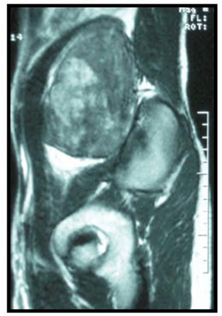
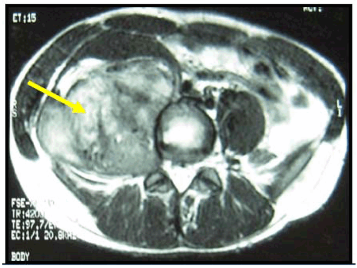
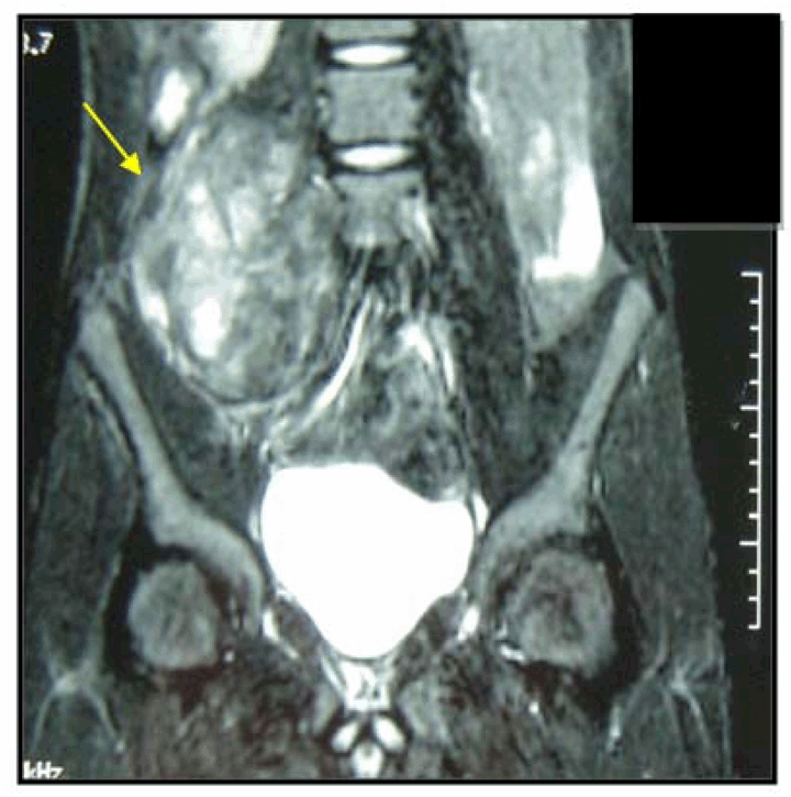
A 19-year-old male with no previous major health problems, presented to our department complaining of right abdominal pain. The condition had started three months before. The physical examination revealed an evident mass in the right upper abdomen. He denied any fever, anorexia, asthenia or weight loss. This pain was non-radiating and did not respond to over the counter analgesics. His initial laboratory reports were within normal limits. Computed tomography (CT) scan revealed a 17-cm heterogeneous retroperitoneal solid mass located on the abdominal aorta and closely attached to the posterior wall of the vertebra. The tumor was surrounded by major abdominal blood vessels, naming the portal vein and its principal tributary, celiac trunk and its principal's branches. Magnetic resonance imaging (MRI) scan of the abdomen showed a hypointense mass on T1, 17x11x8 cm in diameter, in the right para-aortic region, (Figure 1) heterogeneously hyperintense on T2 (Figure 2) and widened paravertebral foramen L4-L5 in the same regions on MRI (Figure 3). The patient had an ultrasound-guided needle biopsy of the mass. Microscopic examination of the biopsy fragments showed spindle cells with blunt nuclei in a fascicular pattern and areas of nuclear atypical cells were noted. The mass was diagnosed pathologically as retroperitoneal schwannoma and confirmed with immunohistochemical examination (positive for S100, negative AML). A right lumbotomy was performed. A large retroperitoneal tumor was found, surrounded by the major abdominal blood vessels such as the MRI results. A bloc resection of the tumor was performed. The patient was discharged 10 days after surgery. Histopathologic examination of the mass confirms a benign schwannoma. After three years of follow-up, the patient was free of disease. | ||||||
| ||||||
| ||||||
| ||||||
|
Discussion
| ||||||
|
Generally, schwannomas are benign tumors that develop from Schwann cells of the peripheral nerve sheath derived from the neuro ectoderm. Retroperitoneal schwannomas are rare, accounting for only 1.2% of all retroperitoneal tumors. [2] These tumors are frequently seen in adult population between the ages of 20 and 50. Women are affected twice as often as men. [3] [4] The symptomatology is nonspecific and depends on the location and size of the lesion, cause of the slowly growth and the anatomical position of the tumor, that is why the diagnosis of retroperitoneal schwannomas is often delayed. Neurologic symptoms are rare. A few cases also present with abdominal complaints or lower back pain such as our patient. [5] MRI is considered as the diagnostic modality of choice in the evaluation of schwannomas, target sign and fascicular sign are two characters of schwannomas showed by MRI, but these typical signs are rare. Generally, MRI showed schwannomas as hypointense on T1 and hyperintense on T2, calcification can be seen in 23% [6], such as the MRI of our patient. The differential diagnosis for retroperitoneal schwannomas includes other neurogenic tumors such as paraganglioma, neuroblastoma and pheochromocytoma. The treatment of schwannomas is only surgical, since, schwannomas are radiotherapy and chemotherapy resistant [7]. However, the necessity for negative soft tissue margins is controversial especially when adjacent tissue or viscera need to be sacrificed. The prognosis of benign schwannomas is good and the most frequent complication is recurrence of the tumor, probably due to incomplete excision, which is reported in 5–10% cases [8] our patient was free of disease after three years of follow-up. | ||||||
|
Conclusion
| ||||||
|
Retroperitoneal schwannoma is a difficult clinical diagnosis and requires a high index of suspicion. Magnetic resonance imaging scan may help in the diagnosis of schwannoma and diagnosis is based on histopathologic examination and immunohistochemistry. Total excision has a therapeutic effect and a good prognosis. | ||||||
|
References
| ||||||
| ||||||

|
[HTML Abstract]
[PDF Full Text]
|
|
Author Contributions
Khaled Ben Ahmed – Substantial contributions to conception and design, Acquisition of data, Drafting the article, Revising it critically for important intellectual content, Final approval of the version to be published Faouzi Mallat – Substantial contributions to conception and design, Acquisition of data, Drafting the article, Final approval of the version to be published Wissem Hmida – Substantial contributions to conception and design, Acquisition of data, Drafting the article, Final approval of the version to be published Sidiya Oueld Chavey – Substantial contributions to conception and design, Drafting the article, Revising it critically for important intellectual content, Final approval of the version to be published Amel Ben Abdallah – Substantial contributions to conception and design, Acquisition of data, Drafting the article, Revising it critically for important intellectual content, Final approval of the version to be published Kalthoum Tlili – Substantial contributions to conception and design, Acquisition of data, Drafting the article, Revising it critically for important intellectual content, Final approval of the version to be published |
|
Guarantor of submission
The corresponding author is the guarantor of submission. |
|
Source of support
None |
|
Conflict of interest
Authors declare no conflict of interest. |
|
Copyright
© 2014 Khaled Ben Ahmed et al. This article is distributed under the terms of Creative Commons Attribution License which permits unrestricted use, distribution and reproduction in any medium provided the original author(s) and original publisher are properly credited. Please see the copyright policy on the journal website for more information. |
|
|
|
About The Authors
| |||
| |||