| |
|
|
|
Clinical Image
| ||||||
| Sternoclavicular hyperostosis | ||||||
| Michael T Flannery1, Kara F Villarreal AA2 | ||||||
|
1MD, FACP, Professor of Medicine, 12901 Bruce B Downs Blvd. MDC Box 19 Room L1041, Tampa, Fl.
2Pasco-Hernando Community College of Nursing. | ||||||
| ||||||
|
[HTML Abstract]
[PDF Full Text]
[Print This Article]
[Similar article in Pumed] [Similar article in Google Scholar] 
|
| How to cite this article |
| Flannery MT, Kara F Villarreal AA. Sternoclavicular hyperostosis. Int J Case Rep Images 2014;5(6):459–461. |
|
Case Report
| ||||||
|
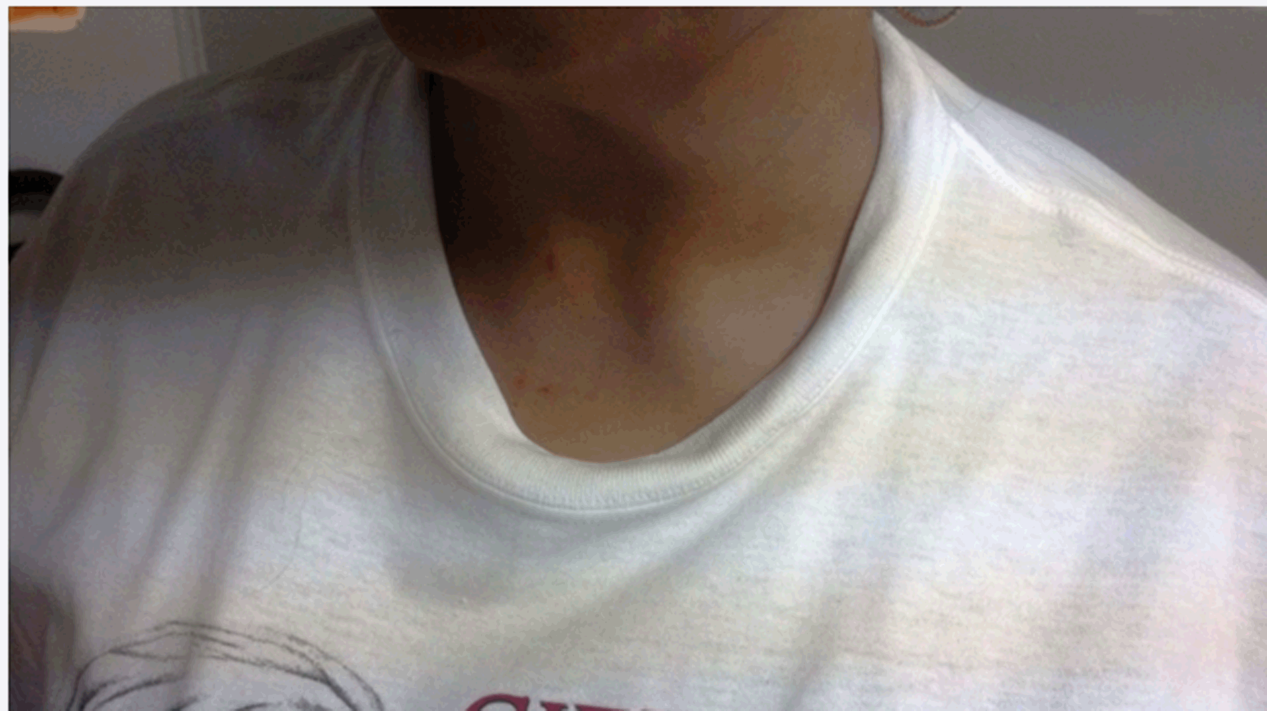
Our patient was a 48-year-old white female with a several month history of a non-enlarging, mildly tender 3×3 cm mass of the medial clavicle and sternum (Figure 1) (Figure 2) with arrow. She had a history of moderately controlled hypertension and poorly controlled hypertriglyceridemia. Medications included: lisinopril 20 mg, hydrochlorothiazide 12.5 mg and fenofibrate 145 mg daily. Tramadol 50 mg every eight hours was used for pain as needed. There was no history of trauma, erythema, fever, skin lesions or any other bone pain. Physical examination confirmed a solid mass with minimal tenderness and no lymphadenopathy of cervical, axillary or inguinal regions. A magnetic resonance imaging study demonstrated no signs of infection or tumor but there was ossification of the sternocostoclavicular ligaments with loss of the inferior margin of the clavicle. Laboratory studies were normal including complete blood counts, complete metabolic profile, serum and urine protein electrophoresis, quantitative immunoglobulins and an erythrocyte sedimentation rate. Based on the classic findings on exam, with negative laboratory studies, classic imaging without infection or tumor the diagnosis of sternoclavicular hyperostosis was suggested. Follow-up to date has demonstrated no changes in size or symptoms. | ||||||
| ||||||
|
Discussion
| ||||||
|
Sternoclavicular hyperostosis is described as bony overgrowth with soft tissue ossification of the medial clavicle, upper ribs, and sternum [1]. It occurs in both sexes with a broad age range from 11 to 88 years. In a review of 251 cases 139 were Japanese and 114 Caucasian [2]. The most common symptom is pain and while the etiology is unknown, SH usually starts with nonspecific inflammation of the sternoclavicular ligaments which may lead to chronic inflammation with progressive hyperostosis and soft tissue ossification. The differential includes osteitis condensans, aseptic necrosis, Tietze's syndrome, osteoarthritis, spontaneous subluxation, osteomyelitis and tumors such as osteosarcoma, metastatic breast and prostate cancer, thyroid cancer or extension from a Pancoast tumor of the lung. Biopsy is planned if further enlargement occurs. Approximately 50% of patients with SH have skin lesions such as palmoplantar pustulosis, pustular psoriasis, hidradenitis suppurativa and psoriasis vulgaris [3]. Patient's may be treated with non-steroidal anti-inflammatory drugs (NSAID's) for relief if needed. Progression can be monitored over time for increased size or symptoms that may justify biopsy or repeat imaging. | ||||||
|
Conclusion
| ||||||
|
Sternoclavicular hyperostosis Sternoclavicular hyperostosis is a relatively rarely described process in literature. It is important to recognize this process as a low likelihood of cancer and infection given the patient's presentation, therefore, avoiding an extensive work-up. However, if further growth occurs over time then a biopsy via a fine needle aspiration of the lesion may be warranted along with additional imaging. | ||||||
|
References
| ||||||
| ||||||
|
[HTML Abstract]
[PDF Full Text]
|
|
Author Contributions
Michael T Flannery – Substantial contributions to conception and design, Acquisition of data, Analysis and interpretation of data, Drafting the article, Revising it critically for important intellectual content, Final approval of the version to be published Kara F Villarreal AA – Analysis and interpretation of data, Drafting the article, Revising it critically for important intellectual content, Final approval of the version to be published |
|
Guarantor of submission
The corresponding author is the guarantor of submission. |
|
Source of support
None |
|
Conflict of interest
Authors declare no conflict of interest. |
|
Copyright
© 2014 Michael T Flannery et al. This article is distributed under the terms of Creative Commons Attribution License which permits unrestricted use, distribution and reproduction in any medium provided the original author(s) and original publisher are properly credited. Please see the copyright policy on the journal website for more information. |
|
|
|
About The Authors
| |||
| |||
| |||