  |

|
 |
|
Clinical Image
| ||||||
| Gallstone ileus in an already cholecystectomized patient | ||||||
| Andrea Rossetti1, Nicolas Christian Buchs2, Vincent Ott2, Philippe Morel2 | ||||||
|
1Clinic for Visceral Surgery and Transplantation, Department of Surgery, University Hospital of Geneva, Switzerland, Clinic for Surgery, Kantonspital St. Gallen, Switzerland.
2Clinic for Visceral Surgery and Transplantation, Department of Surgery, University Hospital of Geneva, Switzerland. | ||||||
| ||||||
|
[HTML Abstract]
[PDF Full Text]
[Print This Article]
[Similar article in Pumed] [Similar article in Google Scholar] 
|


| How to cite this article |
| Rossetti A, Buchs NC, Ott V, Morel P. Gallstone ileus in an already cholecystectomized patient. Int J Case Rep Images 2014;5(6):453–455. |
|
Case Report
| ||||||
|
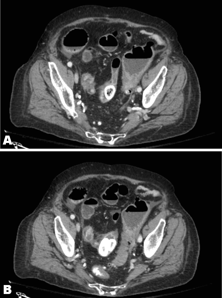
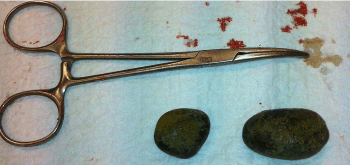
A 91-year-old female was admitted to the emergency department with a three-day history of abdominal pain and vomiting. She reported having similar but lower in intensity episodes in the past. Her past medical history was significant for an open cholecystectomy, performed many years ago. No surgical details were available. She also had numerous comorbidities which included type II diabetes, chronic heart failure, and hypertension. On clinical examination, the patient was non-febrile and hemodynamically stable. Tenderness of lowers quadrants and increased bowel sounds were also reported. Blood tests of the patient showed increase inflammatory parameters, (PCR 67 and with cells count 19.600). Computed tomography (CT) scan showed an image compatible with a foreign body localized in the distal portion of small bowel, not far away from the ileocecal valve (Figure 1A-B). Due to the importance and the persistence of clinical symptoms, we decided to perform an exploratory laparotomy. Finally, two gallstones (3x5 cm and 2x3 cm) (Figure 2) were found 10 cm from the ileocecal valve. An intestinal resection was then performed with direct anastomosis. The postoperative course was remarkable for heart failure with respiratory distress and pulmonary infection. The length of stay was 30 days. | ||||||
| ||||||
|
| ||||||
| ||||||
|
Discussion
| ||||||
|
Gallstone ileus is a rare complication of cholecystitis that occurs in 0.5% of cases [1]. It usually occurs because of a cholecystoenteric fistula, allowing the passage of a large stone in the intestine [2] [3]. It can happen after sphincterotomy as well, but remains an uncommon complication. Unfortunately in our patient, we had no operative details of the previous cholecystectomy, performed many years ago. While uncommon in general population, in patients older than 65 years, it is the cause of 25% of all non-strangulated small bowel obstruction [1]. In the past, the mortality rate due to gallstone ileus was nearly 50%, but in recent years has improved to about 8–18% [1] [3] [4] [5]. This relatively high morality is related to the advanced age of the typical patient. Moreover, in a series of 37 patients showed that 62% of patients had serious concomitant diseases [3]. In addition, the diagnosis can be challenging. At admission, it is usually missed (in up to 54% of patients) and the diagnosis is mostly made during surgery [1] [3] [4] [5] [6]. A plain abdominal radiography can show a calcified mass with obstructive signs. Classically, the Rigler's triad is the presence of an aerobilia, a mechanical bowel obstruction and an ectopic gallstone [2]. In case of doubt, a computed tomography is usually performed. It confirms the diagnosis in most of patients, as it was our case. Our patient had already had a cholecystectomy, the classical triad was not present. Yet, the other signs were clearly noted. The location of the obstruction is classical as well. The distal ileon is the most frequent site involved [3] [6] [7]. The presence of several gallstones is not uncommon as well. In 16% of cases, more than one stone can be found [3]. Concerning the management, surgery is required to relieve the obstruction. The removal of impacted stone is mandatory, and does not necessitate always an intestinal resection. Yet, in our case, the small bowel showed suffering signs and a resection was considered as the safer option. However, cases of gallstone intestinal obstruction without a biliodigestive fistula have been described [8]. A rare occurrence is the gallstone ileus after lost biliary calculi. In literature, two cases of intestinal obstruction when a "lost" calculus after a difficult laparoscopic cholecystectomy migrated through the jejunal wall are reported [9] [10]. In case of cholecystoenteric fistula, different approaches can be considered. A one-stage procedure consisting of the removal of the impacted stone, fistula repair and cholecystectomy can be performed, but may be associated with a higher complications rate. On the other hand, a two-stage procedure consisting of enterolithotomy followed by elective biliary surgery is usually considered as safer [1] [3]. However, in our case, there was only the impacted stone to treat, since the gallbladder was already removed. It would have been interesting to know exactly what happened during the surgery at that time and whether a fistula was present or not. | ||||||
|
Conclusion
| ||||||
|
In conclusion, a cholecystectomy does not prevent the occurrence of a gallstone ileus. This pathology, even if uncommon, has to be considered when the plain abdominal radiography shows a mechanical obstruction with an ectopic calcified mass. The treatment remains prompt surgery, especially in fragile patients, that constitute majority of the population at risk for this pathology. | ||||||
|
References
| ||||||
| ||||||

|
[HTML Abstract]
[PDF Full Text]
|
|
Author Contributions
Andrea Rossetti – Substantial contributions to conception and design, Acquisition of data, or analysis and interpretation of data, Drafting the article or revising it critically for important intellectual content, Final approval of the version to be published Nicolas Christian Buchs – Substantial contributions to conception and design, Acquisition of data, Analysis and interpretation of data, Drafting the article or revising it critically for important intellectual content, Final approval of the version to be published Vincent Ott – Substantial contributions to conception and design, Acquisition of data, Analysis and interpretation of data, Drafting the article or revising it critically for important intellectual content, Final approval of the version to be published Philippe Morel – Substantial contributions to conception and design, Acquisition of data, or analysis and interpretation of data, Drafting the article or revising it critically for important intellectual content, Final approval of the version to be published |
|
Guarantor of submission
The corresponding author is the guarantor of submission. |
|
Source of support
None |
|
Conflict of interest
Authors declare no conflict of interest. |
|
Copyright
© 2014 Andrea Rossetti et al. This article is distributed under the terms of Creative Commons Attribution License which permits unrestricted use, distribution and reproduction in any medium provided the original author(s) and original publisher are properly credited. Please see the copyright policy on the journal website for more information. |
|
|