  |

|
 |
|
Case in Images
| ||||||
| Gigantic peripheral ossifying fibroma of the mandible: A Venezuelan case report | ||||||
| Grimaldo-Carjevschi M1, Villarroel-Dorrego M2, Vargas F3, Romero Y1 | ||||||
|
1DDS, MSc, Dental School, Universidad Central de Venezuela Caracas, Venezuela.
2DDS, MSc, PhD, Dental School, Universidad Central de Venezuela Caracas, Venezuela. 3DDS, Oral & Maxillofacial postgraduate programme, Universidad Gran Mariscal de Ayacucho. Barcelona, Venezuela. | ||||||
| ||||||
|
[HTML Abstract]
[PDF Full Text]
[Print This Article]
[Similar article in Pumed] [Similar article in Google Scholar] 
|


| How to cite this article |
| Grimaldo-Carjevschi M, Villarroel-Dorrego M, Vargas F, Romero Y. Gigantic peripheral ossifying fibroma of the mandible: A Venezuelan case report. Int J Case Rep Images 2014;5(6):444–452. |
|
Abstract
|
|
Introduction:
Peripheral ossifying fibroma is a non-neoplasic, reactive lesion of the gingiva with osseous production that occurs mostly in women. Lesions tend to be "small" in size but there have been reports of uncommon huge peripheral ossifying fibromas.
Case Report: We report a case of a Venezuelan female patient with a gigantic peripheral ossifying fibroma in the anterior mandibular region, sized 5.3x4.5x3.2 cm, arising from the gum between right lateral incisor (42) and right canine (43). Conclusion: Peripheral ossifying fibroma is a reactive lesion which may reach giant dimensions. Management and possible factors implicated in such overgrowth cases are discussed. This is a case of a gigantic reactive lesion that may be mistaken by a true neoplasm. After removal and oral hygiene control, lesion has not recurred on a five-year follow-up period. | |
|
Keywords:
Reactive lesion, Peripheral ossifying fibroma, Periodontal lesion non-associated to dental biofilm
| |
|
Introduction
| ||||||
|
Oral reactive lesions comprise a variety of pathologies that include pyogenic granuloma, peripheral giant cell granuloma, traumatic fibroma and peripheral ossifying fibroma (POF). Peripheral ossifying fibroma is a common reactive lesion located exclusively on the gum and theorically derives from periodontal ligament. As the definition states, POF is a reactive lesion not a neoplasia, it must neither be considered to be the peripheral counterpart of the central ossifying fibroma, which is a real neoplasia, even though they have similar histomorphologic characteristics[1]. At this point, it is convenient to have clear that a reactive lesion is a reparative, inflammatory or traumatic proliferation, generally over a genetically or acquired conditioned terrain, depending of the stimuli that caused it and that some times is reversible [2]. Irritating elements such as dental biofilm and calculus, minor trauma, masticatory forces and others influence the development of POF, reason for which it is considered that the cellular proliferation (hyperplasia) are strictly inflammation related. Depending of the stimuli that caused it and time exposed the lesion is sometimes reversible [1]. Reports have been found in literature of POF surpassing the 'size limit' with no evidence of regression, such as a 'big' POF of 2.2x3 cm [3], were even this last POF could be considered small, next to the 'giants' reviewed and, to the one reported in this article. The aim of this article is to report a giant POF, clinical characteristics, management and evolution. | ||||||
|
Case Report
| ||||||
|
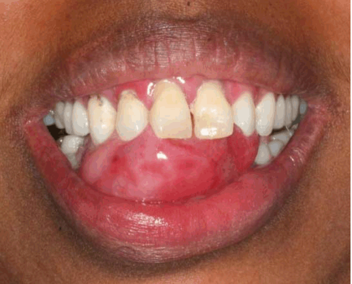
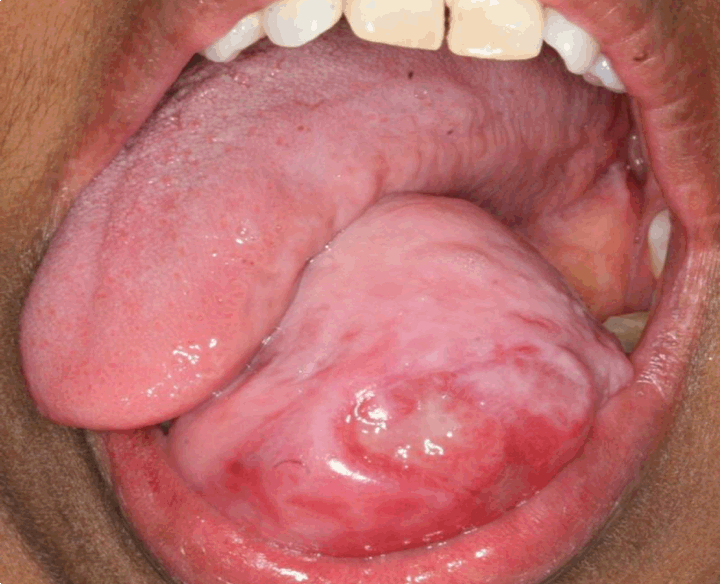
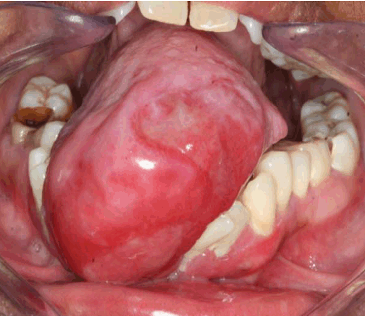
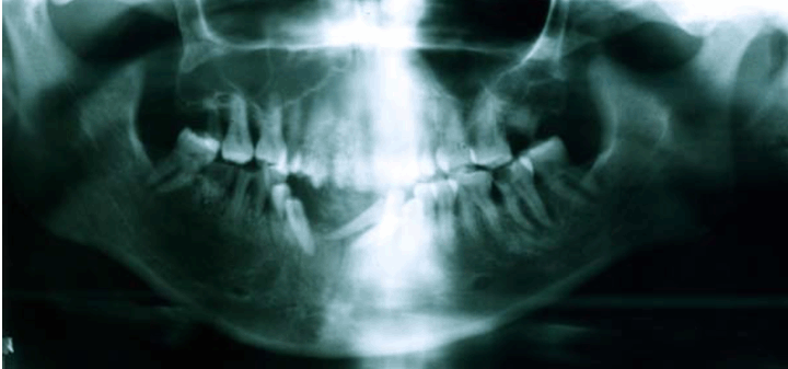
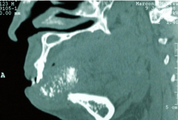
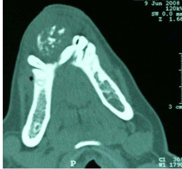
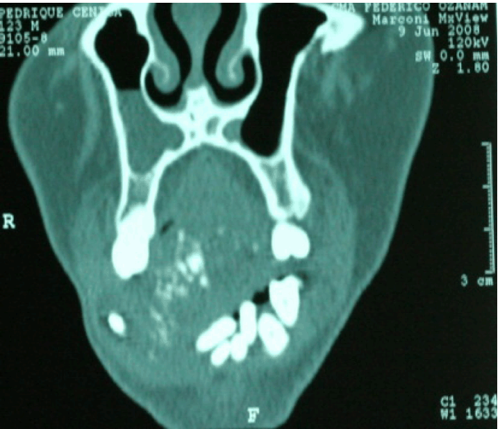
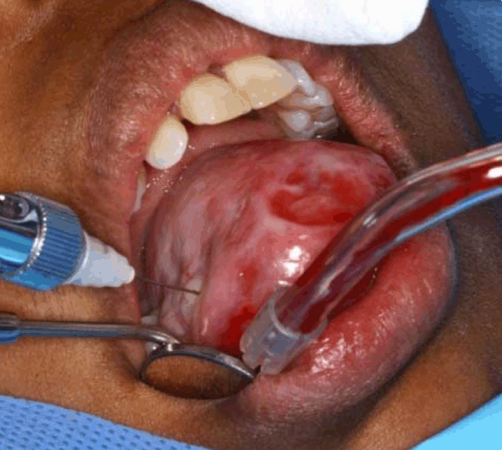
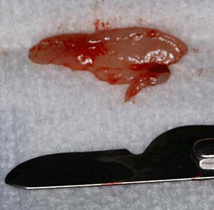
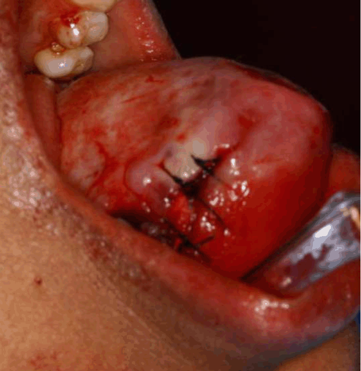
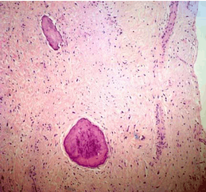
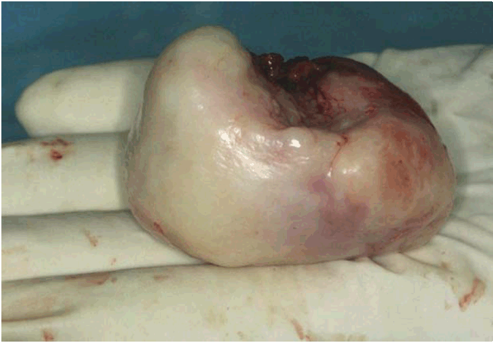
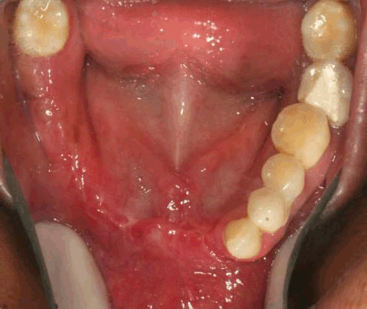
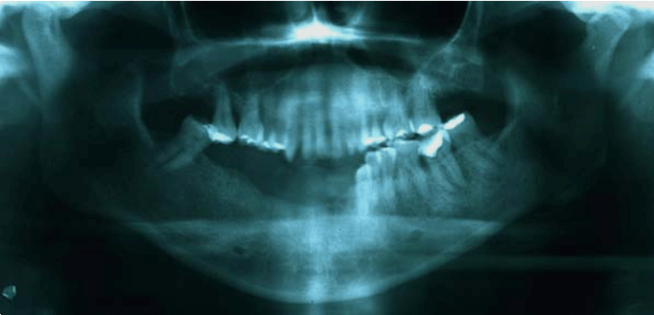
A case of a 28-year-old single Venezuelan female babysitter, natural from Caracas, Venezuela is reported to the Oral Medicine Service by a private practitioner, regarding an asymptomatic exophytic tumorous mass on the mandible of approximately 14 months of evolution. The patient referred it had commenced as a firm lump resembling the color of surrounding mucosa in the vestibular gum of mandibular anterior teeth. She identified two growth phases, a slow growing phase and a fast growing phase, but did not establish the periods of such. Interestingly to be noted, she had not been to a practitioner due to cancerophobia and odontophobia, consenting on assisting by insistence of her family. She also admitted it had been her very first visit to a dentist ever. Concerning function limitations, phonation, mastication and aesthetic, she stated that she had adapted to the mass so well she was able to eat to her satisfaction and speak relatively well and that she was not very concerned of the aesthetic appearance. Personal and familiar background was non contributory to the case. Clinical examination: Extraorally, the upper and middle third of her face were relatively symmetric, the inferior third presents with a remarked protrusion of the maxillary anterior teeth that occlude over a pink colored mass that fails to be fully covered by the lower lip. There is a moderate asymmetry in which a protrusion is seen towards the right side of the lower lip and chin (Figure 1). Intraorally, a giant mass measured 5.3x4.5x3.2 cm, pediculated, clear pink colored in most its surface with bright erythematous zones surrounding ulcerous areas in the front portion of the lesion, of approximately 2.5 cm the largest in the amplest extension, of irregular shape and mostly well defined. Lesion presented also indented areas in the antero-superior zone coinciding with occlusion of maxillary teeth (Figure 2). The lesion was noted to emerge from the vestibular gum and interdental papilla between teeth 4.2 and 4.3, extending lingually and occupying most of the oral cavity, causing mesial displacement of 4.1 and 4.2 which were displaced lingual to 3.1 and 3.2 (which where distally displaced). A great amount of local irritants were present as well as several caries and root remnants (Figure 3). The patient had a remarkable overjet which was though firth to be caused by the lesion, the patient would indicate she had it from long before and it was then attributed to the late digital suction. Imagenologic examination: A panoramic X-ray and a computed tomography (CT) scan were performed, revealing a radiopaque image with a cottony pattern, radically displacing the 4.1, 4.2 mesially and the 4.3 and 4.4 distally (Figure 4). The lesion image overlaps the maxillary teeth from the 1.4 to the middle line (eclipsed by the artefact). Generally, a condition of dental care abandonment was evident with the presence of multiple root remnants, radiolucid images corresponding to caries and pulpar affection. The CT scan revealed a mixed hyperdense image within an isodense matrix. The hyperdense area was formed by numerous "particles" with an equal or similar density to bone (Figure 5) (Figure 6) (Figure 7) . Differential Diagnosis: Considering clinical and radiological presentation some differential diagnosis were suggested: bone tumors as osteoblastoma and periosteal osteosarcoma, reactive lesions as POF. Incisional biopsy: General dental treatment such as oral hygiene was performed prior to the surgical phases. Blood tests were around normal limits. An incisional biopsy was performed under local anesthesia. Mental nerve was anesthetized and then local anesthetic was placed to delimit the operating area (Figure 8). A deep incision was made using a N° 15 blade (Figure 9) . The sensation during incision was like cutting in to a "hard pile of biscuits", getting harder as the cut deepened. Finally, hemostatic sponges were placed in and suture was taken using 3-0 black silk interrupted stitches (Figure 10) as approaching of flanks was also difficult. Postoperative control and stitch removal was done one week after showing adequate cicatrisation. Histopathological study: The histopathological study revealed a fragment of reactive tissue constituted by a great number of collagen fibres irregularly distributed and a moderate number of fibroblasts, scarce number of blood vessels and isolated isles of osteoid with osteocytes immerse within and surrounded by a matrix of osteogenic material with osteoblasts (Figure 11). A definitive diagnosis of POF was concluded, confirming the provisional diagnosis. Excisional biopsy: Once confirming the diagnosis by means of the incisional biopsy, the patient was prepared for exeresis under general anesthetic with nasotracheal intubation and because the difficult airway (Mallampati IV), attributed to the size of the lesion, intubation was assisted with nasofibro-bronchoscopy, provided by the Pneumology Service. A vestibular Newman flap was designed, taking the prevision of including 5 mm lingually to the pediculated border of the lesion. Odontectomy of all teeth with mobility higher to grade III was done and a regularization osteotomy was performed bearing in mind future prosthetic rehabilitation. An immediate prosthesis was not possible due to the impossibility of preoperative impressions. The procedure was concluded with a continuous stitch hermetic mucous closure using Vicryl 3-0 (Figure 12) (Figure 13). The excisional histopathological study confirmed the previous incisional study. Postoperative control: In postoperative control, the mucosa and the ridge showed no clinical evidence of injury. In fact, it was of good quality and height for an implanted prosthetic plan (Figure 14) (Figure 15). A provisional denture was installed with tissue conditioner and perio-orthodontic treatment is being made to finally conclude with dental implants. | ||||||
| ||||||
| ||||||
| ||||||
|
| ||||||
| ||||||
| ||||||
| ||||||
| ||||||
| ||||||
| ||||||
| ||||||
| ||||||
| ||||||
| ||||||
|
| ||||||
| ||||||
|
Discussion
| ||||||
|
Peripheral ossifying fibroma is a reactive lesion which may reach giant dimensions. When injured, body cells can either undergo regeneration or healing and the later, when exposed to a chronic damage, will involve collagen deposition and scar formation. The roll of growth factors must be considered since they can induce gene transcription silent in inactive cells and also can induce entry of the cells in to the cell cycle. As a periodontal lesion, growth factors such as the fibroblast growth factor-2 (FGF-2) are likely to be involved. FGF-2 is present in different periodontal tissues and besides other effects, it has a mitotic action on fibroblasts; increase preosteoblasts which will derive in Osteoblasts and consequently form bone[4]. It is probable that at least the following elements are likely to be involved in the etiopathogenesis of the POF [1][5] [6]:
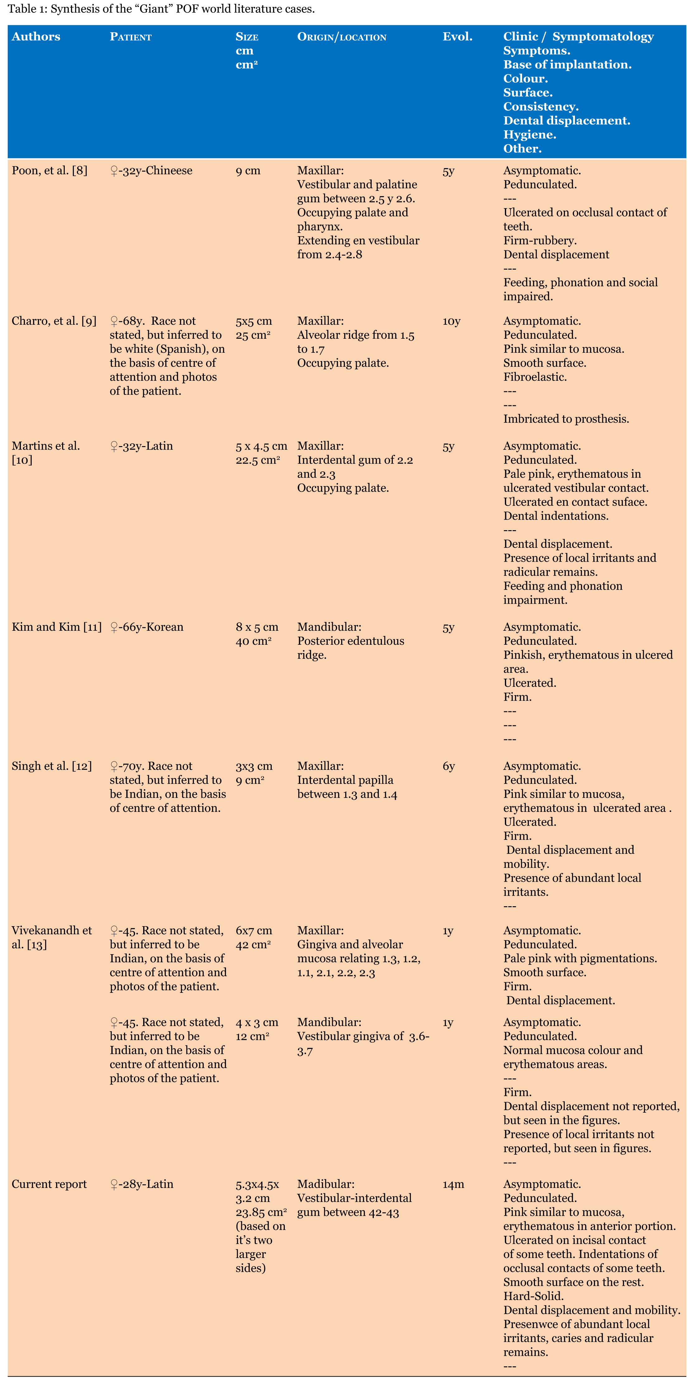
Not only the previous factors described above seem to be responsible for the growth, but a hormonal influence may be considered, as there is a higher incidence in women at the ages of most predominant hormonal changes [1] [7]. The relation of some of the reactive lesions with sex hormones has been suggested, scenario in which the gingiva could be considered a target organ, however, there is no sufficient evident to support this association [7]. For the purpose of the review on Giant POF, only those measuring a minimum of 3 cm in at least 2 dimensions where considered. When data was not expressly stated, it was deducted on the basis of the text and images. Giant POF turn out to be very uncommon, we were only able to find seven more cases in the world literature in six papers plus this case which accounts a total of eight [8] [9] [10] [11] [12] [13] (Table 1). Interestingly, all cases were reported in female patients with an average age of 48.25 years old and peek ages of 70 and 28 years. Exclusive female affectation may suggest a hormone involvement in the development of the diseases. All but one [11] were reported to occur in dentate areas. One Chinese, one inferred to be white (Spanish) based on geographic location and/or photographs, two Latin-Americans (including this case), one Korean, three inferred to be Indian based on geographic location and/or photographs. The average size (using the largest dimension provided) was 5.78 cm with peaks from 9 to 3 cm. The average evolution time was 4.27 years between 14 months to 10 years. Regarding the clinical appearance and symptomatology, all cases were asymptomatic, 5 cases occurred in maxilla and 3 in the mandible. Other features can be seen in Table 1. We propose that for a better study of these rare cases, giant POF or even a giant reactive lesion, should comply with any of the following criteria:
We also want to empathize the importance of a complete description of the case, not only considering the lesion itself, but, the patient as well. The mere fact that reactive lesions have the possibility to regress excludes them as a neoplasm; nevertheless these 'giant' POFs seem to defy this condition and wonder what can cause a reactive lesion to grow at such an excessive and uncommon size. | ||||||
| ||||||
|
| ||||||
|
Conclusion
| ||||||
|
Giant peripheral ossifying fibroma have only been reported in females, probably by a relation between grow factors and hormones. These lesions are extremely uncommon even if an underreporting is expected. Many factors must take place in the growth of these peripheral ossifying fibroma as briefly described; it would be of great interest to study the molecular basis of these lesions, with emphasis on growth factors. A socio-psychological reflection must also be considered, discussing why patients would allow these or other lesions to grow at that extent before looking for assistance. | ||||||
|
References
| ||||||
| ||||||


|
[HTML Abstract]
[PDF Full Text]
|
|
Author Contributions
Grimaldo-Carjevschi M – Substantial contributions to conception and design, Acquisition of data, Analysis and interpretation of data, Drafting the article, Revising it critically for important intellectual content, Final approval of the version to be published Villarroel-Dorrego M – Analysis and interpretation of data, Drafting the article, Final approval of the version to be published Vargas F, Romero Y – Analysis and interpretation of data, Drafting the article, Final approval of the version to be published |
|
Guarantor of submission
The corresponding author is the guarantor of submission. |
|
Source of support
None |
|
Conflict of interest
Authors declare no conflict of interest. |
|
Copyright
© 2014 Grimaldo-Carjevschi M et al. This article is distributed under the terms of Creative Commons Attribution License which permits unrestricted use, distribution and reproduction in any medium provided the original author(s) and original publisher are properly credited. Please see the copyright policy on the journal website for more information. |
|
|