  |

|
 |
|
Case Report
| ||||||
| A rare cause of upper gastrointestinal bleeding: Posttraumatic pseudoaneurysm | ||||||
| Negi RC1, Brij Sharma2, Bhupender3, Gaurav Kapoor3, Bal Beer Verma4, Ashok Sharma5 | ||||||
|
1Senior Resident, Department of Medicine and Gastroenterology, IGMC Shimla, HP, India.
2Assisstant Prof, Department of Medicine and Gastroenterology, IGMC Shimla, HP, India. 3Junior Resident, Department of Medicine and Gastroenterology, IGMC Shimla, HP, India. 4Associate Professor, Department of Medicine and Gastroenterology, IGMC Shimla, HP, India. 5Professor, Department of Medicine and Gastroenterology, IGMC Shimla, HP, India. | ||||||
| ||||||
|
[HTML Abstract]
[PDF Full Text]
[Print This Article]
[Similar article in Pumed] [Similar article in Google Scholar] 
|


| How to cite this article |
| Negi RC, Sharma B, Bhupender, Kapoor G, Verma BB, Sharma A. A rare cause of upper gastrointestinal bleeding: Posttraumatic pseudoaneurysm. International Journal of Case Reports and Images 2014;5(5):387–390. |
|
Abstract
|
|
Introduction:
Hemobilia is a rare cause of upper gastrointestinal bleeding. It needs prompt diagnosis and immediate management to safe the life. Posttraumatic pseudoaneurysm of hepatic artery is seen mostly after abdominal trauma causing liver injury or interventions on hepatobiliary system. On esophagogastroduodenoscopy active bleeding without apparent source is clue to the diagnosis.
Case Report: A 27-year-old male presented with history of one episode of hematemesis and melena. There was history of roadside accident one and a half months before and he had liver injury which was revealed on computed tomography of the abdomen. Patient was managed conservatively and discharged. On examination, patients was anemic and had postural fall in blood pressure. Emergency esophagogastroduodenoscopy revealed blood coming from ampulla. Computed tomography angiography (CTA) revealed pseudoaneurysm in segmental branches of right hepatic artery with active bleeding. Conclusion: Hemobilia is defined as bleeding into the biliary tree from abnormal communication between blood vessel and bile duct. The most common cause of posttraumatic hemobilia is pseudoaneurysm. The classic triad of hemobilia is absent in 70% of cases and in such cases clinical diagnosis is difficult. The CTA is the investigation of choice and embolization is the treatment option. Surgery should be done in cases refractory to embolization. | |
|
Keywords:
Hemobilia, pseudoaneurysm, embolization, Gastrointestinal bleeding.
| |
|
Introduction
| ||||||
|
Hemobilia is a rare cause of gastrointestinal (GI) bleeding which develops as a result of communication between blood vessel and biliary tract. [1] Posttraumatic pseudoaneurysm of hepatic artery is seen in 1% of hepatic injury. [2] It should be considered in patients presenting with upper gastrointestinal bleeding with prior history of abdominal trauma. The patients commonly present with either hematemesis or melena but may also present with abdominal pain or jaundice. High index of suspicion is required for the diagnosis of hemobilia | ||||||
|
Case Report
| ||||||
|
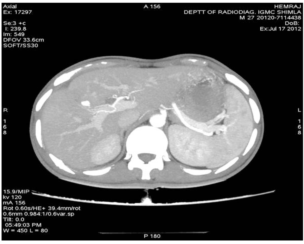
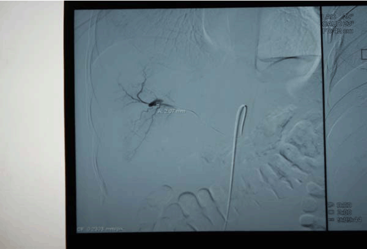
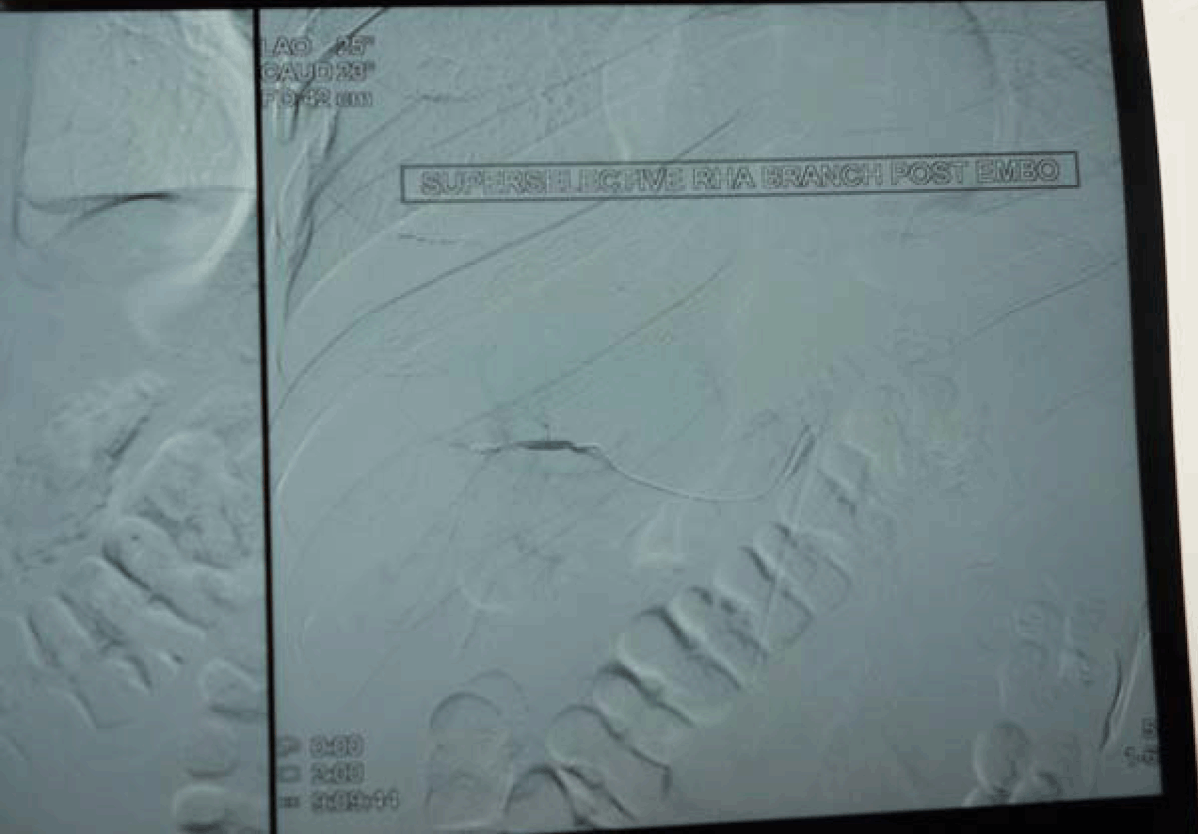
A 27-year-old male presented with complaints of one episode of hematemesis seven days back and melena for seven days. There was a history of roadside accident one and a half months before with grade IV liver injury, revealed on computed tomography (CT) scan of the abdomen. Patient was managed conservatively in Department of Surgery and discharged from hospital after one week in stable condition. On examination, the patient was anemic, pulse 110/min, blood pressure 80/50 mmHg with postural fall in blood pressure. The systemic examination was normal. Clinical possibility of upper GI bleed was kept. Emergency investigation revealed hemoglobin 4.5 g/dL, TLC 4900/mm3. BU 30 mg%, S. cr.-1.0 mg%, and electrolytes Na-135 mEq /L, K-4.1 mEq/L, Cl-105. The nasogastric tube drainage was persistently revealed altered blood. The patient was stabilized with three units of blood transfusion and emergency esophagogastroduodenoscopy was done. The esophagogastroduodenoscopy revealed alerted blood in the stomach with large clot in the fundus but source was not evident, first and second part of duodenum was also normal, but blood was present. On visualizing the ampulla, there was evidence of blood coming from ampulla. Emergency computed tomography angiography revealed pseudoaneurysm in segment VIII of liver involving segmental branches of right hepatic artery with active bleeding with communication with biliary radicals, with blood in gallbladder, common bile duct and second part of duodenum with subcapsular hematoma in segment VIII. (Figure 1) The digital selective arteriography revealed pseudoaneurysm from segmental branches of right hepatic artery. (Figure 2) Embolization was done and post embolization digital selective arteriography revealed complete non opacification of pseudoaneurysm. (Figure 3) | ||||||
| ||||||
| ||||||
| ||||||
|
Discussion
| ||||||
|
Hemobilia is defined as bleeding into the biliary tree from abnormal communication between blood vessel and bile duct. [1] Sandworm and Mirkovitch described hepatic artery pseudoaneurysm formation after blunt trauma or penetrating trauma to liver, after percutaneous diagnostic or therapeutic procedures on hepatobiliary system. [2] The mean time period between traumatic hepatic injury and presentation of patients with hemobilia is reported to be four weeks. [3] The most common cause of posttraumatic hemobilia is a pseudoaneurysm. The classic hemobilia triad described by Quinke in 1871 consists of upper gastrointestinal bleeding (hematemesis 60% or melena 90%) and biliary colic (70%) and obstructive jaundice in 70% of cases. [4] The classic triad is absent in almost 70% of cases and in such cases clinical diagnosis is difficult to suspect. [5] The hemobilia may be minor or major, in minor hemobilia minor bleeding stops spontaneously whereas in hemobilia major the blood flows rapidly into the duodenum presenting as melena or hematemesis. Earlier the most common cause of hemobilia was accidental trauma but nowadays the common cause is iatrogenic injury to liver due to liver biopsy or percutaneous interventions. [6] In a study done by Srivastava et al. from India the predominant cause of hemobilia was liver injury following roadside accidents. [7] The hemobilia should be suspected in patients with prior history of abdominal trauma presenting with upper gastrointestinal bleeding. The presenting case also had a history of abdominal trauma which was managed conservatively. The patient presented with hematemesis and melena after four weeks of abdominal trauma. In such patients esophagogastroduodenoscopy will reveal altered blood and clot in stomach and duodenum but without apparent source of active bleeding. The blood may be seen in ampulla. The gold standard diagnostic investigation is angiography to localize the site of bleeding but in the present era CT scan and magnatic resonance imaging (MRI) scan are also used for diagnostic purpose as non-invasive technique. [7] [8] The selective arterial embolization of pseudoaneurysm is the procedure of choice for management of hemobilia. Surgery is required in case refractory to embolization.[9] | ||||||
|
Conclusion
| ||||||
|
Hemobilia is a rare cause of upper gastrointestinal bleeding due to posttraumatic pseudoaneurysm and it should be diagnosed promptly. High index of suspicion is needed for diagnosis. Angiography is the choice of investigation for localization of bleed and embolization is the treatment of choice. | ||||||
|
References
| ||||||
| ||||||

|
[HTML Abstract]
[PDF Full Text]
|
|
Author Contributions
Negi RC – Substantial contributions to conception and design, Acquisition of data, Analysis and interpretation of data, Drafting the article, Revising it critically for important intellectual content, Final approval of the version to be published Brij Sharma – Analysis and interpretation of data, Drafting the article, Revising it critically for important intellectual content, Final approval of the version to be published Bhupender – Analysis and interpretation of data, Drafting the article, Revising it critically for important intellectual content, Final approval of the version to be published Gaurav Kapoor – Analysis and interpretation of data, Drafting the article, Revising it critically for important intellectual content, Final approval of the version to be published Bal Beer Verma – Analysis and interpretation of data, Drafting the article, Revising it critically for important intellectual content, Final approval of the version to be published Ashok Sharma – Analysis and interpretation of data, Drafting the article, Revising it critically for important intellectual content, Final approval of the version to be published |
|
Guarantor of submission
The corresponding author is the guarantor of submission. |
|
Source of support
None |
|
Conflict of interest
Authors declare no conflict of interest. |
|
Copyright
© 2014 Negi RC et al. This article is distributed under the terms of Creative Commons Attribution License which permits unrestricted use, distribution and reproduction in any medium provided the original author(s) and original publisher are properly credited. Please see the copyright policy on the journal website for more information. |
|
|