| |
|
|
|
Case Report
| ||||||
| Gastric outlet obstruction due to a giant antral polyp which have malignant transformation | ||||||
| Erkan Oymaci1, Ali Coskun1, Deniz Ucar1, Erdem Sari1, Nazif Erkan1, Mehmet Yildirim1, Hale Kizanoğlu2 | ||||||
|
1MD, Department of General Surgery, Izmir Bozyaka Trainig and Research Hospital, Izmir, Turkey.
2MD, Izmir Bozyaka Trainig and Research Hospital, Department of Pathology, Izmir, Turkey. | ||||||
| ||||||
|
[HTML Abstract]
[PDF Full Text]
[Print This Article]
[Similar article in Pumed] [Similar article in Google Scholar] 
|
| How to cite this article |
| Oymaci E, Coskun A, Ucar D, Sari E, Erkan N, Yildirim M, Kizanoglu H. Gastric outlet obstruction due to a giant antral polyp which have malignant transformation. International Journal of Case Reports and Images 2014;5(5):382–386. |
|
Abstract
|
|
Introduction:
Gastric polyps are seen 2–3% in all gastrointestinal endoscopic examinations. Hyperplastic polyps, the most common type of gastric polyps, are present 85–90% of cases. Giant villous tumors of the stomach are somewhat rare and the high incidence of malignant transformation of them requires prompt diagnosis. Giant gastric polyps can also cause gastrointestinal bleeding that leads to iron deficiency anemia or partial gastric outlet obstruction. All giant polyps must be resected endoscopically or surgically and still there are no marked guidelines for the optimal management of gastric polyps at the time of initial examination. The point to be taken into consideration for patients is the need for radical surgery or not.
Case Report: A 62-year-old male was admitted to our clinic with complaints of epigastric pain, nausea and vomiting. Upper gastrointestinal endoscopy revealed that 6x7 cm diameter pedunculated polypoid mass which obstruct the pyloric channel in the gastric antrum. The patient is treated by distal subtotal gastrectomy and gastroenterostomy. The histopathologic examination of the specimen confirmed the occurrence of adenocarcinoma. Conclusion: Giant hyperplastic gastric polyps are fairly uncommon. We presented a case of giant gastric polyp which have malignant transformation and cause gastric outlet obstruction that resected surgically in an attempt to add to the current literature. | |
|
Keywords:
Gastric polyp, Villous adenoma, Obstruction, Surgery
| |
|
Introduction
| ||||||
|
Gastric polyps are seen 2–3% in all gastrointestinal endoscopic examinations. Hyperplastic polyps, the most common type of gastric polyps, are present 85–90% of cases. [1] Hyperplastic polyps, fundic gland polyps, gastric adenomas and some of gastric carcinoid tumors may present as a gastric polyp. Villous adenomas of the stomach are somewhat rare with approximately 100 cases only reported in literature and have tendency to undergo malignant transformation as high as 72%. [2] They are frequently multiple and associated with other gastrointestinal neoplasms. The high incidence of malignant transformation of gastric villous adenoma requires prompt diagnosis of this rare tumor. Polyps greater than 2 cm are at significant risk for malignancy and they require complete resection. [3] Except fundic gland polyps which have a clear typical feature, upper endoscopy cannot reliably distinguish the type of gastric polyp by gross inspection. Thus, histopathological diagnosis is important although whether to biopsy or excise gastric polyps is not always clear. [4] Giant villous tumors of the stomach are somewhat rare and the high incidence of malignant transformation of them requires prompt diagnosis. Giant gastric polyps can also cause gastrointestinal bleeding that lead to iron deficiency anemia or partial gastric outlet obstruction. Gastric outlet obstruction presents with nausea and vomiting and usually develops over weeks to months. It may be complete or incomplete with intermittent symptoms. Gastric villous adenomas have potential for malignant transformation and hence must be excised endoscopically or surgically whichever may be feasible. We presented a case of giant gastric villous adenoma which have malignant transformation and cause gastric outlet obstruction in an attempt to add to the current literature. | ||||||
|
Case Report
| ||||||
|
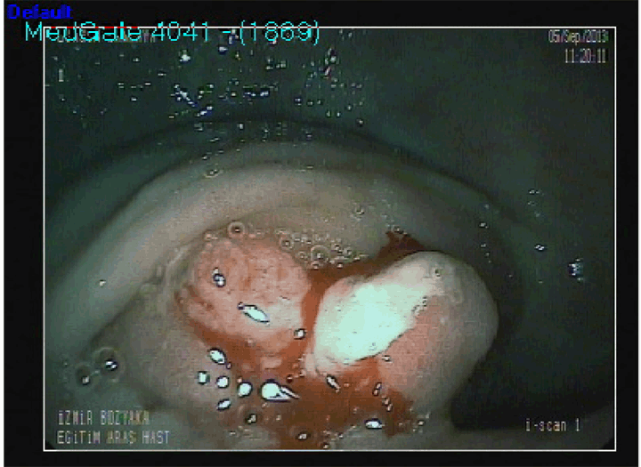
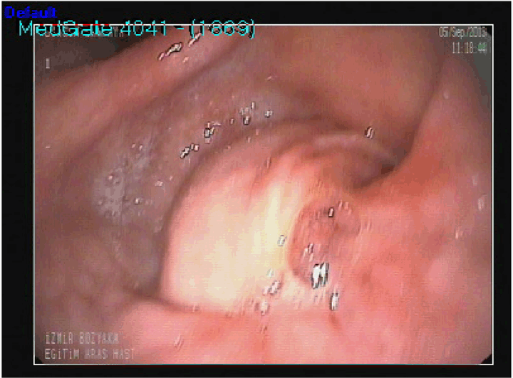
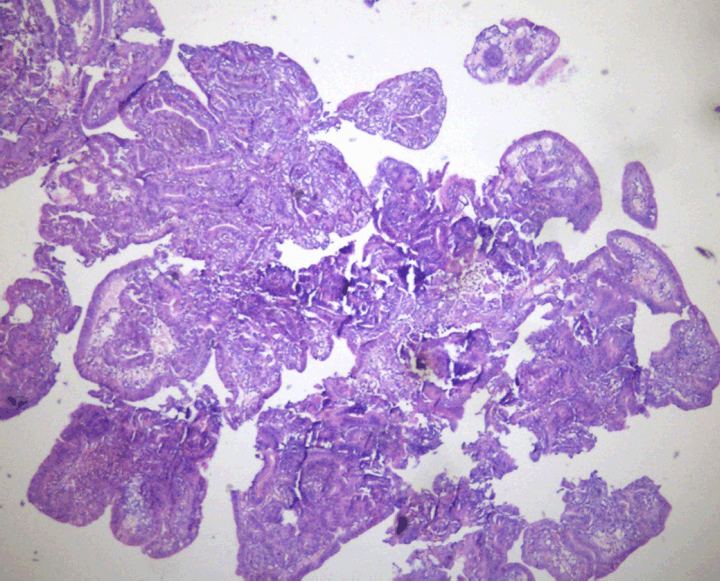
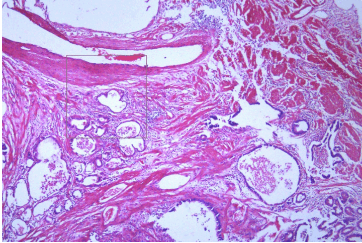
A 62-year-old male was admitted to our clinic with complaints of epigastric pain, nausea and vomiting. He described two pain and vomiting episodes of similar type, each lasting three or four days, in last two months. His medical background and family history were unremarkable. Physical examination revealed that mild epigastric tenderness and normal bowel sounds. At the admission, laboratory tests revealed hemoglobin of 9.5 g/dL with a mean corpuscular volume of 68.9 fl. Serum iron was 30 µg/dL and total iron-binding capacity was 408 µg/dL. The other laboratory results were also within normal limits. Upper gastrointestinal endoscopy revealed a 6x7 cm diameter pedinculated polyp covered with irregular villous mucosa arising from the antrum of the stomach and which totally obstructed the entrance of the pyloric channel. (Figure 1) (Figure 2) This polyp was seen prolapsing into the bulbus of the duodenum causing gastric outlet obstruction. Multiple biopsy were taken from both the polypoid mass and antrum. Histopathologic examination of the biopsy revealed a gastric villous tumor with carcinomatous change. Microscopic findings of the biopsy showed malignant glandular formation invading the lamina propria on the stomach. It also showed finger-like projections covered by dysplastic epithelium. (Figure 3) (Figure 4) The patient underwent a distal subtotal gastrectomy with Billroth II gastrojejunostomy reconstruction and lymph node dissection on the third day of admission. (Figure 5) (Figure 6) No postoperative complications occurred and the patient is discharged on the postoperative sixth day. Evaluation of the specimen showed that the polyp has the largest diameter of 7 cm. The histopathologic examination of the specimen confirmed the presence of villous adenoma with the occurrence of adenocarcinoma with no morphological and histochemical evidence of Helicobacter pylori infection. There was no lymph node metastasis. | ||||||
| ||||||
| ||||||
| ||||||
| ||||||
| ||||||
| ||||||
|
Discussion
| ||||||
|
Most of gastric polyps have as-ymptomatic presentations and are incidentally finding with an incidence of approximately 6% on up-per endoscopic examination. [5] Their pathogenesis is unknown but the majority of polyps occur as multiple lesions which arising from inflamed gastric mucosa. Infection of the gastric mucosa with Helicobacter pylori has been reported in up to 90% of the cases. [6] Giant gastric polyps are fairly uncommon. Patients with hyperplastic polyps, greater than 3 cm in largest diameter are more likely to be symptomatic. These giant hyperplastic polyps represent about 2% of all hyperplastic polyps. [7] Gastric polyps can vary in size from a few millimeters to several centimeters. The median size of polyps removed endoscopically was 3 cm while the median size of surgically removed polyps was 6 cm. [8] [9] Kumar et al. have reported the largest endoscopically treated polyp causing intermittent gastric outlet obstruction to date. [9] They removed an 8-cm polyp: two-thirds of the polyp was snared and the remainder excised at a subsequent visit. Aksel et al. reported that the size of the gastric polyp was 12 cm diameter in their study and made radical gastrectomy for the gastric polyp which is in giant diameter and causes severe bleeding. [7] In our case, we also made radical gastrectomy due to malignant transformation which include high grade dysplasia and giant 6x7 cm diameters of polyp. Symptomatic presentations can range from an ulcerated polyp leading to anemia to complete gastric outlet obstruction. Al-Haddad et al. reported that the incidence of hyperplastic polyps in iron-deficiency patients was 1.4% and the largest polyp was 5 cm in diameter. [10] Anemia is the most frequent clinical manifestation of diffuse gastric polyposis. Our patient has both gastric outlet obstruction and iron-deficiency anemia in laboratory tests at admission. Villous tumors of the gastrointestinal tract are neoplasms that arising from the columnar epithelium because such neoplasms occur infrequently in the upper gastrointestinal tract, the clinical, radiologic, and pathologic features have not been completely defined. Inaba et al. reported that the villous adenoma accompanied by malignant changes was positive by the carcinoembryonic antigen peroxidase-antiperoxidase method. [11] This result showed the biological property of villous adenoma that they can easily change into malignancy. Adenoma of the stomach and duodenum are subject to malignancy more often than colonic adenomas. It might be possible to encounter with dysplasia and carcinoma around the polyp. There is a synchronous adenocarcinoma risk in another part of the stomach after polypectomy in up to 30% of cases. [12] So, it is necessary to examine surrounding tissue and to take multiple biopsies from the gastric mucosa with endoscopic intermittent follow-up. It is quite important because of the possibility of recurrence at the polypectomy site and development of malignancy in the remote gastric mucosa after polypectomy. [13] | ||||||
|
Conclusion
| ||||||
|
As conclusion, we presented a case of giant gastric villous polyp which have malignant transformation and cause gastric outlet obstruction that resected surgically in an attempt to add to the current literature. The point to be taken into consideration for patients is the need for radical surgery or not. The main disadvantage of endoscopic methods is the risk of incomplete tumor resection. So, it seems to be there is no alternative treatment other than radical gastrectomy for the gastric giant polyp which causing obstruction. | ||||||
|
References
| ||||||
| ||||||
|
[HTML Abstract]
[PDF Full Text]
|
|
Author Contributions
Erkan Oymaci – Substantial contributions to conception and design, Acquisition of data, Analysis and interpretation of data, Drafting the article, Revising it critically for important intellectual content, Final approval of the version to be published Ali Coskun – Analysis and interpretation of data, Revising it critically for important intellectual content, Final approval of the version to be published Deniz Ucar – Analysis and interpretation of data, Revising it critically for important intellectual content, Final approval of the version to be published Erdem Sari – Analysis and interpretation of data, Revising it critically for important intellectual content, Final approval of the version to be published Nazif Erkan – Analysis and interpretation of data, Revising it critically for important intellectual content, Final approval of the version to be published Mehmet Yildirim – Analysis and interpretation of data, Revising it critically for important intellectual content, Final approval of the version to be published Hale Kizanoğlu – Analysis and interpretation of data, Revising it critically for important intellectual content, Final approval of the version to be published |
|
Guarantor of submission
The corresponding author is the guarantor of submission. |
|
Source of support
None |
|
Conflict of interest
Authors declare no conflict of interest. |
|
Copyright
© 2014 Erkan Oymaci et al. This article is distributed under the terms of Creative Commons Attribution License which permits unrestricted use, distribution and reproduction in any medium provided the original author(s) and original publisher are properly credited. Please see the copyright policy on the journal website for more information. |
|
|