  |

|
 |
|
Case Report
| ||||||
| A rare case of myositis ossificans progressiva presenting as multiple progressive contracture | ||||||
| Anil Mehtani1, Jatin Prakash2, Suresh Chand3, Abhinav Sinha2, Ajeet Singh2, Harvinder Dev2 | ||||||
|
1Head of Department of Orthopedics, Lady Hardinge Medical College, Shaheed Bhagat Singh Marg, New Delhi, India.
2MBBS, MS(Ortho), DNB, Department of Orthopedics, Lady Hardinge Medical College, Shaheed Bhagat Singh Marg, New Delhi, India. 3Rresident ,Department of Orthopedics, Lady Hardinge Medical College, Shaheed Bhagat Singh Marg, New Delhi, India. | ||||||
| ||||||
|
[HTML Abstract]
[PDF Full Text]
[Print This Article]
[Similar article in Pumed] [Similar article in Google Scholar] 
|


| How to cite this article |
| Mehtani A, Prakash J, Chand S, Sinha A, Singh A, Dev H. A rare case of myositis ossificans progressiva presenting as multiple progressive contracture. International Journal of Case Reports and Images 2014;5(5):345–350. |
|
Abstract
|
|
Introduction:
Myositis ossificans progressiva is a rare disease characterized by formation of areas of calcification in soft tissue such as ligaments, muscles and tendons. There are a few sporadic case reports all over the world. The disease has an incidence of less than 1 in 10,000,000 population. Myositis ossificans progressiva is a disease of early childhood. The disease is often progressive with multiple soft tissue contracture and subsequent death by third or fourth decade of life. There is no effective treatment till date.
Case Report: We herein present a case report of myositis ossificans progressiva presented to us with numerous lumps and shoulder and hip contracture. Patient was treated conservatively on bisphosphonates. No progression of lumps or swelling were seen after one year of follow-up. Conclusion: This presents a case report of a very rare disease. In most cases there is history of any trauma or inciting factors that result in formation of myositis mass. This case, however, presents a very aggressive form of disease with patient developing spontaneous swellings and progressive contractures. | |
|
Keywords:
Myositis ossificans progressive, Multiple contractures, Pediatric
| |
|
Introduction
| ||||||
|
Myositis ossificans progressive also known as fibrodysplasia ossificans, Münchmeyer syndrome, stiff-man syndrome, and progressive ossifying myositis is a very rare and crippling disorder. The disease mostly involves patients in their first decade, progressing rapidly to involve muscles, tendons and ligaments. Patients are generally, confined to wheel chair life and mostly live till fourth to fifth decade. [1] The disease is very rare with an incidence of less than 1 in 10,000,000 population and around 700 cases have been reported in literature to date. We report a young boy who presented with very rapid progression of disabling muscle contractures diagnosed clinicoradiologically as myositis ossificans progressiva. | ||||||
|
Case Report
| ||||||
|
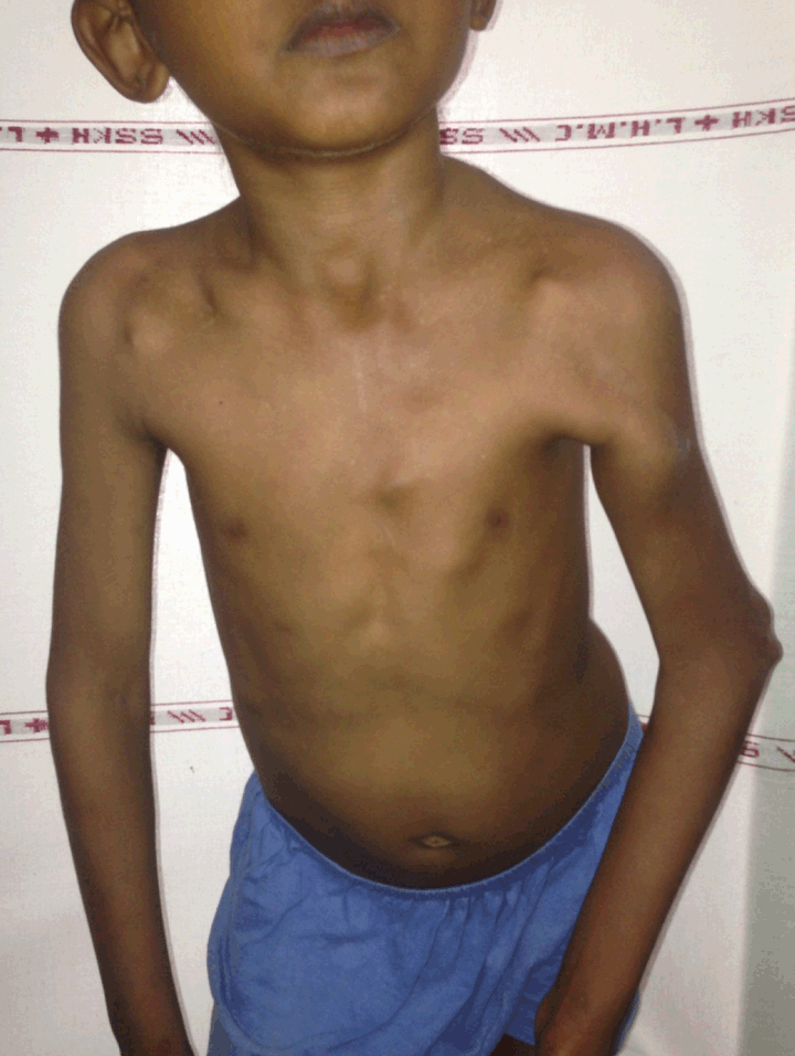
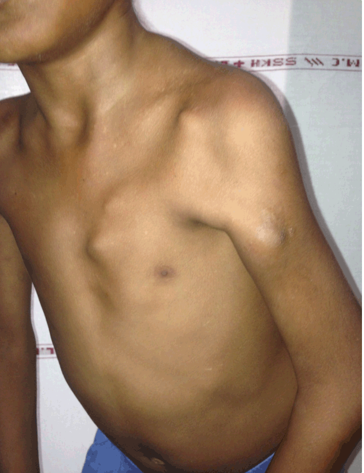
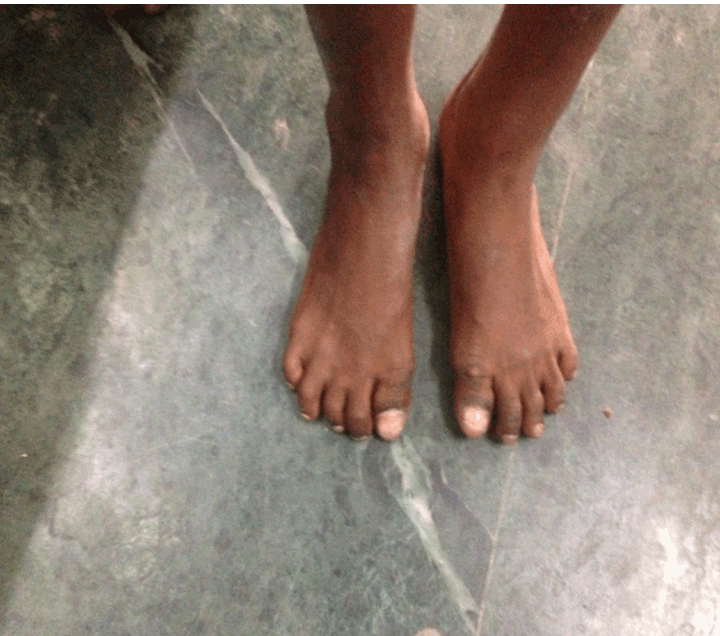
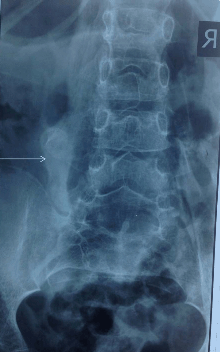
An eight-year-old boy was presented to our outpatient department with multiple lumps all around the body with non- healing ulcers over scapula. On detailed history, patient's parents noticed lump initially in first year of his life and they gave history that the lumps have progressed both in size and number since then. These were not associated with any trauma. He gradually developed restricted mobility of the left shoulder, followed by neck and right shoulder by four years of age. This was followed by more lumps in back and lower legs causing restriction of motion of lower spine and hip joint. Patient developed ulcers in the skin overlying lump at scapular region. (Figure 1) There was no history of fever, bleeding tendencies, hematuria, seizures, deafness, mental retardation, joint swelling, rash, abdominal colic, fractures, thyroid swelling, or any drug intake. Physical examination revealed multiple lumps in the neck, scapula, back, iliac region, knee and trunk. There was kyphotic deformity in dorsal spine and muscle contractures involving the sternocleidomastoids, latissimus dorsi, pectoralis major, (Figure 2) and the cervical muscles, with restricted abduction and internal rotation of both shoulders. There were bilateral flexion deformities of hip joint. He also had short great toes. (Figure 3) Chest expansion was restricted in spirometry testing. Routine laboratory investigations were normal. Serum calcium chemistry was also normal. The spirometry showed moderate restriction and electrocardiogram cardiac echo were essentially normal. X-ray of cervical spine showed calcific strands around both shoulder joints (left more than right) and in the paraspinal regions. (Figure 4) A detailed skeletal survey of the body revealed calcification in the soft tissues surrounding the cervical region, left shoulder, in the anterior chest wall, the thorax, and the paraspinal muscles and knee. (Figure 5) Considering both the clinical and the radiological features, sporadic myositis ossificans progressiva was diagnosed. The child was treated with graded physiotherapy. Bisphosphonates were added. As there was no acute flare-up, steroids were not given. The patient has been followed-up for one year. No new lumps have been noticed after starting of bisphosphonates. | ||||||
| ||||||
| ||||||
| ||||||
| ||||||
| ||||||
|
Discussion
| ||||||
|
Myositis ossificans progressiva is a rare, progressive, crippling disorder, with an incidence of less than 1 in 10,000,000 population. The condition has a male preponderance. This is a mesodermal disorder with defect in reparative process [2] [3] causing heterotopic ossification which usually begin in 5–7 years of life. [4] Our case however, has history of ossifications from first year of life. The case presented late to us with all characteristic features of short great toe, multiple contractures and multiple ossifications. The X-rays were also characteristic in showing the lesion. Based on these characteristic findings, the diagnosis was pretty straightforward. Otherwise diagnostic errors have been documented in up to 87% of myositis ossificans progressiva cases worldwide with cancer being the most common erroneous diagnosis. [5] This is very important to note as error in diagnosis would lead to unwanted biopsies doing more harm than good. Initial symptoms include painful lumps, mostly starting cranially in neck and shoulder region and progressing caudally involving scapula, trunk and hip regions. This is sometimes associated with stiffness and decreased mobility at joint site resulting in progressive contractures. [2] This case also had this characteristic pattern. Mostly these swellings are preceded by local trauma, injection site, biopsy or a venipuncture site, however, no such inciting factor was observed. [6] Associated skeletal features of great diagnostic significance include short hallux with synostosis, hallux valgus (75–90%), and short thumbs. [2] Kyphoscoliosis, with restricted shoulder and pelvic girdle movements and restrictive pulmonary disease, can occur. Mental retardation, alopecia, and cardiac conduction defects are other associations. [4] This case had short great toes, no mental retardation or conductive deafness. Radiological investigations are characteristic in myositis ossificans progressive. There is microdactyly of big toes (90%) and thumbs (50%), progressive fusion of the posterior arches of the cervical spine, narrowed antero-posterior diameter of lumbar vertebral bodies, with or without bony ankylosis with soft tissue calcification at multiple sites. [7] There is no effective treatment till date. Multiple treatments have been tried. Steroids are useful in acute flare-ups, bisphosphonates are thought to decrease ectopic calcifications. This was observed in our case as well when oral bisphosphonates have stopped further progression in a short follow-up of one year. However, longer follow-up would be more helpful. Some newer and investigational drugs include antiangiogenic agents such as squalamine, thalidomide, COX-2 inhibitors, BMP4 antagonists, and noggin and gremlin gene therapy. However none of them has a proven efficacy. Surgeries at large are contraindicated including procedures like biopsy. Surgical release of contractures is recommended only if joint movement is severely impeding movement or there is nerve impingement and this is not without increased risk of further ossification. [8] [9] | ||||||
|
Conclusion
| ||||||
|
This presents a case report of a very rare disease. In most cases there is history of any trauma or inciting factors that result in formation of myositis mass. This case however presents a very aggressive form of disease with patient developing spontaneous swellings and progressive contractures. The disease was controlled on bisphosphonates and no new swelling developed in follow-up of one year. | ||||||
|
References
| ||||||
| ||||||


|
[HTML Abstract]
[PDF Full Text]
|
|
Author Contributions
Anil Mehtani – Substantial contributions to conception and design, Acquisition of data, Analysis and interpretation of data, Drafting the article, Revising it critically for important intellectual content, Final approval of the version to be published Jatin Prakash – Acquisition of data, Revising it critically for important intellectual content, Final approval of the version to be published Suresh Chand – Acquisition of data, Revising it critically for important intellectual content, Final approval of the version to be published Abhinav Sinha – Acquisition of data, Revising it critically for important intellectual content, Final approval of the version to be published Ajeet Singh – Acquisition of data, Revising it critically for important intellectual content, Final approval of the version to be published Harvinder Dev – Acquisition of data, Revising it critically for important intellectual content, Final approval of the version to be published |
|
Guarantor of submission
The corresponding author is the guarantor of submission. |
|
Source of support
None |
|
Conflict of interest
Authors declare no conflict of interest. |
|
Copyright
© 2014 Anil Mehtani et al. This article is distributed under the terms of Creative Commons Attribution License which permits unrestricted use, distribution and reproduction in any medium provided the original author(s) and original publisher are properly credited. Please see the copyright policy on the journal website for more information. |
|
|
|
About The Authors
| |||
| |||
| |||
| |||
| |||
| |||
| |||
