| |
|
|
|
Clinical Image
| ||||||
| Anomalous left vertebral artery | ||||||
| Hussein Ahmed Hassan1, Caroline Edward Ayad1, Tag Eldin Mohamed Ibrahim2, Ikhlas Abdelaziz Hassanand1 | ||||||
|
1PhD, Diagnostic Radiologic Technology, Sudan University of Science and Technology, Radiology Department, College of Medical Radiological Science, Khartoum, Sudan; 2 MD, Consultant Radiologist, Radiology Department, Modern Medical Center, Khartoum, Sudan. | ||||||
| ||||||
|
[HTML Abstract]
[PDF Full Text]
[Print This Article]
[Similar article in Pumed] [Similar article in Google Scholar] 
|
| How to cite this article: |
| Hassan HA, Ayad CE, Ibrahim TEM, Hassanand IA. Anomalous left vertebral artery. International Journal of Case Reports and Images 2014;5(3):247–249. |
|
Case Report
|
|
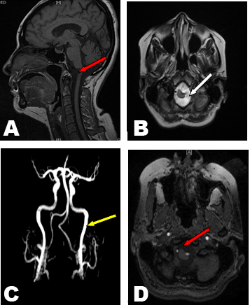
A 40-year-old Sudanese female was referred to the magnetic resonance imaging (MRI) department presented with upper limbs weakness, urine incompetence and general weakness evaluation. Brain MRI axial and sagittal T1, T2, post contrast series, fluid attenuation inversion recovery (FLAIR), magnetic resonance angiography (MRA) were done. Images show anomalous left vertebral artery impressing into the cord substance at cervico–medullary junction causes cervico-medullary stenosis, no evidence of cerebral tonsil herniation or syrinx formation, in right parietal sub cortical white matter; two small high signal intensity foci were seen on T2 and FLAIR images suggestive of ischemic foci. Brain MRA confirmed malformation in the left vertebral artery, images of MRA showed the left vertebral artery entering foramen magna with medial tortuous kinking to the medulla oblongata. Normal MRI scan appearance of the basal ganglia, thalami, internal and external capsule on both sides were detected also the ventricular system and extra axial cerebrospinal fluid (CSF) space are within normal limits, no mass effect or midline shift. Normal configuration of optic chiasm, optic nerves, pituitary gland and its stalk, Normal features of the cerebellar hemispheres, vermis, peduncles and brain stem with no abnormal intensities. Normal MRI features of the cerebello pontine angles and internal auditory canal, no evidence of space occupied lesion (SOL), fresh or sub dural collection was detected. (Figure 1) |
|
|
|
Discussion
|
|
Anatomical variation is defined as the normal flexibility in the topography and morphology of body structures. [1] Anomalous origin of vertebral arteries, is of relation with the embryologic development of the aortic arch and the brachiocephalic arteries. [2] Anomalous origin of the vertebral artery is rare and is mostly seen in the left vertebral artery which originates directly from the aortic arch between the left common carotid artery and the left subclavian artery, [3] with a reported prevalence of 2.4–5.8% in a large autopsy series. [4] Also anomalous origins of both vertebral arteries are uncommon. There are only unreliable cases in the radiology literature which discuss the conventional angiographic findings. [5] The advances in the imaging modalities in recent years have required more accurate knowledge and a greater understanding of the normal anatomy and variation and their clinical importance. [6] Studies were done to ensure the incidence of anomalous origin of the left vertebral artery citied in the radiology literature, and suggest that the current increased frequency of cross-sectional imaging; could elevate the observed incidence of this anomaly in practice. [7] With modern multi-detector computed tomography technology, supra aortic arteries can be well delineated with excellent image quality. Reporting of anomalous origin of the left vertebral artery is important; as the anomaly has significant clinical and surgical implications during endovascular treatment of aortic arch injuries and during angioplasty and stent procedures in conditions. [8] Diagnostic cerebro vascular imaging can be achieved either by catheterization during angiography or by computed tomography angiography (CTA), MRA or Doppler sonography. [9] A thorough identification of anomalous origin of vertebral artery is paramount when performing both diagnostic and interventional angiography. If the vertebral arteries are not identified in their normal position, this can be misinterpreted as the vessel being congenitally absent. This information is important for endovascular or cardiothoracic surgeries in head and neck regions. [10] Anomalous origin of the vertebral artery did not result in clinical symptoms, [11] [12] but In some cases, patients may complain of symptoms of dizziness. [13] Anomalous origin effects hemodynamic and may lead to intracerebral malformation. [10] The knowledge of a potential vertebral artery origin variant appears to be obligatory for planning vascular surgery and endovascular intervention. [14] |
|
Conclusion
|
|
The true value of detecting anomalous origins of vertebral artery is the diagnostic gain prior to the surgery of supra aortic arteries. Using magnetic resonance imaging grants an excellent diagnostic value, as it is noninvasive with expansive imaging appearance of both vessels and soft tissues, it has been anticipated as diagnostic imaging modality for detection of anomalous vertebral artery. |
|
References
|
|
|
[HTML Abstract]
[PDF Full Text]
|
|
Author Contributions
Hussein Ahmed Hassan – Substantial contributions to conception and design, Acquisition of data, Analysis and interpretation of data, Drafting the article, Revising it critically for important intellectual content, Final approval of the version to be published Caroline Edward Ayad – Acquisition of data, Analysis and interpretation of data, Drafting the article, Revising it critically for important intellectual content, Final approval of the version to be published Tag Eldin Mohamed Ibrahim – Acquisition of data, Analysis and interpretation of data, Drafting the article, Revising it critically for important intellectual content, Final approval of the version to be published Ikhlas Abdelaziz Hassanand – Acquisition of data, Analysis and interpretation of data, Drafting the article, Revising it critically for important intellectual content, Final approval of the version to be published |
|
Guarantor of submission
The corresponding author is the guarantor of submission. |
|
Source of support
None |
|
Conflict of interest
Authors declare no conflict of interest. |
|
Copyright
© Hussein Ahmed Hassan et al. 2014; This article is distributed the terms of Creative Commons Attribution License which permits unrestricted use, distribution and reproduction in any means provided the original authors and original publisher are properly credited. (Please see Copyright Policy for more information.) |
|
|