|

|
 |
|
Case Report
| ||||||
| Prolonged aphasia and perfusion computed tomography abnormalities in migraine with aura | ||||||
| Anselm Angermaier1, Soenke Langner2, Michael Kirsch3, Alexander V Khaw4 | ||||||
|
1MD, Resident, Department of Neurology, University Medicine Greifswald, DE–17475 Greifswald (Germany).
2MD, PhD, Consultant, Institute for Diagnostic Radiology and Neuroradiology, University Medicine Greifswald, DE–17475 Greifswald (Germany). 3MD, Consultant, Institute for Diagnostic Radiology and Neuroradiology, University Medicine Greifswald, DE–17475 Greifswald (Germany). 4MD, Consultant, Department of Neurology, University Medicine Greifswald, DE–17475 Greifswald (Germany). | ||||||
| ||||||
|
[HTML Abstract]
[PDF Full Text]
[Print This Article]
[Similar article in Pumed] [Similar article in Google Scholar] 
|


| How to cite this article |
| Angermaier A, Langner S, Kirsch M, Khaw AV. Prolonged aphasia and perfusion computed tomography abnormalities in migraine with aura. International Journal of Case Reports and Images 2014;5(3):222–225. |
|
Abstract
|
|
Introduction:
Migraine with aura is defined as a recurrent disorder manifesting in attacks of reversible focal neurological symptoms that usually develop gradually, last for less than 60 minutes and is followed by characteristic headache and vegetative symptoms. Acute aphasia is a well-known aura symptom. We present a case of an acute focal neurologic deficit in which perfusion imaging proved helpful in rapid decision making for the appropriate treatment by identifying the syndrome as a stroke mimic.
Case Report: A 24-year-old male student was admitted with global aphasia and headache precluding any interview for the patient’s medical history. Initial perfusion computed tomography scan showed hypoperfusion in the entire left hemisphere, pronounced in the left occipitotemporal lobe and Broca’s area. This pattern which was not restricted to a vascular territory and hypoperfusion above critical ischemia guided us in classifying the deficit as a stroke mimic, specifically as migraine aura. Conclusion: In the hyperacute phase of stroke-like symptoms, multimodal computed tomography scan can add valuable information for differentiating ischemic stroke from stroke mimics and support treatment decision-making. | |
|
Keywords:
Migraine with aura, Aphasia, Perfusion computed tomography (PCT) scan, Cortical spreading depression
| |
|
Introduction
|
|
Aphasia is a well-known symptom in migraine with aura, but a major stroke symptom as well. [1] Differentiating between both in the acute setting has tremendous effects on treatment and prognosis. We present a case of a migraine attack with prolonged aphasic aura in which perfusion computed tomography (PCT) scan guided us in classifying the deficit as a stroke mimic. |
|
Case Report
|
|
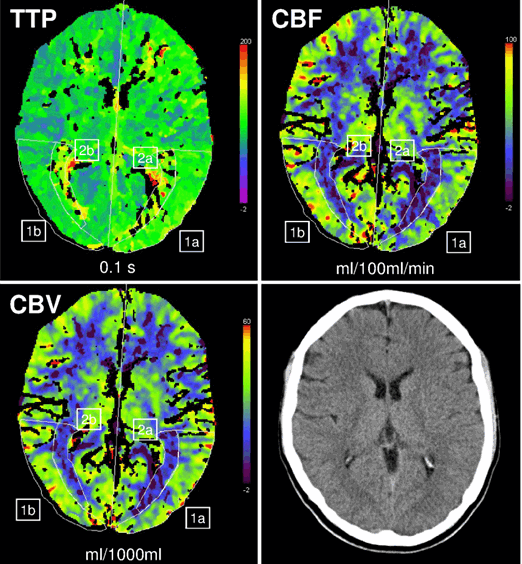
A 24-year-old, right-handed, male student who was living alone, was referred to our emergency ward at 00:30 because of acute speech problems and headache. Exact time of onset could not be determined. The emergency physician reported that the patient called a friend at about midnight indicating speech difficulties. His friend then called an ambulance. He was able to open the door to EMS and walk. Clinical examination demonstrated severe aphasia. In the hospital the patient was unable to speak, read or write, could follow simple commands, was agitated and indicated by gesture that he was having severe headache, photophobia and phonophobia. Suddenly, he started vomiting. No other focal neurological deficits were found. Differential diagnoses included intracranial hemorrhage or ischemic infarction, cerebral venous thrombosis and migraine with aura. Multimodal computed tomography scan was undertaken according to our hyperacute stroke imaging algorithm. Non-contrast computed tomography showed no early signs of cerebral ischemia and ruled out intracranial hemorrhage. Computed tomography angiography (CTA) demonstrated no arterial occlusion or cerebral sinus thrombosis. However, perfusion-CT showed a modest hypoperfusion of the left hemisphere with an area of more severe hypoperfusion in the left Broca and occipitotemporal region which was not restricted to one vascular territory (increase of time to peak (TTP) up to 21%; decrease of cerebral blood flow (CBF) and cerebral blood volume (CBV) up to 38% and 30%, respectively in comparison to the corresponding contralateral area; (Figure 1). Therefore, ischemic infarction was deemed unlikely and consideration of thrombolysis was aborted. At this time the patient’s mother contacted the hospital by phone reporting that her son had called her at 21:30 complaining about blurred vision since 19:00 and a bilateral scotoma wandering from the outside to inside of the visual field for five minutes and paresthesia in his right arm and around the right angle of his mouth. Then a severe pulsating bifrontal headache occurred. At 22:00 during a second telephone call his mother noted he developed progressive problems in finding words. Suddenly, he started vomiting. His mother, father and sister were known to have migraine. He had not been diagnosed as having migraine, but during childhood and adolescence he had about six episodes with visual disorders or numbness in his right arm followed by headaches. However, there had never been vomiting, photophobia or phonophobia. Within 24 hours after analgesics and antiemetic medication aphasia and headache resolved completely. A follow-up magnetic resonance imaging (MRI) scan after four days showed no signs for infarction or perfusion abnormalities. |
|
|
|
Discussion
|
|
Our patient presented with evolving neurologic symptoms of sudden visual and sensory disturbances followed by headache, vomiting and global aphasia which lasted for less than 24 hours. The diagnosis of migraine with prolonged aura was made [2]. A transient ischemic attack was considered unlikely because of the following reasons:
However, in the hyperacute phase of admission the patient’s severe aphasia precluded communication of the first four of the above-mentioned five criteria. Perfusion changes during a migraine attack with aura are well known from single-photon emission computed tomography (SPECT) and positron emission tomography (PET) studies, [3] but there is also rising knowledge from acute imaging with computed tomography (CT) scan or MRI scan. Nieuwkamp et al. presented a first ever migraine where initial PCT showed modest hypoperfusion of the left hemisphere which the authors attributed to migraine aura. [4] Recently, Floery et al. showed that patients with migraine aura had hypoperfusion in more than one vascular territory but no diffusion-weighted Imaging (DWI) abnormality. [5] 133Xe-SPECT studies demonstrated ‘spreading oligemia’ from occipital to frontal which is likely to be a secondary phenomenon to cortical Spreading depression (CSD). [6] The patient´s aphasic aura is consistent with nonischemic hypoperfusion in the cortex supplied by the middle cerebral artery suggesting a correlation between location of reduced CBF and clinical symptoms. The PCT did not indicate infarction, as CBF and CBV values were above critical ischemic levels, [7] and CTA did not show any arterial stenosis as another possible cause of non-critical hypoperfusion. Therefore, we believe that this ictal hypoperfusion occurred secondary to CSD. Another important differential diagnosis for stroke mimics, postictal focal deficit (Todd’s paresis), which would have warranted electroencephalography (EEG) after imaging, was abandoned as the decisive clues became available by the mother’s information. The role of PCT in epileptogenic conditions is yet unclear, as both hypoperfusion and hyperperfusion have been demonstrated in case reports, possibly depending on whether the underlying pathophysiology was truly postictal or focal status epilepticus, respectively. [8] [9] |
|
Conclusion
|
|
Multimodal computed tomography scan can provide important information for differential diagnosis between migraine with aura and ischemic stroke. This can decisively influence hyperacute treatment, especially if taking medical history is hampered or even impossible. |
|
References
|
|

|
[HTML Abstract]
[PDF Full Text]
|
|
Author Contributions
Anselm Angermaier – Conception and design, Acquisition of data, Analysis and interpretation of data, Drafting the article, Critical revision of the article, Final approval of the version to be published SoenkeLangner – Analysis and interpretation of data, Critical revision of the article, Final approval of the version to be published Michael Kirsch – Acquisition of data, Analysis and interpretation of data, Critical revision of the article, Final approval of the version to be published Alexander V Khaw – Conception and design, Acquisition of data, Analysis and interpretation of data, Drafting the article, Critical revision of the article, Final approval of the version to be published |
|
Guarantor of submission
The corresponding author is the guarantor of submission. |
|
Source of support
None |
|
Conflict of interest
Authors declare no conflict of interest. |
|
Copyright
© Anselm Angermaier et al. 2014; This article is distributed the terms of Creative Commons Attribution License which permits unrestricted use, distribution and reproduction in any means provided the original authors and original publisher are properly credited. (Please see Copyright Policy for more information.) |
|
|
|
About The Authors
| |||
| |||
| |||