| |
|
|
|
Case Report
| ||||||
| Pentalogy of Cantrell diagnosed in the first trimester of pregnancy | ||||||
| Yasemin Cekmez1, Tülay Tos1, Zehra Yilmaz1, Nilay Pişkinpaşa1, Tuncay Küçüközkan2 | ||||||
|
1MD, Department of, Obstrectic and Gynecology, Sami Ulus Medical and Research Hospital, Ankara, Turkey.
2Professor, Department of, Obstrectic and Gynecology, Sami Ulus Medical and Research Hospital, Ankara,Turkey. | ||||||
| ||||||
|
[HTML Abstract]
[PDF Full Text]
[Print This Article]
[Similar article in Pumed] [Similar article in Google Scholar] 
|
| How to cite this article: |
| Cekmez Y, Tos T, Yilmaz Z, Pişkinpaşa N, Küçüközkan T. Pentalogy of Cantrell diagnosed in the first trimester of pregnancy. International Journal of Case Reports and Images 2014;5(3):215–217. |
|
Abstract
|
|
Introduction:
Pentalogy of Cantrell is a rare syndrome of unknown etiology characterized by multiple structural congenital anomalies.
Case Report: We aimed to present an early diagnosed case of pentalogy of Cantrell accompanied by craniorachischisis. Conclusion: Pentalogy of Cantrell can be diagnosed with a careful ultrasonography in the first trimester. Potential side effects of pregnancy termination to the mother can be reduced by the early diagnose of the disease. | |
|
Keywords:
Pentalogy of Cantrell, Fetus, Ectopia cordis, Craniorachischisis
| |
|
Introduction
| ||||||
|
Pentalogy of Cantrell is a rare syndrome defined by Cantrell et al. in 1958. The syndrome is characterized by the front upper abdominal midline and sternum defects, the lack of diaphragmatic face of the pericardium and the front face of diaphragm, and a variety of cardiac anomalies with unknown etiology. [1] The incidence has been reported to be 1 per 5.5 million live births. [2] Prognosis is associated with the defects defined and severity of cardiac anomalies. We aimed to present a case of pentalogy of Cantrell accompanied by craniorachischisis. | ||||||
|
Case Report
| ||||||
|
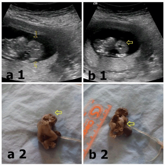
A 26-year-old (G4P1) woman was admitted to our hospital at her 12th week of gestation. In the routine ultrasound examination for first-trimester screening, crown rump length (CRL) was 40 mm in diameter, compatible with the single live fetus of 10 weeks 6 days. The fetal calvarial bone was deformed (exencephaly). (Figure 1a1) The view of herniated organs out of the abdominal wall defect and the defects in the lower end of the sternum was compatible with omphalocele and ectopia cordis. (Figure 1b1) Fetal spine was deformed and short. With these available ultrasonographic findings the diagnosis of Pentalogy of Cantrell was established and the pregnancy was terminated with the consent of mother and father. By the postmortem examination fetal omphalocele, (Figure 1b2) ectopia cordis and diaphragmatic hernia was detected. In addition to these findings, we observed an exencephaly continuing with spinal dysraphism as additional anomaly (craniorachischisis). (Figure 1a2) Fetal spine was deformed and short. | ||||||
| ||||||
|
Discussion
| ||||||
|
Pentalogy of Cantrell is a rare syndrome that causes defects involving diaphragm, abdominal wall, pericardium, heart and lower sternum. In a study consisting of 61 patients with Cantrell pentalogy, Toyama has proposed a classification for this syndrome. [3] According to this classification, patients including all five defects (definite diagnosis) were classified as type 1, patients including 4 defects 2 of which were intra-cardiac and ventral wall abnormalities (moderate diagnosis) were classified as type 2, patients including different combinations of incomplete abnormalities (always accompanied with sternal defects) were classified as type 3. Although the early gestational week of our patient prevented the detailed evaluation of intra-cardiac defects, the presence of ectopia cordis and omphalocele provided us to diagnose the Cantrell pentalogy in the first trimester. The pathogenesis of Cantrell pentalogy is uncertain. The syndrome is thought to arise from an embryological development defect occuring in a segment of lateral mesoderm, 14–18 days after the conception. [1] As the transverse septum of the diaphragm cannot develop, a problem occurs in the ventro-medial migration of upper abdomens mesodermal fold. For this reason, the defects detected in the midline, sternum and diaphragm, consists of the lack of mesoderm migration. Although the pentalogy of Cantrell appears sporadicly, rarely it may be in association with chromosomal abnormalities (trisomy 13, trisomy 18 and triploidy). [4] In addition, as it was seen in two boys from the same family in literature, a familial component of the syndrome can be considered. [5] In the differential diagnosis of Cantrell pentalogy, thoraco-abdominal ectopia cordis, amniotic band syndrome, limb body wall complex (LBWC), and body stalk anomaly should be considered. [6] [7] Craniofacial and central nervous system defects (cleft lip and palate, encephalocele, hydrocephalus, and craniorachischisis), limb defects (clubfoot, absence of the radius and tibia or hypodactyly), abdominal organ defects (gallbladder agenesis and polysplenia) may accompany to Cantrell pentalogy. Craniorachischisis is a related neural tube defect, in which anencephaly is accompanied by a contiguous open defect of the spine (spina bifida totalis). It is an extreme example of defective closure of the neural tube during early embryogenesis, around 20 to 22 days gestation. [8] In our case, the craniorachischisis was detected as an additional anomaly. The prognosis of Cantrell pentalogy is related to the severity of defects identified. Ectopia cordis is a standalone component of the penthalogy that has a change of living although the poor prognosis of the disease. [9] As other symptoms that accompany to Cantrell pentalogy is closely associated with the prognosis they should be screened carefully during the ultrasound scan. The disease was detected at 10 weeks 6 days according to crown-rump length in our patient and the pregnancy was terminated. | ||||||
|
Conclusion
| ||||||
|
In conclusion detection of this fetal disease is possible with a careful ultrasonographic examination in the first trimester and so the mortality and morbidity to the mother with late termination can be reduced. | ||||||
|
References
| ||||||
| ||||||
|
[HTML Abstract]
[PDF Full Text]
|
|
Author Contributions
Yasemin Cekmez – Substantial contributions to conception and design, Acquisition of data, Analysis and interpretation of data, Drafting the article or revising it critically for important intellectual content, Final of the version to be published Tülay Tos – Substantial contributions to conception and design, Acquisition of data, Analysis and interpretation of data, Drafting the article or revising it critically for important intellectual content, Final of the version to be published. Zehra Yilmaz – Substantial contributions to conception and design, Acquisition of data, Analysis and interpretation of data, Drafting the article or revising it critically for important intellectual content, Final of the version to be published Nilay Pişkinpaşa – Substantial contributions to conception and design, Acquisition of data, Analysis and interpretation of data, Drafting the article or revising it critically for important intellectual content, Final of the version to be published Tuncay Küçüközkan – Substantial contributions to conception and design, Acquisition of data, Analysis and interpretation of data, Drafting the article or revising it critically for important intellectual content, Final of the version to be published |
|
Guarantor of submission
The corresponding author is the guarantor of submission. |
|
Source of support
None |
|
Conflict of interest
Authors declare no conflict of interest. |
|
Copyright
© Yasemin Cekmez et al. 2014; This article is distributed the terms of Creative Commons Attribution License which permits unrestricted use, distribution and reproduction in any means provided the original authors and original publisher are properly credited. (Please see Copyright Policy for more information.) |
|
|